

The DOI matches its Crossref record; 27 of the 28 references it cites were found in a registry or an index.
Distinct 18F-AV-1451 tau PET retention patterns in early- and late-onset Alzheimer’s disease
Open accessAbstract
Patients with Alzheimer's disease can present with different clinical phenotypes. Individuals with late-onset Alzheimer's disease (>65 years) typically present with medial temporal lobe neurodegeneration and predominantly amnestic symptomatology, while patients with early-onset Alzheimer's disease (<65 years) exhibit greater neocortical involvement associated with a clinical presentation including dyspraxia, executive dysfunction, or visuospatial impairment. We recruited 20 patients with early-onset Alzheimer's disease, 21 with late-onset Alzheimer's disease, three with prodromal early-onset Alzheimer's disease and 13 with prodromal late-onset Alzheimer's disease, as well as 30 cognitively healthy elderly controls, that had undergone 18F-AV-1451 tau positron emission tomography and structural magnetic resonance imaging to explore whether early- and late-onset Alzheimer's disease exhibit differential regional tau pathology and atrophy patterns. Strong associations of lower age at symptom onset with higher 18F-AV-1451 uptake were observed in several neocortical regions, while higher age did not yield positive associations in neither patient group. Comparing patients with early-onset Alzheimer's disease with controls resulted in significantly higher 18F-AV-1451 retention throughout the neocortex, while comparing healthy controls with late-onset Alzheimer's disease patients yielded a distinct pattern of higher 18F-AV-1451 retention, predominantly confined to temporal lobe regions. When compared against each other, the early-onset Alzheimer's disease group exhibited greater uptake than the late-onset group in prefrontal and premotor, as well as in inferior parietal cortex. These preliminary findings indicate that age may constitute an important contributor to Alzheimer's disease heterogeneity highlighting the potential of tau positron emission tomography to capture phenotypic variation across patients with Alzheimer's disease.
Overview
The claims and statistics of this paper haven’t been checked yet.
Run the check — free, no account needed. It reads the open-access text and checks the sources it cites.
Integrity signals
Registry
Cites
Statistics
Figures
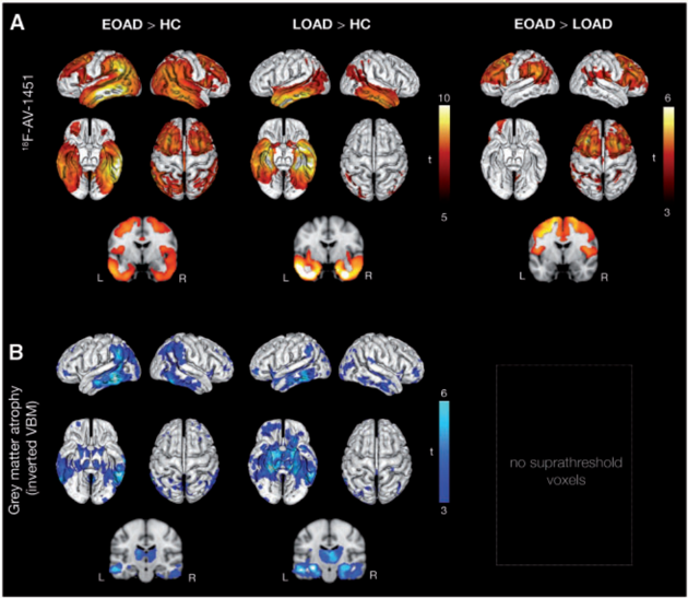
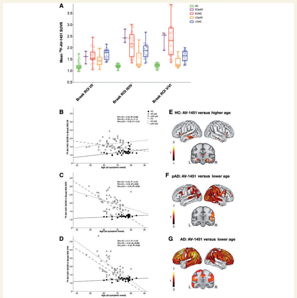
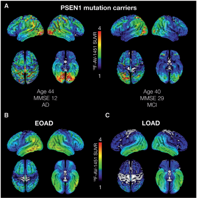
Verification
Claims the paper makes
The claims and statistics of this paper haven’t been checked yet.
Run the check — free, no account needed. It reads the open-access text and checks the sources it cites.
Claims other papers make about this one
Each is checked against this paper’s own text: does it say what the citing paper says it does?
In the citing paper
“Elevated tau PET binding in both medial temporal lobe structures and the neocortex is strongly associated with positive amyloid PET scans and with clinical impairment across the normal aging to dementia clinical spectrum [82,130–141].”
From NIA‐AA Research Framework: Toward a biological definition of Alzheimer's disease (2018) · cites this paper as [137]
In the citing paper
“Recent tau PET studies with [18F]flortaucipir showed that EOAD had greater tau burden in the neocortex compared to LOAD patients (Cho et al., 2017; Schöll et al., 2017).”
From Effects of Alzheimer’s and Vascular Pathologies on Structural Connectivity in Early- and Late-Onset Alzheimer’s Disease (2021) · cites this paper as Schöll et al., 2017
Self-check
Percentages, sums and reported differences re-computed from the paper’s own numbers.
The self-check needs the paper’s full text.
Citations
Cites
Folstein, M. F., Folstein, S. E., & McHugh, P. R. (1975) · Journal of Psychiatric Research · doi:10.1016/0022-3956(75)90026-6 · Cited by 88,282
McKhann, G., Drachman, D., Folstein, M., Katzman, R., Price, D., & Stadlan, E. M. (1984) · Neurology · doi:10.1212/wnl.34.7.939 · Cited by 27,915
Neuropathological stageing of Alzheimer-related changes.
Braak, H., & Braak, E. (1991) · Acta Neuropathologica · doi:10.1007/bf00308809 · Cited by 16,123
Voxel-Based Morphometry—The Methods
Ashburner, J., & Friston, K. J. (2000) · NeuroImage · doi:10.1006/nimg.2000.0582 · Cited by 8,676
Automatically Parcellating the Human Cerebral Cortex
Fischl, B. R. (2003) · Cerebral Cortex · doi:10.1093/cercor/bhg087 · Cited by 4,208
Cognitive reserve in ageing and Alzheimer's disease
Stern, Y. (2012) · The Lancet Neurology · doi:10.1016/s1474-4422(12)70191-6 · Cited by 3,557
Scheltens, P., Blennow, K., Breteler, M. M. B., de Strooper, B., Frisoni, G. B., Salloway, S., & Van der Flier, W. M. (2016) · The Lancet · doi:10.1016/s0140-6736(15)01124-1 · Cited by 3,125
A/T/N: An unbiased descriptive classification scheme for Alzheimer disease biomarkers
Jack, C. R., Bennett, D. A., Blennow, K., Carrillo, M. C., Feldman, H. H., Frisoni, G. B., Hampel, H., Jagust, W. J., Johnson, K. A., Knopman, D. S., et al. (2016) · Neurology · doi:10.1212/wnl.0000000000002923 · Cited by 1,864
Prevalence of Cerebral Amyloid Pathology in Persons Without Dementia: A Meta-analysis
Jansen, W. J., Ossenkoppele, R., Knol, D. L., Tijms, B. M., Scheltens, P., Verhey, F. R. J., Visser, P. J., Aalten, P., Aarsland, D., Alcolea, D., et al. (2015) · JAMA · doi:10.1001/jama.2015.4668 · Cited by 1,610
Self-citationPET Imaging of Tau Deposition in the Aging Human Brain
Schöll, M., Lockhart, S. N., Schonhaut, D. R., O’Neil, J. P., Janabi, M., Ossenkoppele, R., Baker, S. L., Vogel, J. W., Faria, J., Schwimmer, H. D., et al. (2016) · Neuron · doi:10.1016/j.neuron.2016.01.028 · Cited by 1,214
Self-citationTau PET patterns mirror clinical and neuroanatomical variability in Alzheimer’s disease
Ossenkoppele, R., Schonhaut, D. R., Schöll, M., Lockhart, S. N., Ayakta, N., Baker, S. L., O’Neil, J. P., Janabi, M., Lazaris, A., Cantwell, A., et al. (2016) · Brain · doi:10.1093/brain/aww027 · Cited by 1,185
Self-citationCorrection for partial volume effects in PET: principle and validation.
ROUSSET, O. G., Ma, Y., & Evans, A. C. (1998) · PubMed · Cited by 919
Early Clinical PET Imaging Results with the Novel PHF-Tau Radioligand [F18]-T808
Chien, D. T., Szardenings, A. K., Bahri, S., Walsh, J. C., Mu, F., Xia, C. A., Shankle, W. R., Lerner, A. J., Su, M., Elizarov, A. M., et al. (2013) · Journal of Alzheimer s Disease · doi:10.3233/jad-130098 · Cited by 683
Berg, L., McKeel, D. W., Miller, J. P., Storandt, M., Rubin, E. H., Morris, J. C., Baty, J., Coats, M., Norton, J., Goate, A. M., et al. (1998) · Archives of Neurology · doi:10.1001/archneur.55.3.326 · Cited by 657
Early clinical PET imaging results with the novel PHF-tau radioligand [F-18]-T807
Chien, D. T., Bahri, S., Szardenings, A. K., Walsh, J. C., Mu, F., Su, M.-Y., Shankle, W. R., Elizarov, A., & Kolb, H. C. (2013) · Journal of Alzheimer's Disease · doi:10.3233/jad-122059 · Cited by 651
The topography of grey matter involvement in early and late onset Alzheimer's disease
Frisoni, G. B., Pievani, M., Testa, C., Sabattoli, F., Bresciani, L. C., Bonetti, M., Beltramello, A., Hayashi, K. M., Toga, A. W., & Thompson, P. M. (2007) · Brain · doi:10.1093/brain/awl377 · Cited by 444
Palmqvist, S., Mattsson, N., Hansson, O., & Initiative, F. T. A. D. N. (2016) · Brain · doi:10.1093/brain/aww015 · Cited by 400
Self-citationAFNI: What a long strange trip it's been
Cox, R. W. (2011) · NeuroImage · doi:10.1016/j.neuroimage.2011.08.056 · Cited by 344
Rabinovici, G. D., Furst, A. J., Alkalay, A., Racine, C. A., O’Neil, J. P., Janabi, M., Baker, S. L., Agarwal, N., Bonasera, S. J., Mormino, E. C., et al. (2010) · Brain · doi:10.1093/brain/awp326 · Cited by 289
The significance of medial temporal lobe atrophy: A postmortem MRI study in the very old
Barkhof, F., Polvikoski, T. M., van Straaten, E. C. W., Kalaria, R. N., Sulkava, R., Aronen, H. J., Niinistö, L., Rastas, S., Oinas, M., Scheltens, P., et al. (2007) · Neurology · doi:10.1212/01.wnl.0000277459.83543.99 · Cited by 204
Koss, E., Edland, S., Fillenbaum, G., Mohs, R., Clark, C., Galasko, D., & Morris, J. C. (1996) · Neurology · doi:10.1212/wnl.46.1.136 · Cited by 150
Amyloid burden and metabolic function in early-onset Alzheimer’s disease: parietal lobe involvement.
Ossenkoppele, R., Zwan, M. D., Tolboom, N., van Assema, D. M. E., Adriaanse, S. F., Kloet, R. W., Boellaard, R., Windhorst, A. D., Barkhof, F., Lammertsma, A. A., et al. (2012) · Brain · doi:10.1093/brain/aws113 · Cited by 123
Self-citationMild to moderate Alzheimer dementia with insufficient neuropathological changes
Serrano‐Pozo, A., Qian, J., Monsell, S. E., Blacker, D., Gómez‐Isla, T., Betensky, R. A., Growdon, J. H., Johnson, K. A., Frosch, M. P., Sperling, R. A., et al. (2014) · Annals of Neurology · doi:10.1002/ana.24125 · Cited by 120
Early-Onset Dementia Is Associated with Higher Mortality
Koedam, E., Pijnenburg, Y. A. L., Deeg, D. J. H., Baak, M. M. E., van der Vlies, A. E., Scheltens, P., & van der Flier, W. M. (2008) · Dementia and Geriatric Cognitive Disorders · doi:10.1159/000149585 · Cited by 106
Age at onset is associated with disease severity in Lewy body variant and Alzheimer's disease
Ho, G., Hansen, L., Alford, M., Foster, K., Salmon, D., Galasko, D., Thal, L., & Masliah, E. (2002) · NeuroReport · doi:10.1097/00001756-200210070-00028 · Cited by 69
Modeling Strategies for Quantification of In Vivo 18F-AV-1451 Binding in Patients with Tau Pathology
Hahn, A., Schain, M., Erlandsson, M., Sjölin, P., James, G. M., Strandberg, O. T., Hägerström, D., Lanzenberger, R. R., Jögi, J., Olsson, T. G., et al. (2016) · Journal of Nuclear Medicine · doi:10.2967/jnumed.116.174508 · Cited by 57
Self-citationSmith, R. A., Wibom, M., Olsson, T. G., Hägerström, D., Jögi, J., Rabinovici, G. D., & Hansson, O. (2016) · Journal of Alzheimer s Disease · doi:10.3233/jad-151004 · Cited by 30
Self-citation
Cited by
Sorted by citations
NIA‐AA Research Framework: Toward a biological definition of Alzheimer's disease
Jack, C. R., Bennett, D. A., Blennow, K., Carrillo, M. C., Dunn, B., Haeberlein, S. B., Holtzman, D. M., Jagust, W., Jessen, F., Karlawish, J., et al. (2018) · Alzheimer's & Dementia · doi:10.1016/j.jalz.2018.02.018 · Cited by 10,537
Johnson, E. C. B., Dammer, E. B., Duong, D. M., Ping, L., Zhou, M., Yin, L., Higginbotham, L. A., Guajardo, A., White, B., Troncoso, J. C., et al. (2020) · Nature Medicine · doi:10.1038/s41591-020-0815-6 · Cited by 1,001
Imaging the evolution and pathophysiology of Alzheimer disease
Jagust, W. (2018) · Nature Reviews Neuroscience · doi:10.1038/s41583-018-0067-3 · Cited by 604
Amyloid-β-independent regulators of tau pathology in Alzheimer disease
van der Kant, R., Goldstein, L. S. B., & Ossenkoppele, R. (2020) · Nature reviews. Neuroscience · doi:10.1038/s41583-019-0240-3 · Cited by 580
Self-citationOssenkoppele, R., Rabinovici, G. D., Smith, R., Cho, H., Schöll, M., Strandberg, O., Palmqvist, S., Mattsson, N., Janelidze, S., Santillo, A., et al. (2018) · JAMA · doi:10.1001/jama.2018.12917 · Cited by 472
Self-citation
Metadata
- Authors
- Schöll, M., Ossenkoppele, R., Strandberg, O., Palmqvist, S., study, T. S. B., Jögi, J., Ohlsson, T., Smith, R., & Hansson, O.
- Affiliations
- Lund University (SE)
- Amsterdam Neuroscience (NL)
- Skåne University Hospital (SE)
- Corresponding author
- Schöll, Michael
- Venue
- Brain
- Publisher
- Oxford University Press (OUP)
- Published
- 2017-07-16
- Volume
- 140
- Issue
- 9
- Pages
- 2286-2294
- ISSN
- 0006-8950 (ISSN-L)
- 1460-2156
- OpenAlex
- W2751819353
- Cited by
- 180
- References
- 28
- Citations by year
- 2026: 8 · 2025: 19 · 2024: 13 · 2023: 24 · 2022: 17 · 2021: 30 · 2020: 29 · 2019: 23 · 2018: 16 · 2017: 1
- FWCI
- 15.50
- Type
- journal article
- Language
- en
- Open access
- hybrid
- doi.org
- Licence
- Locations
- Brain (publishedVersion, cc-by-nc)
- PubMed (publishedVersion)
- Pure Amsterdam UMC (publishedVersion, other-oa)
- Amsterdam UMC (VU Amsterdam) - Institutional Repository (publishedVersion)
- UCL Discovery (University College London) (submittedVersion)
- Lund University Publications (Lund University) (submittedVersion)
- Data Archiving and Networked Services (DANS) (submittedVersion)
- Funders
- European Research Council
- Swedish Research Council
- Swedish Brain Foundation
- Topics
- Health Sciences › Medicine › Psychiatry and Mental health › Dementia and Cognitive Impairment Research
- Life Sciences › Neuroscience › Cellular and Molecular Neuroscience › Alzheimer's disease research and treatments
- Health Sciences › Medicine › Radiology, Nuclear Medicine and Imaging › Medical Imaging Techniques and Applications
- Keywords
- Alzheimer's disease, early-onset Alzheimer's disease, late-onset Alzheimer's disease, 18F-AV-1451, tau PET, age at symptom onset, prodromal Alzheimer's disease, temporal lobe atrophy, prefrontal cortex, inferior parietal cortex, disease heterogeneity, clinical phenotype
- MeSH terms
- Aged, Aging, Alzheimer Disease, Atrophy, Carbolines, Cerebral Cortex, Female, Humans, Magnetic Resonance Imaging, Male, Middle Aged, Case-Control Studies, tau Proteins, Age of Onset, Tauopathies, Positron-Emission Tomography, Neuroimaging, Prodromal Symptoms
About these checks
Registry, retraction, Cites and statistics results are computed, not judged, with one exception: a printed reference that no index lists is read once by a model before it counts as a citation error. Claims are extracted by a model and kept only when their quote appears verbatim in the text; each cited source is then checked, with a model’s help, for whether it says what the claim relies on. Those verdicts can be wrong — read the claim’s quote and the cited source before relying on one.
Statistics and arithmetic are recomputed, not judged.
Confidence bands are shown only for fields calibrated on our annotated test set. None is calibrated yet, so no band is shown.
What counts as a claim ›How we measure confidence ›How we check this ›
Are you an author, or do you see a problem with this page? Request a review
