

The DOI matches its Crossref record; 54 of the 54 references it cites were found in a registry or an index.
Longitudinal tau PET in ageing and Alzheimer’s disease
Open accessAbstract
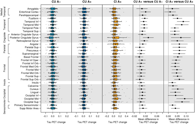
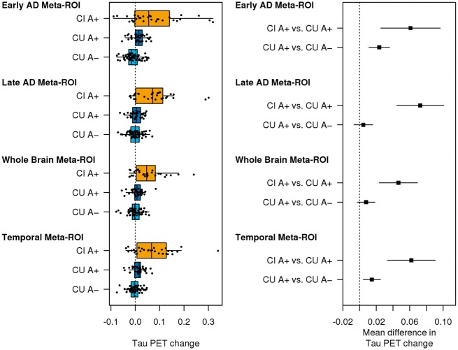
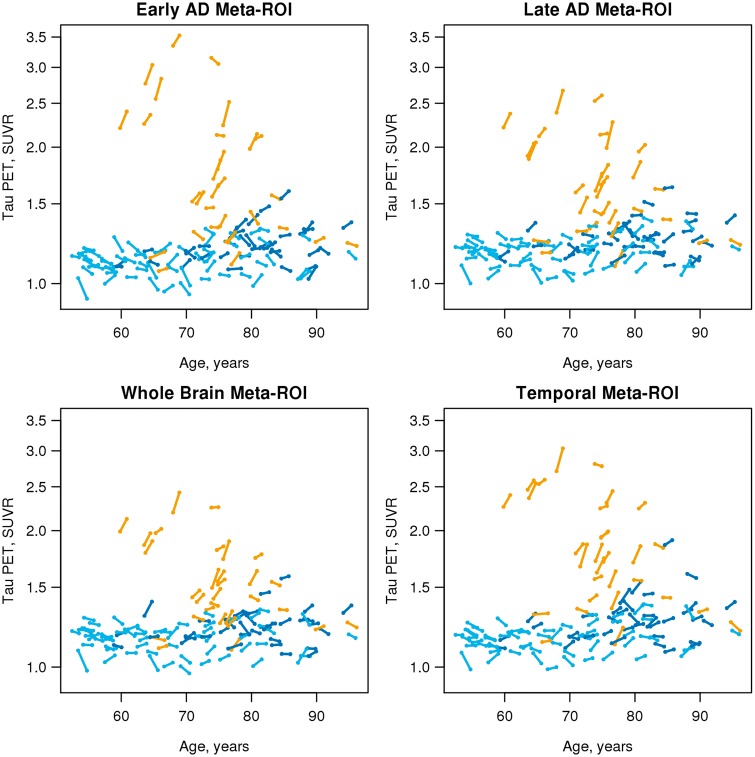
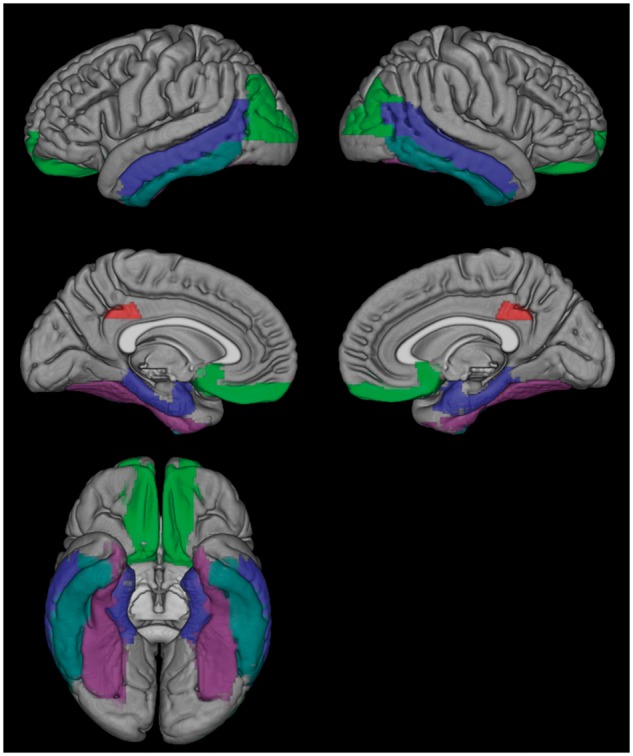
See Hansson and Mormino (doi:10.1093/brain/awy065) for a scientific commentary on this article.Our objective was to compare different whole-brain and region-specific measurements of within-person change on serial tau PET and evaluate its utility for clinical trials. We studied 126 individuals: 59 cognitively unimpaired with normal amyloid, 37 cognitively unimpaired with abnormal amyloid, and 30 cognitively impaired with an amnestic phenotype and abnormal amyloid. All had baseline amyloid PET and two tau PET, MRI, and clinical assessments. We compared the topography across all cortical regions of interest of tau PET accumulation rates and the rates of four different whole-brain or region-specific meta-regions of interest among the three clinical groups. We computed sample size estimates for change in tau PET, cortical volume, and memory/mental status indices for use as outcome measures in clinical trials. The cognitively unimpaired normal amyloid group had no observable tau accumulation throughout the brain. Tau accumulation rates in cognitively unimpaired abnormal amyloid were low [0.006 standardized uptake value ratio (SUVR), 0.5%, per year] but greater than rates in the cognitively unimpaired normal amyloid group in the basal and mid-temporal, retrosplenial, posterior cingulate, and entorhinal regions of interest. Thus, the earliest elevation in accumulation rates was widespread and not confined to the entorhinal cortex. Tau accumulation rates in the cognitively impaired abnormal amyloid group were 0.053 SUVR (3%) per year and greater than rates in cognitively unimpaired abnormal amyloid in all cortical areas except medial temporal. Rates of accumulation in the four meta-regions of interest differed but only slightly from one another. Among all tau PET meta-regions of interest, sample size estimates were smallest for a temporal lobe composite within cognitively unimpaired abnormal amyloid and for the late Alzheimer's disease meta-region of interest within cognitively impaired abnormal amyloid. The ordering of the sample size estimates by outcome measure was MRI < tau PET < cognitive measures. At a group-wise level, observable rates of short-term serial tau accumulation were only seen in the presence of abnormal amyloid. As disease progressed to clinically symptomatic stages (cognitively impaired abnormal amyloid), observable rates of tau accumulation were seen uniformly throughout the brain providing evidence that tau does not accumulate in one area at a time or in start-stop, stepwise sequence. The information captured by rate measures in different meta-regions of interest, even those with little topographic overlap, was similar. The implication is that rate measurements from simple meta-regions of interest, without the need for Braak-like staging, may be sufficient to capture progressive within-person accumulation of pathologic tau. Tau PET SUVR measures should be an efficient outcome measure in disease-modifying clinical trials.
Overview
The claims and statistics of this paper haven’t been checked yet.
Run the check — free, no account needed. It reads the open-access text and checks the sources it cites.
Integrity signals
Registry
Cites
Statistics
Figures
Verification
Claims the paper makes
The claims and statistics of this paper haven’t been checked yet.
Run the check — free, no account needed. It reads the open-access text and checks the sources it cites.
Claims other papers make about this one
Each is checked against this paper’s own text: does it say what the citing paper says it does?
The source backs part of the claim, not all of it — or backs it for a different population, condition or comparison.
In the citing paper
“Longitudinal deposition in tau PET is associated with baseline levels tau and Aβ deposition is a necessary antecedent for spread of tau beyond the temporal lobe in AD [189, 190] (Fig. 3).”
From The use of neuroimaging techniques in the early and differential diagnosis of dementia (2023) · cites this paper as [189]
Experimental — accuracy not yet measuredThe source backs part of the claim, not all of it — or backs it for a different population, condition or comparison.
In the citing paper
“Brain imaging has helped to improve the biological classification of AD by allowing proposed classifications based on pathology, like the ATN staging, to be applied in vivo and opened a new window of therapeutic intervention at preclinical stages.”
From The use of neuroimaging techniques in the early and differential diagnosis of dementia (2023) · cites this paper as [189]
Experimental — accuracy not yet measuredThe cited source says what the claim attributes to it.
In the citing paper
“Longitudinal deposition in tau PET is associated with baseline levels tau and Aβ deposition is a necessary antecedent for spread of tau beyond the temporal lobe in AD [189, 190] (Fig. 3).”
From The use of neuroimaging techniques in the early and differential diagnosis of dementia (2023) · cites this paper as [189]
The check wasn’t confident either way, so it gives no verdict — never disputed.
In the citing paper
“Such imaging has also improved our understanding of the role of mixed pathologies in dementia [102, 175, 189, 247].”
From The use of neuroimaging techniques in the early and differential diagnosis of dementia (2023) · cites this paper as [189]
Experimental — accuracy not yet measuredIn the citing paper
“In contrast, cross-sectional and longitudinal human cohort studies of biomarkers support a scenario in which amyloid pathology (Aβ42 lowering in CSF, positive amyloid PET imaging) precedes tau pathology (tau-PET and phospho-Tau elevation in CSF) and then neurodegeneration 126-128 .”
From The amyloid hypothesis in Alzheimer disease: new insights from new therapeutics (2022) · cites this paper as [127]
In the citing paper
“Antemortem diagnosis also relies heavily on the presence of Aβ and tau, either as measured in the spinal fluid or imaged using positron emission tomography (PET) [7–10].”
From Systems-based proteomics to resolve the biology of Alzheimer’s disease beyond amyloid and tau (2021) · cites this paper as [7]
Self-check
Percentages, sums and reported differences re-computed from the paper’s own numbers.
The self-check needs the paper’s full text.
Citations
Cites
The Elements of Statistical Learning
Hastie, T., Tibshirani, R., & Friedman, J. (2009) · Springer Series in Statistics · doi:10.1007/978-0-387-84858-7 · Cited by 25,970
The Elements of Statistical Learning
Hastie, T., Friedman, J., & Tibshirani, R. (2001) · Springer Series in Statistics · doi:10.1007/978-0-387-21606-5 · Cited by 24,162
McKhann, G. M., Knopman, D. S., Chertkow, H., Hyman, B. T., Jack, C. R., Kawas, C. H., Klunk, W. E., Koroshetz, W. J., Manly, J. J., Mayeux, R., et al. (2011) · Alzheimer's & Dementia · doi:10.1016/j.jalz.2011.03.005 · Cited by 18,657
Self-citationNeuropathological stageing of Alzheimer-related changes.
Braak, H., & Braak, E. (1991) · Acta Neuropathologica · doi:10.1007/bf00308809 · Cited by 16,123 · cited 2×
“However, that fact that elevated rates in CU A + relative to CU A were not confined to the entorhinal cortex suggests that early tau accumulation may not be as spatially restricted as implied in Braak staging (Braak and Braak, 1991).”
BackgroundResultMild cognitive impairment as a diagnostic entity
Petersen, R. C. (2004) · Journal of Internal Medicine · doi:10.1111/j.1365-2796.2004.01388.x · Cited by 7,864
Self-citationImaging brain amyloid in Alzheimer's disease with Pittsburgh Compound‐B
Klunk, W. E., Engler, H., Nordberg, A., Wang, Y., Blomqvist, G., Holt, D. P., Bergström, M., Savitcheva, I., Huang, G., Estrada, S., et al. (2004) · Annals of Neurology · doi:10.1002/ana.20009 · Cited by 4,496
Jack, C. R., Knopman, D. S., Jagust, W. J., Petersen, R. C., Weiner, M. W., Aisen, P. S., Shaw, L. M., Vemuri, P., Wiste, H. J., Weigand, S. D., et al. (2013) · The Lancet Neurology · doi:10.1016/s1474-4422(12)70291-0 · Cited by 4,396
Self-citationThe antibody aducanumab reduces Aβ plaques in Alzheimer’s disease
Sevigny, J., Chiao, P., Bussière, T., Weinreb, P. H., Williams, L., Maier, M., Dunstan, R., Salloway, S., Chen, T., Ling, Y., et al. (2016) · Nature · doi:10.1038/nature19323 · Cited by 3,059
Frequency of Stages of Alzheimer-Related Lesions in Different Age Categories
Braak, H., & Braak, E. (1997) · Neurobiology of Aging · doi:10.1016/s0197-4580(97)00056-0 · Cited by 1,656
Propagation of Tau Pathology in a Model of Early Alzheimer's Disease
de Calignon, A., Polydoro, M., Suárez-Calvet, M., William, C., Adamowicz, D. H., Kopeikina, K. J., Pitstick, R., Sahara, N., Ashe, K. H., Carlson, G. A., et al. (2012) · Neuron · doi:10.1016/j.neuron.2011.11.033 · Cited by 1,427
Research works printed but not linked: 3
- Bejanin, Do neurodegeneration or amyloid pathology contribute to the relationship between AV-1451 and cognitive symptoms in Alzhimer's disease?, 252
- La Joie, Relationships between AV1451-PET and CSF biomarkers in a heterogeneous clinical sample, 258
- Lowe, 2018, Widespread brain Tau and its association with aging, braak stage, and Alzheimer’s dementia, Brain, 147, 271, 10.1093/brain/awx320 · DOI
Cited by
Sorted by citations
Items 201–300 of 442
Longitudinal Tau Positron Emission Tomography in Dementia with Lewy Bodies
Chen, Q., Przybelski, S. A., Senjem, M. L., Schwarz, C. G., Lesnick, T. G., Botha, H., Knopman, D. S., Graff‐Radford, J., Savica, R., Jones, D. T., et al. (2022) · Movement Disorders · doi:10.1002/mds.28973 · Cited by 23
Self-citationLongitudinal predictive modeling of tau progression along the structural connectome
Yang, F., Chowdhury, S. R., Jacobs, H. I. L., Sepulcre, J., Wedeen, V. J., Johnson, K. A., & Dutta, J. (2021) · NeuroImage · doi:10.1016/j.neuroimage.2021.118126 · Cited by 24
Hojjati, S. H., Feiz, F., Ozoria, S., Razlighi, Q. R., & Initiative, F. T. A. D. N. (2021) · Journal of Alzheimer s Disease · doi:10.3233/jad-210419 · Cited by 22
Two-Year Prognostic Utility of Plasma p217+tau across the Alzheimer's Continuum
Feizpour, A., Doré, V., Doecke, J. D., Saad, Z. S., Triana‐Baltzer, G., Slemmon, R., Maruff, P. T., Krishnadas, N., Bourgeat, P., Huang, K., et al. (2023) · The Journal of Prevention of Alzheimer s Disease · doi:10.14283/jpad.2023.83 · Cited by 21
Triana‐Baltzer, G., Kolen, K. V., Theunis, C., Moughadam, S., Slemmon, R., Mercken, M. H., Galpern, W. R., Sun, H. F., & Kolb, H. C. (2020) · Journal of Alzheimer s Disease · doi:10.3233/jad-200463 · Cited by 23
Distinct Factors Drive the Spatiotemporal Progression of Tau Pathology in Older Adults
Adams, J. N., Harrison, T. M., Maass, A., Baker, S. L., & Jagust, W. J. (2021) · Journal of Neuroscience · doi:10.1523/jneurosci.1601-21.2021 · Cited by 22
APOE ε4 influences medial temporal atrophy and tau deposition in atypical Alzheimer's disease
Singh, N. A., Tosakulwong, N., Graff‐Radford, J., Machulda, M. M., Pham, N. T. T., Sintini, I., Weigand, S. D., Schwarz, C. G., Senjem, M. L., Carrasquillo, M. M., et al. (2022) · Alzheimer s & Dementia · doi:10.1002/alz.12711 · Cited by 21
Self-citationPawlik, D., Leuzy, A., Strandberg, O. T., & Smith, R. A. (2020) · NeuroImage · doi:10.1016/j.neuroimage.2020.117193 · Cited by 23
“Exceptional brain aging” without Alzheimer’s disease: triggers, accelerators, and the net sum game
Vemuri, P. (2018) · Alzheimer s Research & Therapy · doi:10.1186/s13195-018-0373-z · Cited by 25
Self-citationMacedo, A. C., Therriault, J., Tissot, C., Aumont, É., Servaes, S., Rahmouni, N., Arias, J. F., Lussier, F. Z., Wang, Y.-T., Ng, K. P., et al. (2024) · Neurobiology of Aging · doi:10.1016/j.neurobiolaging.2024.09.009 · Cited by 23
Higuchi, M. (2019) · Advances in experimental medicine and biology · doi:10.1007/978-981-32-9358-8_18 · Cited by 20
Neshan, M., Malakouti, S. K., Kamalzadeh, L., Makvand, M., Campbell, A., & Ahangari, G. (2021) · Journal of Alzheimer s Disease · doi:10.3233/jad-210480 · Cited by 19
Evaluating Plasma p-tau217 as an Endpoint for Alzheimer Disease Clinical Trials
Ferreira, P. C. L., Bellaver, B., Povala, G., Bauer‐Negrini, G., Aguzzoli, C. S., Ferrari‐Souza, J. P., Leffa, D. T., Soares, C., Lussier, F. Z., Medeiros, M. S., et al. (2025) · Neurology · doi:10.1212/wnl.0000000000214441 · Cited by 18
Xu, Y., Zheng, F., Zhong, Q., & Zhu, Y. (2023) · Journal of Alzheimer s Disease · doi:10.3233/jad-230002 · Cited by 20
Tau: a biomarker of Huntington’s disease
Lepinay, E., & Cicchetti, F. (2023) · Molecular Psychiatry · doi:10.1038/s41380-023-02230-9 · Cited by 19
An Open Question: Is the A2A Adenosine Receptor a Novel Target for Alzheimer’s Disease Treatment?
Merighi, S., Poloni, T. E., Pelloni, L., Pasquini, S., Varani, K., Vincenzi, F., Borea, P. A., & Gessi, S. (2021) · Frontiers in Pharmacology · doi:10.3389/fphar.2021.652455 · Cited by 18
Recent Advances in Imaging of Preclinical, Sporadic, and Autosomal Dominant Alzheimer's Disease
Buckley, R. F. (2021) · Neurotherapeutics · doi:10.1007/s13311-021-01026-5 · Cited by 18
Distinct and joint effects of low and high levels of Aβ and tau deposition on cortical thickness
Hojjati, S. H., Butler, T., Chiang, G. C.-Y., Habeck, C. G., RoyChoudhury, A., Feiz, F., Shteingart, J., Nayak, S., Ozoria, S., Guerrero, A. F., et al. (2023) · NeuroImage Clinical · doi:10.1016/j.nicl.2023.103409 · Cited by 17
Yoon, S. H., Kim, H., Lee, J., Chun, J., Sohn, Y. H., Lee, P. H., Ryu, Y. H., Cho, H., Yoo, H. S., & Lyoo, C. H. (2023) · Neurology · doi:10.1212/wnl.0000000000207917 · Cited by 18
Longitudinal synaptic loss versus tau Braak staging in amnestic mild cognitive impairment
Vanderlinden, G., Koole, M. J. B., Michiels, L., Lemmens, R., Vandenbulcke, M., & Laere, K. V. (2024) · Alzheimer s & Dementia · doi:10.1002/alz.14412 · Cited by 18
Shojaie, M., Tabarestani, S., Cabrerizo, M., DeKosky, S. T., Vaillancourt, D. E., Loewenstein, D., Duara, R., & Adjouadi, M. (2021) · Journal of Alzheimer s Disease · doi:10.3233/jad-210064 · Cited by 17
Planche, V., Coupé, P., Helmer, C., Goff, M. L., Amieva, H., Tison, F., Dartigues, J., & Catheline, G. (2019) · Neurobiology of Aging · doi:10.1016/j.neurobiolaging.2019.03.006 · Cited by 17
Moscoso, A., Grothe, M. J., Schöll, M., & Initiative, A. D. N. (2021) · European Journal of Nuclear Medicine and Molecular Imaging · doi:10.1007/s00259-021-05195-5 · Cited by 16
Doering, S., McCullough, A. A., Gordon, B. A., Chen, C. D., McKay, N. S., Hobbs, D. A., Keefe, S., Flores, S. E., Scott, J., Smith, H. D., et al. (2024) · EBioMedicine · doi:10.1016/j.ebiom.2024.105080 · Cited by 19
Pursuit of precision medicine: Systems biology approaches in Alzheimer's disease mouse models
Gurdon, B., & Kaczorowski, C. C. (2021) · Neurobiology of Disease · doi:10.1016/j.nbd.2021.105558 · Cited by 16
Deep learning improves utility of tau PET in the study of Alzheimer's disease
Zou, J., Park, D., Johnson, A., Feng, X., Pardo, M., France, J., Tomljanovic, Z., Brickman, A. M., Devanand, D. P., Luchsinger, J. A., et al. (2021) · Alzheimer's & Dementia: Diagnosis, Assessment & Disease Monitoring · doi:10.1002/dad2.12264 · Cited by 15
Harnessing cognitive trajectory clusterings to examine subclinical decline risk factors
Du, L., Hermann, B. P., Jonaitis, E. M., Cody, K. A., Rivera‐Rivera, L. A., Rowley, H. A., Field, A. S., Eisenmenger, L. B., Christian, B. T., Betthauser, T. J., et al. (2023) · Brain Communications · doi:10.1093/braincomms/fcad333 · Cited by 15
The AROMHA brain health test is a remote olfactory assessment to screen for cognitive impairment
Jobin, B., Magdamo, C., Delphus, D., Runde, A., Reineke, S., Alejandro-Soto, A., Ergun, B., Mukhija, S., Albers, A. D., & Albers, M. W. (2025) · Scientific Reports · doi:10.1038/s41598-025-92826-8 · Cited by 15
Sadleir, K. R., Gómez, K., Edwards, A. E., Patel, A. J., Ley, M. L., Khatri, A. W., Guo, J., Mahesh, S., Watkins, E. A., Popović, J., et al. (2025) · Acta Neuropathologica · doi:10.1007/s00401-025-02888-1 · Cited by 18
Utility of plasma GFAP as a secondary endpoint for clinical trials in Alzheimer’s disease
Abbas, S., Ferreira, P. C. L., Bellaver, B., Povala, G., Rohden, F., Aguzzoli, C. S., Zalzale, H., Ferrari‐Souza, J. P., Leffa, D. T., Lussier, F. Z., et al. (2025) · The Journal of Prevention of Alzheimer s Disease · doi:10.1016/j.tjpad.2025.100205 · Cited by 15
Okafor, M. O., Nye, J. A., Shokouhi, M., Shaw, L. M. J., Goldstein, F. C., & Hajjar, I. M. (2020) · Journal of Alzheimer s Disease · doi:10.3233/jad-191330 · Cited by 16
Chen, Q., Abrigo, J., Liu, W., Han, E. Y., Yeung, D. K. W., Shi, L., Au, L. W. C., Deng, M., Chen, S., Leung, E. Y. L., et al. (2022) · Brain Sciences · doi:10.3390/brainsci12060722 · Cited by 15
Polygenic Effect on Tau Pathology Progression in Alzheimer's Disease
Rubinski, A., Frerich, S., Malik, R., Franzmeier, N., Ramı́rez, A., Dichgans, M., Ewers, M., & Initiative, A. D. N. (2022) · Annals of Neurology · doi:10.1002/ana.26588 · Cited by 15
Tau Tangles in Parkinson’s Disease: A 2-Year Follow-Up Flortaucipir PET Study
Hansen, A. K., Parbo, P., Ismail, R., Østergaard, K., Brooks, D. J., & Borghammer, P. (2019) · Journal of Parkinson s Disease · doi:10.3233/jpd-191774 · Cited by 17
Montandon, M., Herrmann, F. R., Garibotto, V., Rodriguez, C. K., Haller, S., & Giannakopoulos, P. (2019) · Neurobiology of Aging · doi:10.1016/j.neurobiolaging.2019.12.002 · Cited by 15
Therapeutic news in Alzheimer’s disease: Soon a disease-modifying therapy?
Villain, N. (2022) · Revue Neurologique · doi:10.1016/j.neurol.2022.02.456 · Cited by 14
A Lipid-Raft Theory of Alzheimer's Disease
Rappoport, A. (2024) · Annual Review of Biochemistry · doi:10.1146/annurev-biochem-052024-115455 · Cited by 15
Plasma GFAP for populational enrichment of clinical trials in preclinical Alzheimer's disease
Bellaver, B., Povala, G., Ferreira, P. C. L., Bauer‐Negrini, G., Lussier, F. Z., Leffa, D. T., Ferrari‐Souza, J. P., Rodrigues, M. S., Amaral, L., Oliveira, M. S., et al. (2025) · Alzheimer s & Dementia · doi:10.1002/alz.70209 · Cited by 15
High Fat Diet Aggravates AD-Related Pathogenic Processes in APP/PS1 Mice
Gong, X., Liang, Z., Liu, W. D., Zhao, Y., Yang, Y., Wu, M., Shang, J., Xiao, Y., Mei, Y., Su, Q., et al. (2021) · Current Alzheimer Research · doi:10.2174/1567205018666210628100812 · Cited by 14
Jiang, C., Wang, Q., Xie, S., Chen, Z., Fu, L., Peng, Q., Liang, Y., Guo, H., Guo, T., & Initiative, A. D. N. (2022) · Brain Communications · doi:10.1093/braincomms/fcac084 · Cited by 14
Verbal memory formation across PET-based Braak stages of tau accumulation in Alzheimer’s disease
Arias, J. F., Therriault, J., Thomas, E. M., Lussier, F. Z., Bezgin, G., Tissot, C., Servaes, S., Mathotaarachchi, S. S., Schoemaker, D., Stevenson, J., et al. (2023) · Brain Communications · doi:10.1093/braincomms/fcad146 · Cited by 17
Tau accumulation in autosomal dominant Alzheimer’s disease: a longitudinal [18F]flortaucipir study
O’Connor, A., Cash, D. M., Poole, T., Markiewicz, P. J., Fraser, M. R., Malone, I. B., Jiao, J., Weston, P. S. J., Flores, S. E., Hornbeck, R. C., et al. (2023) · Alzheimer s Research & Therapy · doi:10.1186/s13195-023-01234-5 · Cited by 15
AD molecular: Imaging tau aggregates with positron emissions tomography
Betthauser, T. J. (2019) · Progress in molecular biology and translational science · doi:10.1016/bs.pmbts.2019.07.007 · Cited by 14
Kourti, M., & Metaxas, A. (2024) · Neurobiology of Disease · doi:10.1016/j.nbd.2024.106427 · Cited by 17
Pragmatic algorithm for visual assessment of 4-Repeat tauopathies in [18F]PI-2620 PET Scans
Bauer, T., Brendel, M., Zaganjori, M., Bernhardt, A. M., Jäck, A., Stöcklein, S., Scheifele, M., Levin, J., van Eimeren, T., Drzezga, A. E., et al. (2025) · NeuroImage · doi:10.1016/j.neuroimage.2025.121001 · Cited by 13
Lagarde, J., Olivieri, P., Tonietto, M., Rodrigo, S., Gervais, P., Caillé, F., Moussion, M., Bottlaender, M., & Sarazin, M. (2023) · Alzheimer s Research & Therapy · doi:10.1186/s13195-023-01237-2 · Cited by 14
EFFECTIVENESS OF LEARNING RATE IN DEMENTIA SEVERITY PREDICTION USING VGG16
Torghabeh, F. A., Modaresnia, Y., & Khalilzadeh, M. M. (2023) · Biomedical Engineering Applications Basis and Communications · doi:10.4015/s1016237223500060 · Cited by 13
Supervised pathway analysis of blood gene expression profiles in Alzheimer's disease
Moradi, E., Marttinen, M., Häkkinen, T., Hiltunen, M. O., & Nykter, M. (2019) · Neurobiology of Aging · doi:10.1016/j.neurobiolaging.2019.07.004 · Cited by 13
Kim, M., Kim, S. J., Park, J. E., Yun, J., Shim, W. H., Oh, J. S., Oh, M., Roh, J. H., Seo, S. W., Oh, S. J., et al. (2021) · Scientific Reports · doi:10.1038/s41598-021-89797-x · Cited by 13
Josephs, K. A., Tosakulwong, N., Gatto, R. G., Weigand, S. D., Ali, F., Botha, H., Graff‐Radford, J., Machulda, M. M., Savica, R., Schwarz, C. G., et al. (2022) · Annals of Neurology · doi:10.1002/ana.26479 · Cited by 13
Self-citationCircadian rest‐activity rhythm and longitudinal brain changes underlying late‐life cognitive decline
Jeon, S. Y., Byun, M. S., Yi, D., Jung, G., Lee, J., Kim, Y. K., Sohn, C., Kang, K. M., Lee, Y. J., & Lee, D. Y. (2022) · Psychiatry and Clinical Neurosciences · doi:10.1111/pcn.13521 · Cited by 12
Association of Basal Forebrain Volume with Amyloid, Tau, and Cognition in Alzheimer’s Disease
Yoo, H. S., Kim, H., Lee, J., Chun, J., Lee, H. S., Grothe, M. J., Teipel, S. J., Cavedo, E., Vergallo, A., Hampel, H. J., et al. (2024) · Journal of Alzheimer s Disease · doi:10.3233/jad-230975 · Cited by 12
Carvalho, D. Z., Křemen, V., Mívalt, F., Louis, E. K. S., McCarter, S. J., Bukartyk, J., Przybelski, S. A., Kamykowski, M. G., Spychalla, A. J., Machulda, M. M., et al. (2024) · Brain Communications · doi:10.1093/braincomms/fcae354 · Cited by 12
Self-citationGiorgio, J., Jonson, C., Wang, Y., Yokoyama, J. S., Wang, J., Jagust, W. J., & Initiative, O. B. O. T. A. D. N. (2025) · Nature Communications · doi:10.1038/s41467-025-60370-8 · Cited by 14
Trudel, L., Therriault, J., Macedo, A. C., Servaes, S., Hosseini, S. A., Bezgin, G., Aumont, É., Rahmouni, N., Arias, J. F., Zheng, Y., et al. (2025) · Neurology · doi:10.1212/wnl.0000000000214111 · Cited by 13
Self-citationHeterogeneity of Tau Deposition and Microvascular Involvement in MCI and AD
Bryant, A. G., Manhard, M. K., Salat, D. H., Rosen, B. R., Hyman, B. T., Johnson, K. A., Huang, S. Y., Bennett, R. E., & Yen, Y. (2021) · Current Alzheimer Research · doi:10.2174/1567205018666211126113904 · Cited by 12
Image-level trajectory inference of tau pathology using variational autoencoder for Flortaucipir PET
Hong, J., Kang, S. K., Alberts, I., Lu, J., Sznitman, R., Lee, J. S., Rominger, A., Choi, H., Shi, K., & Initiative, T. A. D. N. (2022) · European Journal of Nuclear Medicine and Molecular Imaging · doi:10.1007/s00259-021-05662-z · Cited by 13
Protocol Report on the Transcranial Photobiomodulation for Alzheimer’s Disease (TRAP-AD) Study
Iosifescu, D. V., Song, X., Gersten, M. B., Adib, A., Cho, Y., Collins, K. A., Yates, K. F., Puerto, A. M. H., McEachern, K. M., Osorio, R. S., et al. (2023) · Healthcare · doi:10.3390/healthcare11142017 · Cited by 11
Partial volume correction in longitudinal tau PET studies: is it really needed?
Costoya‐Sánchez, A., Moscoso, A., Sobrino, T., Ruibal, Á., Grothe, M. J., Schöll, M., Silva‐Rodríguez, J., & Aguiar, P. (2024) · NeuroImage · doi:10.1016/j.neuroimage.2024.120537 · Cited by 11
Microglia either promote or restrain TRAIL-mediated excitotoxicity caused by Aβ1−42 oligomers
Zou, J., McNair, E. M., DeCastro, S., Lyons, S. P., Mordant, A. L., Herring, L. E., Vetreno, R. P., & Coleman, L. G. (2024) · Journal of Neuroinflammation · doi:10.1186/s12974-024-03208-2 · Cited by 13
Tonietto, M., Sotolongo‐Grau, Ó., Roé-Vellvé, N., Bullich, S., Tartari, J. P., Sanabria, Á., García‐Sánchez, A., Borroni, E., Galli, C., Pérez-Martínez, E., et al. (2024) · Alzheimer s Research & Therapy · doi:10.1186/s13195-024-01622-5 · Cited by 12
Imaging Synaptic Density in Aging and Alzheimer Disease with [ 18 F]SynVesT-1
Giorgio, J., Soleimani‐Meigooni, D. N., Janabi, M., Baker, S. L., Chen, X., Toueg, T. N., Weimer, R. M., Zinnhardt, B., Green, A., Rabinovici, G. D., et al. (2025) · Journal of Nuclear Medicine · doi:10.2967/jnumed.124.269005 · Cited by 12
Tau biomarkers for neurodegenerative diseases: Current state and perspectives
Vacchi, E., Ruíz‐Barrio, I., & Melli, G. (2025) · Parkinsonism & Related Disorders · doi:10.1016/j.parkreldis.2025.107772 · Cited by 11
Accuracy of plasma biomarkers to detect Alzheimer's disease proteinopathy prior to dementia
Cody, K. A., Du, L., Studer, R., Jonaitis, E. M., Asthana, S., Christian, B. T., Chin, N. A., Kirmess, K. M., Meyer, M. R., Yarasheski, K. E., et al. (2025) · Alzheimer s & Dementia · doi:10.1002/alz.14570 · Cited by 11
Depressive Symptoms Correlate With Tau Accumulation Rates in Amyloid Positive Adults
Talmasov, D., Johnson, A. S., Brown, P. J., Provenzano, F. A., Lao, P. J., Marder, K. S., & Miller, J. M. (2025) · American Journal of Geriatric Psychiatry · doi:10.1016/j.jagp.2025.04.207 · Cited by 11
Bonarota, S., Caruso, G., Domenico, C. D., Sperati, S., Tamigi, F. M., Giulietti, G., Giove, F., Caltagirone, C., & Serra, L. (2025) · NeuroImage · doi:10.1016/j.neuroimage.2025.121264 · Cited by 11
Mazer, N. A., Hofmann, C., Lott, D., Gieschke, R., Klein, G., Boess, F., Grimm, H. P., Kerchner, G. A., Baudler, M., Smith, J., et al. (2022) · Alzheimer s & Dementia · doi:10.1002/alz.12877 · Cited by 13
Sintini, I., Graff‐Radford, J., Schwarz, C. G., Machulda, M. M., Singh, N. A., Carlos, A. F., Senjem, M. L., Jack, C. R., Lowe, V. J., Josephs, K. A., et al. (2023) · Alzheimer s & Dementia · doi:10.1002/alz.13396 · Cited by 11
Self-citation[18F]PI-2620 Tau PET signal across the aging and Alzheimer’s disease clinical spectrum
Young, C. B., Vossler, H. A., Romero, A., Smith, V., Park, J., Trelle, A. N., Winer, J. R., Wilson, E. N., Zeineh, M., Sha, S. J., et al. (2024) · Imaging Neuroscience · doi:10.1162/imag_a_00329 · Cited by 10
Ziontz, J., Harrison, T. M., Fonseca, C. S., Giorgio, J., Han, F., Lee, J., Jagust, W. J., & Initiative, A. D. N. (2024) · Human Brain Mapping · doi:10.1002/hbm.70083 · Cited by 10
Premenopausal bilateral oophorectomy and Alzheimer's disease imaging biomarkers later in life
Kantarci, K., Kapoor, E., Geske, J. R., Castillo, A. M., Fields, J. A., Kara, F., Knyazhanskaya, E. E., Schwarz, C. G., Senjem, M. L., Bailey, K. R., et al. (2024) · Alzheimer s & Dementia · doi:10.1002/alz.14469 · Cited by 10
Self-citationLongitudinal tau aggregation, atrophy, and cognitive decline in Alzheimer's disease
Singleton, E. H., Mattsson, N., Binette, A. P., Stomrud, E., Strandberg, O. T., Palmqvist, S., Ossenkoppele, R., & Hansson, O. (2025) · Alzheimer s & Dementia · doi:10.1002/alz.70435 · Cited by 11
Fischer, L., Parker, D. M., Maboudian, S. A., Fonseca, C. S., Tato-Fernández, C., Annen, L., Arunachalam, P., Bacci, J. R., Barboure, M. C., Capelli, S., et al. (2025) · Alzheimer s Research & Therapy · doi:10.1186/s13195-025-01920-6 · Cited by 13
Macedo, A. C., Trudel, L., Therriault, J., Rahmouni, N., Aumont, É., Bezgin, G., Hosseini, S. A., Tissot, C., Chan, T., Servaes, S., et al. (2026) · Nature Aging · doi:10.1038/s43587-026-01143-w · Cited by 10
Bourgeat, P., Krishnadas, N., Doré, V., Mulligan, R. S., Tyrrell, R., Bozinovski, S., Huang, K., Fripp, J., Villemagne, V. L., & Rowe, C. C. (2023) · The Journal of Prevention of Alzheimer s Disease · doi:10.14283/jpad.2023.17 · Cited by 10
Schneider, T. R., Stöckli, L., Felbecker, A., & Nirmalraj, P. N. (2024) · Brain Communications · doi:10.1093/braincomms/fcae180 · Cited by 10
Flortaucipir (tau) PET in LGI1 antibody encephalitis
Day, G. S., Gordon, B. A., McCullough, A. A., Bucelli, R. C., Perrin, R. J., Benzinger, T. L. S., & Ances, B. M. (2021) · Annals of Clinical and Translational Neurology · doi:10.1002/acn3.51297 · Cited by 10
Risacher, S. L. (2024) · CONTINUUM Lifelong Learning in Neurology · doi:10.1212/con.0000000000001509 · Cited by 11
Is longitudinal tau PET ready for use in Alzheimer’s disease clinical trials?
Hansson, O., & Mormino, E. C. (2018) · Brain · doi:10.1093/brain/awy065 · Cited by 10
Coevolution of Atomic Resolution and Whole-Brain Imaging for Tau Neurofibrillary Tangles
Dalton, R. M., Krishnan, H. S., Parker, V. S., Catanese, M. C., & Hooker, J. M. (2020) · ACS Chemical Neuroscience · doi:10.1021/acschemneuro.0c00426 · Cited by 10
Multiple markers contribute to risk of progression from normal to mild cognitive impairment
Rabin, J. S., Neal, T. E., Nierle, H. E., Sikkes, S. A. M., Buckley, R. F., Amariglio, R. E., Papp, K. V., Rentz, D. M., Schultz, A. P., Johnson, K. A., et al. (2020) · NeuroImage Clinical · doi:10.1016/j.nicl.2020.102400 · Cited by 12
Dan, T., Dere, M., Kim, W. H., Kim, M., & Guorong Wu, A. (2024) · Medical Image Analysis · doi:10.1016/j.media.2024.103210 · Cited by 9
Survey among experts on the future role of tau‐PET in clinical practice and trials
Vermeiren, M. R., Calandri, I. L., van der Flier, W. M., van de Giessen, E., & Ossenkoppele, R. (2024) · Alzheimer s & Dementia Diagnosis Assessment & Disease Monitoring · doi:10.1002/dad2.70033 · Cited by 10
Sheng, J., Trelle, A. N., Romero, A., Park, J., Tran, T., Sha, S. J., Andreasson, K. I., Wilson, E. N., Mormino, E. C., & Wagner, A. D. (2025) · Science Advances · doi:10.1126/sciadv.ads4206 · Cited by 9
Rajan, S., Brettschneider, J. A., & Collingwood, J. F. (2020) · Journal of Neuroscience Methods · doi:10.1016/j.jneumeth.2020.108870 · Cited by 9
Yang, S., Liu, H.-C., & Chen, W.-P. (2020) · Neurology and Therapy · doi:10.1007/s40120-020-00215-2 · Cited by 9
Visser, D., Verfaillie, S. C. J., Bosch, I., Brouwer, I., Tuncel, H., Coomans, E. M., Rikken, R. M., Mastenbroek, S. E., Golla, S. S. V., Barkhof, F., et al. (2023) · European Journal of Nuclear Medicine and Molecular Imaging · doi:10.1007/s00259-023-06196-2 · Cited by 9
Recent advances in nanoenzymes for Alzheimer's disease treatment
Bi, X., Cao, N., & He, J. (2024) · Colloids and Surfaces B Biointerfaces · doi:10.1016/j.colsurfb.2024.114139 · Cited by 10
Wang, Y., Cai, L., Zhou, K., Cui, M., & Yao, S. (2022) · Frontiers in Bioengineering and Biotechnology · doi:10.3389/fbioe.2021.812818 · Cited by 9
Clear‐headed into old age: Resilience and resistance against brain aging—A PET imaging perspective
Hoenig, M. C., & Drzezga, A. E. (2022) · Journal of Neurochemistry · doi:10.1111/jnc.15598 · Cited by 9
Visser, D., Tuncel, H., Ossenkoppele, R., Yaqub, M. M., Wolters, E. E., Timmers, T., Weltings, E., Coomans, E. M., den Hollander, M. E., van der Flier, W. M., et al. (2022) · Journal of Nuclear Medicine · doi:10.2967/jnumed.122.263926 · Cited by 9
Translational Scoring of Candidate Treatments for Alzheimer’s Disease: A Systematic Approach
Cummings, J. L. (2020) · Dementia and Geriatric Cognitive Disorders · doi:10.1159/000507569 · Cited by 9
Tau Imaging with 18 F-MK6240 across the Alzheimer’s Disease spectrumPreprint
Rowe, C. C., Doré, V., Krishnadas, N., Burnham, S. C., Lamb, F., Mulligan, R. S., Bozinovski, S., Laws, S. M., Tyrrell, R., Huang, K., et al. (2022) · medRxiv · doi:10.1101/2022.02.13.22270894 · Cited by 9
Gatto, R. G., Youssef, H., Reichard, R. R., Lowe, V. J., Whitwell, J. L., & Josephs, K. A. (2024) · Alzheimer s & Dementia · doi:10.1002/alz.14330 · Cited by 8
Self-citationButts, A. M., Machulda, M. M., Martin, P. R., Przybelski, S. A., Duffy, J. R., Graff‐Radford, J., Knopman, D. S., Petersen, R. C., Jack, C. R., Lowe, V. J., et al. (2022) · Journal of Alzheimer s Disease Reports · doi:10.3233/adr-220010 · Cited by 8
Self-citationTosun, D., Thropp, P., Southekal, S. S., Spottiswoode, B. S., Fahmi, R., & Initiative, A. D. N. (2023) · Alzheimer s & Dementia · doi:10.1002/alz.13164 · Cited by 9
Gatto, R. G., Pham, N. T. T., Duffy, J. R., Clark, H. M., Utianski, R. L., Botha, H., Machulda, M. M., Lowe, V. J., Schwarz, C. G., Jack, C. R., et al. (2024) · Human Brain Mapping · doi:10.1002/hbm.26704 · Cited by 10
Self-citationEligibility for donanemab trial in a population-based study of cognitive aging
Jones, K. E., Aakre, J. A., Castillo, A. M., Ramanan, V. K., Kremers, W. K., Jack, C. R., Vemuri, P., Schwarz, C. G., Lowe, V. J., Knopman, D. S., et al. (2025) · The Journal of Prevention of Alzheimer s Disease · doi:10.1016/j.tjpad.2025.100088 · Cited by 8
Self-citationLeveraging molecular biomarkers to make the common diagnosis in the uncommon patient
Day, G. S., Gordon, B. A., Bucelli, R. C., Perrin, R. J., Lopez-Chiriboga, A. S., & Ances, B. M. (2021) · Journal of Neuroimmunology · doi:10.1016/j.jneuroim.2021.577474 · Cited by 8
The Overlap Index as a means of evaluating early tau-PET signal reliability
Lee, J., Burkett, B. J., Min, H.-K. P., Lundt, E. S., Albertson, S. M., Botha, H., Senjem, M. L., Gunter, J. L., Schwarz, C. G., Jones, D. G., et al. (2022) · Journal of Nuclear Medicine · doi:10.2967/jnumed.121.263136 · Cited by 8
Self-citation
Metadata
- Authors
- Jack, C. R., Wiste, H. J., Schwarz, C. G., Lowe, V. J., Senjem, M. L., Vemuri, P., Weigand, S. D., Therneau, T. M., Knopman, D. S., Gunter, J. L., Jones, D. T., Graff-Radford, J., Kantarci, K., Roberts, R. O., Mielke, M. M., Machulda, M. M., & Petersen, R. C.
- Affiliations
- Mayo Clinic (US)
- Department of Neurology, Mayo Clinic, 200 First Street SW, Rochester, MN 55905, USA
- Corresponding author
- Jack, Clifford R
- ORCID
- Venue
- Brain
- Publisher
- Oxford University Press (OUP)
- Published
- 2018-03-12
- Volume
- 141
- Issue
- 5
- Pages
- 1517-1528
- ISSN
- 0006-8950 (ISSN-L)
- 1460-2156
- PMC
- PMC5917767
- OpenAlex
- W2791135917
- Cited by
- 452
- References
- 54
- Citations by year
- 2026: 36 · 2025: 56 · 2024: 61 · 2023: 49 · 2022: 55 · 2021: 75 · 2020: 56 · 2019: 46 · 2018: 18
- FWCI
- 37.75
- Type
- journal article
- Language
- en
- Open access
- hybrid
- doi.org
- Licence
- Locations
- Brain (publishedVersion, cc-by-nc)
- Europe PMC (PubMed Central) (publishedVersion, cc-by-nc)
- PubMed Central (publishedVersion, cc-by-nc)
- PubMed (publishedVersion)
- Funders
- NIH (R01 AG011378, R01 AG041851, U01 AG006786, R01 AG034676, R01 NS097495, P50 AG016574)
- Topics
- Health Sciences › Medicine › Psychiatry and Mental health › Dementia and Cognitive Impairment Research
- Life Sciences › Neuroscience › Cellular and Molecular Neuroscience › Alzheimer's disease research and treatments
- Health Sciences › Medicine › Radiology, Nuclear Medicine and Imaging › Medical Imaging Techniques and Applications
- Keywords
- tau PET, Alzheimer's disease, amyloid PET, within-person change, sample size determination, clinical trial outcome measures, amyloid-beta, entorhinal cortex, temporal lobe, cognitive impairment, cortical volume, memory decline, brain atrophy
- MeSH terms
- Aged, Aged, 80 and over, Aging, Alzheimer Disease, Brain, Female, Humans, Longitudinal Studies, Magnetic Resonance Imaging, Male, Middle Aged, Psychiatric Status Rating Scales, tau Proteins, Positron-Emission Tomography, Cognitive Dysfunction
About these checks
Registry, retraction, Cites and statistics results are computed, not judged, with one exception: a printed reference that no index lists is read once by a model before it counts as a citation error. Claims are extracted by a model and kept only when their quote appears verbatim in the text; each cited source is then checked, with a model’s help, for whether it says what the claim relies on. Those verdicts can be wrong — read the claim’s quote and the cited source before relying on one.
Statistics and arithmetic are recomputed, not judged.
Confidence bands are shown only for fields calibrated on our annotated test set. None is calibrated yet, so no band is shown.
What counts as a claim ›How we measure confidence ›How we check this ›
Are you an author, or do you see a problem with this page? Request a review
