

The DOI matches its Crossref record; 54 of the 54 references it cites were found in a registry or an index.
Longitudinal tau PET in ageing and Alzheimer’s disease
Open accessAbstract
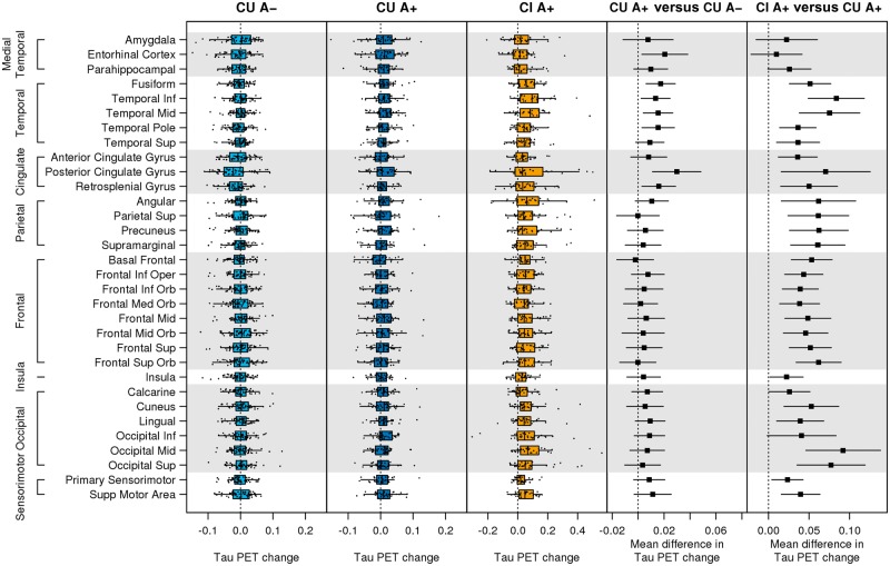
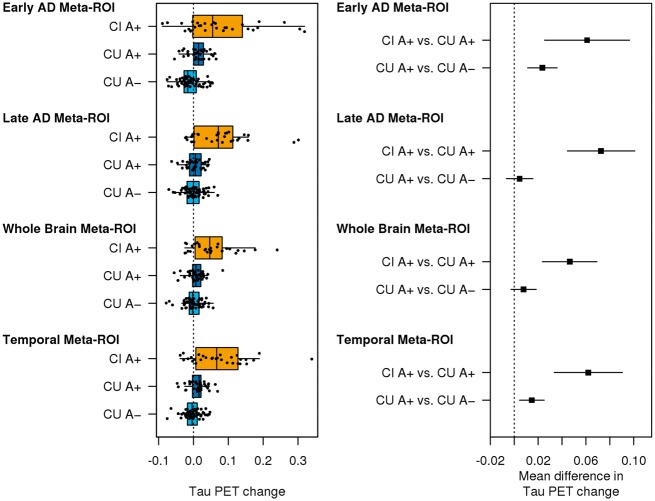
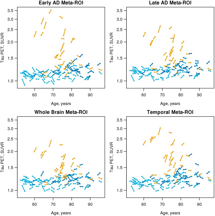
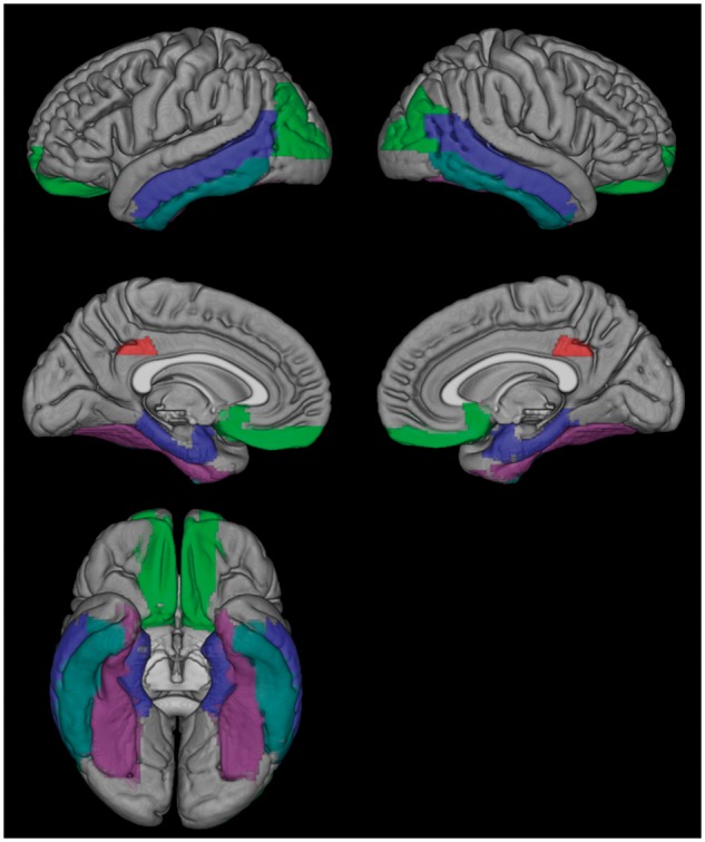
See Hansson and Mormino (doi:10.1093/brain/awy065) for a scientific commentary on this article.Our objective was to compare different whole-brain and region-specific measurements of within-person change on serial tau PET and evaluate its utility for clinical trials. We studied 126 individuals: 59 cognitively unimpaired with normal amyloid, 37 cognitively unimpaired with abnormal amyloid, and 30 cognitively impaired with an amnestic phenotype and abnormal amyloid. All had baseline amyloid PET and two tau PET, MRI, and clinical assessments. We compared the topography across all cortical regions of interest of tau PET accumulation rates and the rates of four different whole-brain or region-specific meta-regions of interest among the three clinical groups. We computed sample size estimates for change in tau PET, cortical volume, and memory/mental status indices for use as outcome measures in clinical trials. The cognitively unimpaired normal amyloid group had no observable tau accumulation throughout the brain. Tau accumulation rates in cognitively unimpaired abnormal amyloid were low [0.006 standardized uptake value ratio (SUVR), 0.5%, per year] but greater than rates in the cognitively unimpaired normal amyloid group in the basal and mid-temporal, retrosplenial, posterior cingulate, and entorhinal regions of interest. Thus, the earliest elevation in accumulation rates was widespread and not confined to the entorhinal cortex. Tau accumulation rates in the cognitively impaired abnormal amyloid group were 0.053 SUVR (3%) per year and greater than rates in cognitively unimpaired abnormal amyloid in all cortical areas except medial temporal. Rates of accumulation in the four meta-regions of interest differed but only slightly from one another. Among all tau PET meta-regions of interest, sample size estimates were smallest for a temporal lobe composite within cognitively unimpaired abnormal amyloid and for the late Alzheimer's disease meta-region of interest within cognitively impaired abnormal amyloid. The ordering of the sample size estimates by outcome measure was MRI < tau PET < cognitive measures. At a group-wise level, observable rates of short-term serial tau accumulation were only seen in the presence of abnormal amyloid. As disease progressed to clinically symptomatic stages (cognitively impaired abnormal amyloid), observable rates of tau accumulation were seen uniformly throughout the brain providing evidence that tau does not accumulate in one area at a time or in start-stop, stepwise sequence. The information captured by rate measures in different meta-regions of interest, even those with little topographic overlap, was similar. The implication is that rate measurements from simple meta-regions of interest, without the need for Braak-like staging, may be sufficient to capture progressive within-person accumulation of pathologic tau. Tau PET SUVR measures should be an efficient outcome measure in disease-modifying clinical trials.
Overview
The claims and statistics of this paper haven’t been checked yet.
Run the check — free, no account needed. It reads the open-access text and checks the sources it cites.
Integrity signals
Registry
Cites
Statistics
Figures
Verification
Claims the paper makes
The claims and statistics of this paper haven’t been checked yet.
Run the check — free, no account needed. It reads the open-access text and checks the sources it cites.
Claims other papers make about this one
Each is checked against this paper’s own text: does it say what the citing paper says it does?
The source backs part of the claim, not all of it — or backs it for a different population, condition or comparison.
In the citing paper
“Longitudinal deposition in tau PET is associated with baseline levels tau and Aβ deposition is a necessary antecedent for spread of tau beyond the temporal lobe in AD [189, 190] (Fig. 3).”
From The use of neuroimaging techniques in the early and differential diagnosis of dementia (2023) · cites this paper as [189]
Experimental — accuracy not yet measuredThe source backs part of the claim, not all of it — or backs it for a different population, condition or comparison.
In the citing paper
“Brain imaging has helped to improve the biological classification of AD by allowing proposed classifications based on pathology, like the ATN staging, to be applied in vivo and opened a new window of therapeutic intervention at preclinical stages.”
From The use of neuroimaging techniques in the early and differential diagnosis of dementia (2023) · cites this paper as [189]
Experimental — accuracy not yet measuredThe cited source says what the claim attributes to it.
In the citing paper
“Longitudinal deposition in tau PET is associated with baseline levels tau and Aβ deposition is a necessary antecedent for spread of tau beyond the temporal lobe in AD [189, 190] (Fig. 3).”
From The use of neuroimaging techniques in the early and differential diagnosis of dementia (2023) · cites this paper as [189]
The check wasn’t confident either way, so it gives no verdict — never disputed.
In the citing paper
“Such imaging has also improved our understanding of the role of mixed pathologies in dementia [102, 175, 189, 247].”
From The use of neuroimaging techniques in the early and differential diagnosis of dementia (2023) · cites this paper as [189]
Experimental — accuracy not yet measuredIn the citing paper
“In contrast, cross-sectional and longitudinal human cohort studies of biomarkers support a scenario in which amyloid pathology (Aβ42 lowering in CSF, positive amyloid PET imaging) precedes tau pathology (tau-PET and phospho-Tau elevation in CSF) and then neurodegeneration 126-128 .”
From The amyloid hypothesis in Alzheimer disease: new insights from new therapeutics (2022) · cites this paper as [127]
In the citing paper
“Antemortem diagnosis also relies heavily on the presence of Aβ and tau, either as measured in the spinal fluid or imaged using positron emission tomography (PET) [7–10].”
From Systems-based proteomics to resolve the biology of Alzheimer’s disease beyond amyloid and tau (2021) · cites this paper as [7]
Self-check
Percentages, sums and reported differences re-computed from the paper’s own numbers.
The self-check needs the paper’s full text.
Citations
Cites
The Elements of Statistical Learning
Hastie, T., Tibshirani, R., & Friedman, J. (2009) · Springer Series in Statistics · doi:10.1007/978-0-387-84858-7 · Cited by 25,970
The Elements of Statistical Learning
Hastie, T., Friedman, J., & Tibshirani, R. (2001) · Springer Series in Statistics · doi:10.1007/978-0-387-21606-5 · Cited by 24,162
McKhann, G. M., Knopman, D. S., Chertkow, H., Hyman, B. T., Jack, C. R., Kawas, C. H., Klunk, W. E., Koroshetz, W. J., Manly, J. J., Mayeux, R., et al. (2011) · Alzheimer's & Dementia · doi:10.1016/j.jalz.2011.03.005 · Cited by 18,657
Self-citationNeuropathological stageing of Alzheimer-related changes.
Braak, H., & Braak, E. (1991) · Acta Neuropathologica · doi:10.1007/bf00308809 · Cited by 16,123 · cited 2×
“However, that fact that elevated rates in CU A + relative to CU A were not confined to the entorhinal cortex suggests that early tau accumulation may not be as spatially restricted as implied in Braak staging (Braak and Braak, 1991).”
BackgroundResultMild cognitive impairment as a diagnostic entity
Petersen, R. C. (2004) · Journal of Internal Medicine · doi:10.1111/j.1365-2796.2004.01388.x · Cited by 7,864
Self-citationImaging brain amyloid in Alzheimer's disease with Pittsburgh Compound‐B
Klunk, W. E., Engler, H., Nordberg, A., Wang, Y., Blomqvist, G., Holt, D. P., Bergström, M., Savitcheva, I., Huang, G., Estrada, S., et al. (2004) · Annals of Neurology · doi:10.1002/ana.20009 · Cited by 4,496
Jack, C. R., Knopman, D. S., Jagust, W. J., Petersen, R. C., Weiner, M. W., Aisen, P. S., Shaw, L. M., Vemuri, P., Wiste, H. J., Weigand, S. D., et al. (2013) · The Lancet Neurology · doi:10.1016/s1474-4422(12)70291-0 · Cited by 4,396
Self-citationThe antibody aducanumab reduces Aβ plaques in Alzheimer’s disease
Sevigny, J., Chiao, P., Bussière, T., Weinreb, P. H., Williams, L., Maier, M., Dunstan, R., Salloway, S., Chen, T., Ling, Y., et al. (2016) · Nature · doi:10.1038/nature19323 · Cited by 3,059
Frequency of Stages of Alzheimer-Related Lesions in Different Age Categories
Braak, H., & Braak, E. (1997) · Neurobiology of Aging · doi:10.1016/s0197-4580(97)00056-0 · Cited by 1,656
Propagation of Tau Pathology in a Model of Early Alzheimer's Disease
de Calignon, A., Polydoro, M., Suárez-Calvet, M., William, C., Adamowicz, D. H., Kopeikina, K. J., Pitstick, R., Sahara, N., Ashe, K. H., Carlson, G. A., et al. (2012) · Neuron · doi:10.1016/j.neuron.2011.11.033 · Cited by 1,427
Research works printed but not linked: 3
- Bejanin, Do neurodegeneration or amyloid pathology contribute to the relationship between AV-1451 and cognitive symptoms in Alzhimer's disease?, 252
- La Joie, Relationships between AV1451-PET and CSF biomarkers in a heterogeneous clinical sample, 258
- Lowe, 2018, Widespread brain Tau and its association with aging, braak stage, and Alzheimer’s dementia, Brain, 147, 271, 10.1093/brain/awx320 · DOI
Cited by
Sorted by citations
Scheltens, P., De Strooper, B., Kivipelto, M., Holstege, H., Chételat, G., Teunissen, C. E., Cummings, J., & van der Flier, W. M. (2021) · The Lancet · doi:10.1016/s0140-6736(20)32205-4 · Cited by 5,465
Alzheimer Disease: An Update on Pathobiology and Treatment Strategies
Long, J. M., & Holtzman, D. M. (2019) · Cell · doi:10.1016/j.cell.2019.09.001 · Cited by 3,204
Revised criteria for diagnosis and staging of Alzheimer's disease: Alzheimer's Association Workgroup
Jack, C. R., Andrews, J. S., Beach, T. G., Buracchio, T., Dunn, B., Graf, A., Hansson, O., Ho, C., Jagust, W., McDade, E., et al. (2024) · Alzheimer's & Dementia · doi:10.1002/alz.13859 · Cited by 2,803
Self-citationDiagnosis and Management of Dementia: Review
Arvanitakis, Z., Shah, R. C., & Bennett, D. A. (2019) · JAMA · doi:10.1001/jama.2019.4782 · Cited by 1,561
Biomarkers for neurodegenerative diseases
Hansson, O. (2021) · Nature Medicine · doi:10.1038/s41591-021-01382-x · Cited by 1,159
Clinical diagnosis of Alzheimer’s disease: Recommendations of the International Working Group
Dubois, B., Villain, N., Frisoni, G. B., Rabinovici, G. D., Sabbagh, M., Cappa, S., Bejanin, A., Bombois, S., Epelbaum, S., Teichmann, M., et al. (2021) · The Lancet Neurology · doi:10.1016/s1474-4422(21)00066-1 · Cited by 1,051
“…studies showed no or only minimal acceleration of tau tracer uptake in the following 1 or 2 years in A(+) versus A(-) cognitive unimpaired elderly 101 *****. ___________________________________________________________________________ * Primary Age Related Tauopathy (PART) has emerged in the…”
BackgroundAssociation of Amyloid and Tau With Cognition in Preclinical Alzheimer Disease: A Longitudinal Study
Hanseeuw, B. J., Betensky, R. A., Jacobs, H. I. L., Schultz, A. P., Sepulcre, J., Becker, J. A., Cosio, D. M. O., Farrell, M., Quiroz, Y. T., Mormino, E. C., et al. (2019) · JAMA Neurology · doi:10.1001/jamaneurol.2019.1424 · Cited by 865
Advancing Research and Treatment for Frontotemporal Lobar Degeneration (ARTFL) investigators, Thijssen, E. H., La Joie, R., Wolf, A., Strom, A., Wang, P., Iaccarino, L., Bourakova, V., Cobigo, Y., Heuer, H., et al. (2020) · Nature Medicine · doi:10.1038/s41591-020-0762-2 · Cited by 761
The amyloid hypothesis in Alzheimer disease: new insights from new therapeutics
Karran, E., & De Strooper, B. (2022) · Nature Reviews Drug Discovery · doi:10.1038/s41573-022-00391-w · Cited by 742
Interaction between Aβ and Tau in the Pathogenesis of Alzheimer’s Disease
Zhang, H., Wei, W., Zhao, M., Ma, L., Jiang, X., Pei, H., Cao, Y., & Li, H. (2021) · International Journal of Biological Sciences · doi:10.7150/ijbs.57078 · Cited by 689
Tau PET imaging in neurodegenerative tauopathies—still a challenge
Leuzy, A., Chiotis, K., Lemoine, L., Gillberg, P.-G., Almkvist, O., Rodriguez-Vieitez, E., & Nordberg, A. (2019) · Molecular Psychiatry · doi:10.1038/s41380-018-0342-8 · Cited by 628
Imaging the evolution and pathophysiology of Alzheimer disease
Jagust, W. (2018) · Nature Reviews Neuroscience · doi:10.1038/s41583-018-0067-3 · Cited by 604
Amyloid-β-independent regulators of tau pathology in Alzheimer disease
van der Kant, R., Goldstein, L. S. B., & Ossenkoppele, R. (2020) · Nature reviews. Neuroscience · doi:10.1038/s41583-019-0240-3 · Cited by 580
Tau biomarkers in Alzheimer's disease: towards implementation in clinical practice and trials
Ossenkoppele, R., van der Kant, R., & Hansson, O. (2022) · The Lancet Neurology · doi:10.1016/s1474-4422(22)00168-5 · Cited by 557
The China Alzheimer Report 2022
Ren, R., Qi, J., Lin, S., Liu, X., Yin, P., Wang, Z., Tang, R., Wang, J., Huang, Q., Li, J., et al. (2022) · General Psychiatry · doi:10.1136/gpsych-2022-100751 · Cited by 357
Functional brain architecture is associated with the rate of tau accumulation in Alzheimer’s disease
Franzmeier, N., Neitzel, J., Rubinski, A., Smith, R. A., Strandberg, O. T., Ossenkoppele, R., Hansson, O., Ewers, M., Initiative, A. D. N., Weiner, M. W., et al. (2020) · Nature Communications · doi:10.1038/s41467-019-14159-1 · Cited by 358
Self-citationOssenkoppele, R., Smith, R., Mattsson-Carlgren, N., Groot, C., Leuzy, A., Strandberg, O., Palmqvist, S., Olsson, T., Jögi, J., Stormrud, E., et al. (2021) · JAMA Neurology · doi:10.1001/jamaneurol.2021.1858 · Cited by 311
Biomarker modeling of Alzheimer’s disease using PET-based Braak staging
Therriault, J., Pascoal, T. A., Lussier, F. Z., Tissot, C., Chamoun, M., Bezgin, G., Servaes, S., Benedet, A. L., Ashton, N. J., Karikari, T. K., et al. (2022) · Nature Aging · doi:10.1038/s43587-022-00204-0 · Cited by 308
van Oostveen, W. M., & de Lange, E. C. M. (2021) · International Journal of Molecular Sciences · doi:10.3390/ijms22042110 · Cited by 297
A walk through tau therapeutic strategies
Jadhav, S., Ávila, J., Schöll, M., Kovács, G. G., Kövari, E., Škrabana, R., Evans, L., Kontseková, E., Malawska, B., de Silva, R., et al. (2019) · Acta Neuropathologica Communications · doi:10.1186/s40478-019-0664-z · Cited by 299
Tagai, K., Ono, M., Kubota, M., Kitamura, S., Takahata, K., Seki, C., Takado, Y., Shinotoh, H., Sano, Y., Yamamoto, Y., et al. (2020) · Neuron · doi:10.1016/j.neuron.2020.09.042 · Cited by 276
Longitudinal tau accumulation and atrophy in aging and alzheimer disease
Harrison, T. M., Joie, R. L., Maaß, A., Baker, S. L., Swinnerton, K. N., Fenton, L. E., Mellinger, T. J., Edwards, L., Pham, J. Q., Miller, B. L., et al. (2018) · Annals of Neurology · doi:10.1002/ana.25406 · Cited by 262
Pontecorvo, M. J., Devous, M. D., Kennedy, I., Navitsky, M., Lu, M., Galante, N., Salloway, S., Doraiswamy, P. M., Southekal, S., Arora, A. K., et al. (2019) · Brain · doi:10.1093/brain/awz090 · Cited by 249
Schöll, M., Maaß, A., Mattsson, N., Ashton, N. J., Blennow, K., Zetterberg, H., & Jagust, W. J. (2018) · Molecular and Cellular Neuroscience · doi:10.1016/j.mcn.2018.12.001 · Cited by 248
Morris, G. P., Clark, I. A., & Vissel, B. (2018) · Acta Neuropathologica · doi:10.1007/s00401-018-1918-8 · Cited by 212
Meilandt, W. J., Ngu, H., Gogineni, A., Lalehzadeh, G., Lee, S.-H., Srinivasan, K., Imperio, J., Wu, T., Weber, M., Kruse, A. J., et al. (2020) · The Journal of Neuroscience · doi:10.1523/jneurosci.1871-19.2019 · Cited by 210
“…model that -amyloid pathology accelerates the accumulation of tau pathology or enhances its spreading (Pooler et al., 2015; He et al., 2018; Jack et al., 2018; Jacobs et al., 2018), and a new report indicates that this effect is further magnified in Trem2-deficient mice (Leyns et al.,…”
BackgroundBiomarker-based staging of Alzheimer disease: rationale and clinical applications
Therriault, J., Schindler, S. E., Salvadó, G., Pascoal, T. A., Benedet, A. L., Ashton, N. J., Karikari, T. K., Apostolova, L. G., Murray, M. E., Verberk, I. M. W., et al. (2024) · Nature Reviews Neurology · doi:10.1038/s41582-024-00942-2 · Cited by 225
Self-citationSafety and Efficacy of Semorinemab in Individuals With Prodromal to Mild Alzheimer Disease
Teng, E., Manser, P. T., Pickthorn, K., Brunstein, F., Blendstrup, M., Bohorquez, S. M. S., Wildsmith, K. R., Toth, B., Dolton, M. J., Ramakrishnan, V., et al. (2022) · JAMA Neurology · doi:10.1001/jamaneurol.2022.1375 · Cited by 212
Tau-positron emission tomography correlates with neuropathology findings.
Lowe, V. J., Lundt, E. S., Albertson, S. M., Min, H., Fang, P., Przybelski, S. A., Senjem, M. L., Schwarz, C. G., Kantarci, K., Boeve, B., et al. (2020) · Alzheimer's & Dementia · doi:10.1016/j.jalz.2019.09.079 · Cited by 203
Self-citationGonzales, M. M., Garbarino, V. R., Marques Zilli, E., Petersen, R. C., Kirkland, J. L., Tchkonia, T., Musi, N., Seshadri, S., Craft, S., & Orr, M. E. (2022) · The Journal of Prevention of Alzheimer's Disease · doi:10.14283/jpad.2021.62 · Cited by 203
Self-citationSanchez, J. S., Becker, J. A., Jacobs, H. I. L., Hanseeuw, B. J., Jiang, S., Schultz, A. P., Properzi, M. J., Katz, S. R., Beiser, A., Satizabal, C. L., et al. (2021) · Science Translational Medicine · doi:10.1126/scitranslmed.abc0655 · Cited by 200
Microglial activation and tau burden predict cognitive decline in Alzheimer’s disease
Malpetti, M., Kievit, R. A., Passamonti, L., Jones, P. S., Tsvetanov, K. A., Rittman, T., Mak, E., Nicastro, N., Bevan-Jones, W. R., Su, L., et al. (2020) · Brain · doi:10.1093/brain/awaa088 · Cited by 194
Mueller, A., Bullich, S., Barret, O., Madonia, J., Berndt, M., Papin, C., Perrotin, A., Koglin, N., Kroth, H., Pfeifer, A., et al. (2019) · Journal of Nuclear Medicine · doi:10.2967/jnumed.119.236224 · Cited by 195
The use of neuroimaging techniques in the early and differential diagnosis of dementia
Chouliaras, L., & O’Brien, J. T. (2023) · Molecular Psychiatry · doi:10.1038/s41380-023-02215-8 · Cited by 221
“Such imaging has also improved our understanding of the role of mixed pathologies in dementia [102, 175, 189, 247].”
BackgroundIn vivo quantification of neurofibrillary tangles with [18F]MK-6240
Pascoal, T. A., Shin, M., Kang, M. S., Chamoun, M., Chartrand, D. A., Mathotaarachchi, S. S., Bennacef, I., Therriault, J., Ng, K. P., Hopewell, R., et al. (2018) · Alzheimer s Research & Therapy · doi:10.1186/s13195-018-0402-y · Cited by 185
Gordon, B. A., Blazey, T. M., Christensen, J., Dincer, A., Flores, S., Keefe, S., Chen, C., Su, Y., McDade, E. M., Wang, G., et al. (2019) · Brain · doi:10.1093/brain/awz019 · Cited by 180
Self-citationCSF tau microtubule binding region identifies tau tangle and clinical stages of Alzheimer’s disease
Horie, K., Barthélemy, N. R., Sato, C., & Bateman, R. J. (2021) · Brain · doi:10.1093/brain/awaa373 · Cited by 180
Patient-centered connectivity-based prediction of tau pathology spread in Alzheimer’s disease
Franzmeier, N., Dewenter, A., Frontzkowski, L. K., Dichgans, M., Rubinski, A., Neitzel, J., Smith, R. A., Strandberg, O. T., Ossenkoppele, R., Büerger, K., et al. (2020) · Science Advances · doi:10.1126/sciadv.abd1327 · Cited by 178
Ossenkoppele, R., Lyoo, C. H., Jester-Broms, J., Sudre, C. H., Cho, H., Ryu, Y. H., Choi, J. Y., Smith, R., Strandberg, O., Palmqvist, S., et al. (2020) · JAMA Neurology · doi:10.1001/jamaneurol.2019.5154 · Cited by 161
Eligibility for Anti-Amyloid Treatment in a Population-Based Study of Cognitive Aging
Pittock, R. R., Aakre, J. A., Castillo, A. M., Ramanan, V. K., Kremers, W. K., Jack, C. R., Vemuri, P., Lowe, V. J., Knopman, D. S., Petersen, R. C., et al. (2023) · Neurology · doi:10.1212/wnl.0000000000207770 · Cited by 151
Self-citationRabinovici, G. D., Knopman, D. S., Arbizu, J., Benzinger, T. L. S., Donohoe, K. J., Hansson, O., Herscovitch, P., Kuo, P. H., Lingler, J. H., Minoshima, S., et al. (2025) · Alzheimer s & Dementia · doi:10.1002/alz.14338 · Cited by 163
Self-citationFerretti, M. T., Martínková, J., Biskup, E. M., Benke, T., Gialdini, G., Nedelská, Z., Rauen, K., Mantua, V., Religa, D., Hort, J., et al. (2020) · European Journal of Neurology · doi:10.1111/ene.14174 · Cited by 152
Recent update on the heterogeneity of the Alzheimer’s disease spectrum
Jellinger, K. A. (2021) · Journal of Neural Transmission · doi:10.1007/s00702-021-02449-2 · Cited by 155
Biomarker-Based Prediction of Longitudinal Tau Positron Emission Tomography in Alzheimer Disease
Leuzy, A., Smith, R., Cullen, N. C., Strandberg, O., Vogel, J. W., Binette, A. P., Borroni, E., Janelidze, S., Ohlsson, T., Jögi, J., et al. (2022) · JAMA Neurology · doi:10.1001/jamaneurol.2021.4654 · Cited by 149
Plasma markers predict changes in amyloid, tau, atrophy and cognition in non-demented subjects
Pereira, J. B., Janelidze, S., Stomrud, E., Palmqvist, S., van Westen, D., Dage, J. L., Mattsson, N., & Hansson, O. (2021) · Brain · doi:10.1093/brain/awab163 · Cited by 147
Sex Mediates Relationships Between Regional Tau Pathology and Cognitive Decline
Buckley, R. F., Scott, M. R., Jacobs, H. I. L., Schultz, A. P., Properzi, M. J., Amariglio, R. E., Hohman, T. J., Mayblyum, D. V., Rubinstein, Z. B., Manning, L. K., et al. (2020) · Annals of Neurology · doi:10.1002/ana.25878 · Cited by 137
The accumulation rate of tau aggregates is higher in females and younger amyloid-positive subjects
Smith, R. A., Strandberg, O. T., Mattsson, N., Leuzy, A., Palmqvist, S., Pontecorvo, M. J., Devous, M. D., Ossenkoppele, R., & Hansson, O. (2020) · Brain · doi:10.1093/brain/awaa327 · Cited by 141
(2022) · doi:10.1038/s41467-022-34129-4 · Cited by 128
Self-citationOssenkoppele, R., Reimand, J., Smith, R. A., Leuzy, A., Strandberg, O. T., Palmqvist, S., Stomrud, E., Zetterberg, H., Initiative, T. A. D. N., Scheltens, P., et al. (2021) · EMBO Molecular Medicine · doi:10.15252/emmm.202114398 · Cited by 130
Distinct tau PET patterns in atrophy‐defined subtypes of Alzheimer's disease
Ossenkoppele, R., Lyoo, C. H., Sudre, C. H., van Westen, D., Cho, H., Ryu, Y. H., Choi, J. Y., Smith, R. A., Strandberg, O. T., Palmqvist, S., et al. (2019) · Alzheimer s & Dementia · doi:10.1016/j.jalz.2019.08.201 · Cited by 119
Systems-based proteomics to resolve the biology of Alzheimer’s disease beyond amyloid and tau
Rayaprolu, S., Higginbotham, L., Bagchi, P., Watson, C. M., Zhang, T., Levey, A. I., Rangaraju, S., & Seyfried, N. T. (2021) · Neuropsychopharmacology · doi:10.1038/s41386-020-00840-3 · Cited by 118
Predicting future rates of tau accumulation on PET
Jack, C. R., Wiste, H. J., Weigand, S. D., Therneau, T. M., Lowe, V. J., Knopman, D. S., Botha, H., Graff-Radford, J., Jones, D. T., Ferman, T. J., et al. (2020) · Brain · doi:10.1093/brain/awaa248 · Cited by 117
Self-citationEdwards, A. L., Collins, J. A., Junge, C., Kordasiewicz, H., Mignon, L., Wu, S., Li, Y., Lin, L., DuBois, J., Hutchison, R. M., et al. (2023) · JAMA Neurology · doi:10.1001/jamaneurol.2023.3861 · Cited by 113
Tau Accumulation in Clinically Normal Older Adults Is Associated with Hippocampal Hyperactivity
Huijbers, W., Schultz, A. P., Papp, K. V., LaPoint, M. R., Hanseeuw, B. J., Chhatwal, J. P., Hedden, T., Johnson, K. A., & Sperling, R. A. (2018) · Journal of Neuroscience · doi:10.1523/jneurosci.1397-18.2018 · Cited by 118
Longitudinal 18F-MK-6240 tau tangles accumulation follows Braak stages
Pascoal, T. A., Benedet, A. L., Tudorascu, D., Therriault, J., Mathotaarachchi, S. S., Savard, M., Lussier, F. Z., Tissot, C., Chamoun, M., Kang, M. S., et al. (2021) · Brain · doi:10.1093/brain/awab248 · Cited by 122
Correlation of In Vivo [18F]Flortaucipir With Postmortem Alzheimer Disease Tau Pathology
Smith, R., Wibom, M., Pawlik, D., Englund, E., & Hansson, O. (2019) · JAMA Neurology · doi:10.1001/jamaneurol.2018.3692 · Cited by 111
Ayalon, G., Lee, S.-H., Adolfsson, O., Foo‐Atkins, C., Atwal, J. K., Blendstrup, M., Booler, H. S., Bravo, J., Brendza, R. P., Brunstein, F., et al. (2021) · Science Translational Medicine · doi:10.1126/scitranslmed.abb2639 · Cited by 112
Ossenkoppele, R., Iaccarino, L., Schonhaut, D. R., Brown, J. A., Joie, R. L., O’Neil, J. P., Janabi, M., Baker, S. L., Kramer, J. H., Gorno‐Tempini, M., et al. (2019) · NeuroImage Clinical · doi:10.1016/j.nicl.2019.101848 · Cited by 109
Entorhinal cortex tau, amyloid-β, cortical thickness and memory performance in non-demented subjects
Knopman, D. S., Lundt, E. S., Therneau, T. M., Vemuri, P., Lowe, V. J., Kantarci, K., Gunter, J. L., Senjem, M. L., Mielke, M. M., Machulda, M. M., et al. (2019) · Brain · doi:10.1093/brain/awz025 · Cited by 104
Self-citationGuo, T., Korman, D., Baker, S. L., Landau, S. M., & Jagust, W. J. (2020) · Biological Psychiatry · doi:10.1016/j.biopsych.2020.06.029 · Cited by 103
Direct Comparison of the Tau PET Tracers18F-Flortaucipir and18F-MK-6240 in Human Subjects
Gogola, A., Minhas, D. S., Villemagne, V. L., Cohen, A. D., Mountz, J. M., Pascoal, T. A., Laymon, C. M., Mason, N. S., Ikonomović, M. D., Mathis, C. A., et al. (2021) · Journal of Nuclear Medicine · doi:10.2967/jnumed.120.254961 · Cited by 104
Donanemab for Alzheimer’s Disease: A Systematic Review of Clinical Trials
Rashad, A., Rasool, A., Shaheryar, M., Sarfraz, A., Sarfraz, Z., Robles‐Velasco, K., & Chérrez-Ojeda, I. (2022) · Healthcare · doi:10.3390/healthcare11010032 · Cited by 102
Sequence of clinical and neurodegeneration events in Parkinson’s disease progression
Oxtoby, N. P., Leyland, L., Aksman, L., Thomas, G. E. C., Bunting, E. L., Wijeratne, P. A., Young, A. L., Zarkali, A., Tan, M. M. X., Bremner, F., et al. (2021) · Brain · doi:10.1093/brain/awaa461 · Cited by 102
Progressive Tau Accumulation in Alzheimer Disease: 2-Year Follow-up Study
Cho, H., Choi, J. Y., Lee, H. S., Lee, J. H., Ryu, Y. H., Lee, M. S., Jack, C. R., & Lyoo, C. H. (2019) · Journal of Nuclear Medicine · doi:10.2967/jnumed.118.221697 · Cited by 94
Self-citationPropagation of Tau Pathology: Integrating Insights From Postmortem and In Vivo Studies
Vogels, T., Leuzy, A., Cicognola, C., Ashton, N. J., Smolek, T., Novák, M., Blennow, K., Zetterberg, H., Hromádka, T., Žilka, N., et al. (2019) · Biological Psychiatry · doi:10.1016/j.biopsych.2019.09.019 · Cited by 99
Longitudinal neuroimaging biomarkers differ across Alzheimer’s disease phenotypes
Sintini, I., Graff‐Radford, J., Senjem, M. L., Schwarz, C. G., Machulda, M. M., Martin, P. R., Jones, D. T., Boeve, B. F., Knopman, D. S., Kantarci, K., et al. (2020) · Brain · doi:10.1093/brain/awaa155 · Cited by 99
Self-citationRelationship between amyloid and tau levels and its impact on tau spreading
Doré, V., Krishnadas, N., Bourgeat, P., Huang, K., Li, S., Burnham, S. C., Masters, C. L., Fripp, J., Villemagne, V. L., & Rowe, C. C. (2021) · European Journal of Nuclear Medicine and Molecular Imaging · doi:10.1007/s00259-021-05191-9 · Cited by 93
Guzmán‐Vélez, E., Diez, I. P., Schoemaker, D., Pardilla‐Delgado, E., Vila‐Castelar, C., Fox‐Fuller, J. T., Baena, A. Y., Sperling, R. A., Johnson, K. A., Lopera, F. J., et al. (2022) · Proceedings of the National Academy of Sciences · doi:10.1073/pnas.2113641119 · Cited by 95
Plasma p‐tau immunoassays in clinical research for Alzheimer's disease
Teunissen, C. E., Kolster, R., Triana‐Baltzer, G., Janelidze, S., Zetterberg, H., & Kolb, H. C. (2024) · Alzheimer s & Dementia · doi:10.1002/alz.14397 · Cited by 96
Frontzkowski, L. K., Ewers, M., Brendel, M., Biel, D., Ossenkoppele, R., Hager, P., Steward, A., Dewenter, A., Roemer, S. N., Rubinski, A., et al. (2022) · Nature Communications · doi:10.1038/s41467-022-32592-7 · Cited by 100
Head-to-head comparison of tau positron emission tomography tracers [18F]flortaucipir and [18F]RO948
Smith, R., Schöll, M., Leuzy, A., Jögi, J., Ohlsson, T., Strandberg, O., & Hansson, O. (2020) · European Journal of Nuclear Medicine and Molecular Imaging · doi:10.1007/s00259-019-04496-0 · Cited by 88
Regional tau pathology and loneliness in cognitively normal older adults
Uquillas, F. D., Jacobs, H. I. L., Biddle, K. D., Properzi, M. J., Hanseeuw, B. J., Schultz, A. P., Rentz, D. M., Johnson, K. A., Sperling, R. A., & Donovan, N. J. (2018) · Translational Psychiatry · doi:10.1038/s41398-018-0345-x · Cited by 86
Is tau in the absence of amyloid on the Alzheimer’s continuum?: A study of discordant PET positivity
Weigand, A. J., Bangen, K. J., Thomas, K. R., Delano‐Wood, L., Gilbert, P. E., Brickman, A. M., Bondi, M. W., & Initiative, A. D. N. (2019) · Brain Communications · doi:10.1093/braincomms/fcz046 · Cited by 93
Leuzy, A., Pascoal, T. A., Strandberg, O. T., Insel, P. S., Smith, R. A., Mattsson, N., Benedet, A. L., Cho, H., Lyoo, C. H., Joie, R. L., et al. (2021) · European Journal of Nuclear Medicine and Molecular Imaging · doi:10.1007/s00259-021-05401-4 · Cited by 89
Tau Imaging in Neurodegenerative Diseases Using Positron Emission Tomography
Wang, Y. T., & Edison, P. (2019) · Current Neurology and Neuroscience Reports · doi:10.1007/s11910-019-0962-7 · Cited by 86
Neuropathological assessment of the Alzheimer spectrum
Jellinger, K. A. (2020) · Journal of Neural Transmission · doi:10.1007/s00702-020-02232-9 · Cited by 85
Rabinovici, G. D., Knopman, D. S., Arbizu, J., Benzinger, T. L. S., Donohoe, K. J., Hansson, O., Herscovitch, P., Kuo, P. H., Lingler, J. H., Minoshima, S., et al. (2025) · Journal of Nuclear Medicine · doi:10.2967/jnumed.124.268756 · Cited by 87
Self-citationLongitudinal tau-PET uptake and atrophy in atypical Alzheimer's disease
Sintini, I., Martin, P. R., Graff‐Radford, J., Senjem, M. L., Schwarz, C. G., Machulda, M. M., Spychalla, A. J., Drubach, D. A., Knopman, D. S., Petersen, R. C., et al. (2019) · NeuroImage Clinical · doi:10.1016/j.nicl.2019.101823 · Cited by 87
Self-citationSyaifullah, A. H., Shiino, A., Kitahara, H., Ito, R., Ishida, M., & Tanigaki, K. (2021) · Frontiers in Neurology · doi:10.3389/fneur.2020.576029 · Cited by 81
Villemagne, V. L., Harada, R., Doré, V., Furumoto, S., Mulligan, R. S., Kudo, Y., Burnham, S. C., Krishnadas, N., Bozinovski, S., Huang, K., et al. (2022) · Journal of Nuclear Medicine · doi:10.2967/jnumed.121.263254 · Cited by 77
Comparing tau status determined via plasma pTau181, pTau231 and [18F]MK6240 tau-PET
Tissot, C., Therriault, J., Kunach, P., Benedet, A. L., Pascoal, T. A., Ashton, N. J., Karikari, T. K., Servaes, S., Lussier, F. Z., Chamoun, M., et al. (2022) · EBioMedicine · doi:10.1016/j.ebiom.2022.103837 · Cited by 78
Data-driven neuropathological staging and subtyping of TDP-43 proteinopathies
Young, A. L., Vogel, J. W., Robinson, J. L., McMillan, C. T., Ossenkoppele, R., Wolk, D. A., Irwin, D. J., Elman, L. B., Grossman, M., Lee, V. M. ‐., et al. (2023) · Brain · doi:10.1093/brain/awad145 · Cited by 84
Mastenbroek, S. E., Vogel, J. W., Collij, L. E., Serrano, G. E., Tremblay, C., Young, A. L., Arce, R. A., Shill, H. A., Driver‐Dunckley, E. D., Mehta, S. H., et al. (2024) · Nature Communications · doi:10.1038/s41467-024-49402-x · Cited by 82
Associations of longitudinal plasma p-tau181 and NfL with tau-PET, Aβ-PET and cognition
Rauchmann, B. S., Schneider-Axmann, T., & Perneczky, R. (2021) · Journal of Neurology, Neurosurgery & Psychiatry · doi:10.1136/jnnp-2020-325537 · Cited by 74
Liang, S., & Gu, Y. (2020) · Sensors · doi:10.3390/s21010220 · Cited by 73
Sintini, I., Graff‐Radford, J., Jones, D. T., Botha, H., Martin, P. R., Machulda, M. M., Schwarz, C. G., Senjem, M. L., Gunter, J. L., Jack, C. R., et al. (2020) · Cerebral Cortex · doi:10.1093/cercor/bhaa319 · Cited by 74
Self-citationCenTauR: Toward a universal scale and masks for standardizing tau imaging studies
Villemagne, V. L., Leuzy, A., Bohorquez, S. M. S., Bullich, S., Shimada, H., Rowe, C. C., Bourgeat, P., Lopresti, B. J., Huang, K., Krishnadas, N., et al. (2023) · Alzheimer s & Dementia Diagnosis Assessment & Disease Monitoring · doi:10.1002/dad2.12454 · Cited by 72
Tabarestani, S., Aghili, M., Eslami, M., Cabrerizo, M., Barreto, A. B., Rishe, N. D., Curiel, R. E., Loewenstein, D., Duara, R., & Adjouadi, M. (2019) · NeuroImage · doi:10.1016/j.neuroimage.2019.116317 · Cited by 72
Sex differences in longitudinal tau-PET in preclinical Alzheimer disease: a meta-analysis
Coughlan, G. T., Klinger, H. M., Boyle, R., Betthauser, T. J., Binette, A. P., Christenson, L., Chadwick, T., Hansson, O., Harrison, T. M., Healy, B., et al. (2025) · JAMA Neurology · doi:10.1001/jamaneurol.2025.0013 · Cited by 69
Self-citationCharacterizing brain tau and cognitive decline along the amyloid timeline in Alzheimer’s disease
Cody, K. A., Langhough, R. E., Zammit, M. D., Clark, L. R., Chin, N. A., Christian, B. T., Betthauser, T. J., & Johnson, S. C. (2024) · Brain · doi:10.1093/brain/awae116 · Cited by 71
Guo, T., Korman, D., Joie, R. L., Shaw, L. M. J., Trojanowski, J. Q., Jagust, W. J., Landau, S. M., & Initiative, A. D. N. (2020) · Alzheimer s Research & Therapy · doi:10.1186/s13195-020-00665-8 · Cited by 67
Tau pathology in cognitively normal older adults
Ziontz, J., Bilgel, M., Shafer, A. T., Moghekar, A., Elkins, W., Helphrey, J. H., Gómez, G., June, D., McDonald, M. A., Dannals, R. F., et al. (2019) · Alzheimer s & Dementia Diagnosis Assessment & Disease Monitoring · doi:10.1016/j.dadm.2019.07.007 · Cited by 67
Tau PET imaging with 18F-PI-2620 in aging and neurodegenerative diseases
Mormino, E. C., Toueg, T. N., Azevedo, E. C., Castillo, J. B., Guo, W., Nadiadwala, A., Corso, N. K., Hall, J. N., Fan, A. P., Trelle, A. N., et al. (2020) · European Journal of Nuclear Medicine and Molecular Imaging · doi:10.1007/s00259-020-04923-7 · Cited by 67
Molecular mechanisms of amyloid formation in living systems
Sinnige, T. (2022) · Chemical Science · doi:10.1039/d2sc01278b · Cited by 67
Alzheimer's disease neuroimaging
Whitwell, J. L. (2018) · Current Opinion in Neurology · doi:10.1097/wco.0000000000000570 · Cited by 67
Fichou, Y., Al‐Hilaly, Y. K., Devred, F., Smet‐Nocca, C., Tsvetkov, P. O., Verelst, J., Winderickx, J., Geukens, N., Vanmechelen, E., Perrotin, A., et al. (2019) · Acta Neuropathologica Communications · doi:10.1186/s40478-019-0682-x · Cited by 67
Tissot, C., Therriault, J., Pascoal, T. A., Chamoun, M., Lussier, F. Z., Savard, M., Mathotaarachchi, S. S., Benedet, A. L., Thomas, E. M., Parsons, M., et al. (2021) · Alzheimer s & Dementia Translational Research & Clinical Interventions · doi:10.1002/trc2.12154 · Cited by 63
Assessing Reactive Astrogliosis with 18 F-SMBT-1 Across the Alzheimer Disease Spectrum
Villemagne, V. L., Harada, R., Doré, V., Furumoto, S., Mulligan, R. S., Kudo, Y., Burnham, S. C., Krishnadas, N., Bourgeat, P., Xia, Y., et al. (2022) · Journal of Nuclear Medicine · doi:10.2967/jnumed.121.263255 · Cited by 63
A data-driven study of Alzheimer's disease related amyloid and tau pathology progression
Aksman, L., Oxtoby, N. P., Scelsi, M. A., Wijeratne, P. A., Young, A. L., Alves, I. L., Collij, L. E., Vogel, J. W., Barkhof, F., Alexander, D. C., et al. (2023) · Brain · doi:10.1093/brain/awad232 · Cited by 69
Widespread distribution of tauopathy in preclinical Alzheimer's disease
Schultz, S. A., Gordon, B. A., Mishra, S., Su, Y., Perrin, R. J., Cairns, N. J., Morris, J. C., Ances, B. M., & Benzinger, T. L. S. (2018) · Neurobiology of Aging · doi:10.1016/j.neurobiolaging.2018.08.022 · Cited by 65
Metadata
- Authors
- Jack, C. R., Wiste, H. J., Schwarz, C. G., Lowe, V. J., Senjem, M. L., Vemuri, P., Weigand, S. D., Therneau, T. M., Knopman, D. S., Gunter, J. L., Jones, D. T., Graff-Radford, J., Kantarci, K., Roberts, R. O., Mielke, M. M., Machulda, M. M., & Petersen, R. C.
- Affiliations
- Mayo Clinic (US)
- Department of Neurology, Mayo Clinic, 200 First Street SW, Rochester, MN 55905, USA
- Corresponding author
- Jack, Clifford R
- ORCID
- Venue
- Brain
- Publisher
- Oxford University Press (OUP)
- Published
- 2018-03-12
- Volume
- 141
- Issue
- 5
- Pages
- 1517-1528
- ISSN
- 0006-8950 (ISSN-L)
- 1460-2156
- PMC
- PMC5917767
- OpenAlex
- W2791135917
- Cited by
- 452
- References
- 54
- Citations by year
- 2026: 36 · 2025: 56 · 2024: 61 · 2023: 49 · 2022: 55 · 2021: 75 · 2020: 56 · 2019: 46 · 2018: 18
- FWCI
- 37.75
- Type
- journal article
- Language
- en
- Open access
- hybrid
- doi.org
- Licence
- Locations
- Brain (publishedVersion, cc-by-nc)
- Europe PMC (PubMed Central) (publishedVersion, cc-by-nc)
- PubMed Central (publishedVersion, cc-by-nc)
- PubMed (publishedVersion)
- Funders
- NIH (R01 AG011378, R01 AG041851, U01 AG006786, R01 AG034676, R01 NS097495, P50 AG016574)
- Topics
- Health Sciences › Medicine › Psychiatry and Mental health › Dementia and Cognitive Impairment Research
- Life Sciences › Neuroscience › Cellular and Molecular Neuroscience › Alzheimer's disease research and treatments
- Health Sciences › Medicine › Radiology, Nuclear Medicine and Imaging › Medical Imaging Techniques and Applications
- Keywords
- tau PET, Alzheimer's disease, amyloid PET, within-person change, sample size determination, clinical trial outcome measures, amyloid-beta, entorhinal cortex, temporal lobe, cognitive impairment, cortical volume, memory decline, brain atrophy
- MeSH terms
- Aged, Aged, 80 and over, Aging, Alzheimer Disease, Brain, Female, Humans, Longitudinal Studies, Magnetic Resonance Imaging, Male, Middle Aged, Psychiatric Status Rating Scales, tau Proteins, Positron-Emission Tomography, Cognitive Dysfunction
About these checks
Registry, retraction, Cites and statistics results are computed, not judged, with one exception: a printed reference that no index lists is read once by a model before it counts as a citation error. Claims are extracted by a model and kept only when their quote appears verbatim in the text; each cited source is then checked, with a model’s help, for whether it says what the claim relies on. Those verdicts can be wrong — read the claim’s quote and the cited source before relying on one.
Statistics and arithmetic are recomputed, not judged.
Confidence bands are shown only for fields calibrated on our annotated test set. None is calibrated yet, so no band is shown.
What counts as a claim ›How we measure confidence ›How we check this ›
Are you an author, or do you see a problem with this page? Request a review
