

Objective Morphological Classification of Neocortical Pyramidal Cells
Open accessPublished version of a preprint
The DOI matches its Crossref record; 88 of the 88 references it cites were found in a registry or an index.
Abstract
A consensus on the number of morphologically different types of pyramidal cells (PCs) in the neocortex has not yet been reached, despite over a century of anatomical studies, due to the lack of agreement on the subjective classifications of neuron types, which is based on expert analyses of neuronal morphologies. Even for neurons that are visually distinguishable, there is no common ground to consistently define morphological types. The objective classification of PCs can be achieved with methods from algebraic topology, and the dendritic arborization is sufficient for the reliable identification of distinct types of cortical PCs. Therefore, we objectively identify 17 types of PCs in the rat somatosensory cortex. In addition, we provide a solution to the challenging problem of whether 2 similar neurons belong to different types or to a continuum of the same type. Our topological classification does not require expert input, is stable, and helps settle the long-standing debate on whether cell-types are discrete or continuous morphological variations of each other.
Overview
The claims and statistics of this paper haven’t been checked yet.
Free: it reads the open-access text and checks the first 5 claims that cite a source we can check. Create a free account to check every claim.
Integrity signals
Registry
Cites
Statistics
Figures




Verification
Claims the paper makes
The claims and statistics of this paper haven’t been checked yet.
Free: it reads the open-access text and checks the first 5 claims that cite a source we can check. Create a free account to check every claim.
Claims other papers make about this one
Each is checked against this paper’s own text: does it say what the citing paper says it does?
The check wasn’t confident either way, so it gives no verdict — never disputed.
In the citing paper
“these cells are expected to have the Sst-Chodl t-type, linked to deep long-range projecting interneurons12,14,36,37 (Fig. 7h).”
From Classification of electrophysiological and morphological neuron types in the mouse visual cortex (2019) · cites this paper as [37]
Experimental — accuracy not yet measured
Self-check
Percentages, sums and reported differences re-computed from the paper’s own numbers.
The self-check needs the paper’s full text.
Citations
Cites
Topics in Optimal Transportation
Villani, C. (2003) · Graduate Studies in Mathematics · doi:10.1090/gsm/058 · Cited by 4,412 · cited 1×
“To evaluate TMD-classification performance, we computed the bottleneck distance (Edelsbrunner and Harer 2008), the Wasserstein distance (Villani 2003, Edelsbrunner and Harer 2010), the sliced-Wasserstein distance (Carrière et al. 2017), and a number of distances on the vectorized persistence…”
MethodRegulation of Synaptic Efficacy by Coincidence of Postsynaptic APs and EPSPs
Markram, H., Lübke, J., Frotscher, M., & Sakmann, B. (1997) · Science · doi:10.1126/science.275.5297.213 · Cited by 3,832 · cited 2×
“2003; Spruston 2008), and spike-timing dependent plasticity (Markram 1997; Sjöström et al. 2001; Froemke et al. 2005).”
BackgroundSelf-citationCarlsson, G. (2009) · Bulletin of the American Mathematical Society · doi:10.1090/s0273-0979-09-01249-x · Cited by 2,302 · cited 2×
“We have developed an alternative representation of morphologies based on persistent homology (Carlsson 2009) that provides a standardized quantification of neuronal branching structure.”
BackgroundMethodSelecting and interpreting measures of thematic classification accuracy
Stehman, S. V. (1997) · Remote Sensing of Environment · doi:10.1016/s0034-4257(97)00083-7 · Cited by 1,729
Computational topology an introduction
Edelsbrunner, H., & Harer, J. (2010) · Cited by 1,718
Pyramidal neurons: dendritic structure and synaptic integration
Spruston, N. (2008) · Nature Reviews Neuroscience · doi:10.1038/nrn2286 · Cited by 1,688 · cited 3×
“1995), integration of synaptic inputs from different cortical layers (Larkum et al. 1999, 2001; Schaefer et al. 2003; Spruston 2008), and spike-timing dependent plasticity (Markram 1997; Sjöström et al.”
BackgroundReconstruction and Simulation of Neocortical Microcircuitry
Markram, H., Muller, E., Ramaswamy, S., Reimann, M. W., Abdellah, M., Sanchez, C. A., Ailamaki, A., Alonso-Nanclares, L., Antille, N., Arsever, S., et al. (2015) · Cell · doi:10.1016/j.cell.2015.09.029 · Cited by 1,557 · cited 2×
“Therefore, it appears that the geometry of basal dendrites does not generalize across cortical layers, regions, and species (Ramaswamy and Markram 2015).”
BackgroundSelf-citationPetilla terminology: nomenclature of features of GABAergic interneurons of the cerebral cortex
The Petilla Interneuron Nomenclature Group (PING) (2008) · Nature Reviews Neuroscience · doi:10.1038/nrn2402 · Cited by 1,491
Active propagation of somatic action potentials into neocortical pyramidal cell dendrites
Stuart, G. J., & Sakmann, B. (1994) · Nature · doi:10.1038/367069a0 · Cited by 1,289 · cited 1×
“2007, van Elburg and van Ooyen 2010, Cuntz 2012, Ooyen and Elburg 2013, Bird and Cuntz 2016) and synaptic events, such as back-propagating action potentials (Stuart and Sakmann 1994), calcium transients in dendrites (Markram and Sakmann 1994; Schiller et al.”
BackgroundMarkram, H. (2006) · Nature Reviews Neuroscience · doi:10.1038/nrn1848 · Cited by 1,185
Self-citation
Cited by
Classification of electrophysiological and morphological neuron types in the mouse visual cortex
Gouwens, N. W., Sorensen, S. A., Berg, J., Lee, C., Jarsky, T., Ting, J., Sunkin, S. M., Feng, D., Anastassiou, C. A., Barkan, E., et al. (2019) · Nature Neuroscience · doi:10.1038/s41593-019-0417-0 · Cited by 456
Phenotypic variation of transcriptomic cell types in mouse motor cortex
Scala, F., Kobak, D., Bernabucci, M., Bernaerts, Y., Cadwell, C. R., Castro, J. R., Hartmanis, L., Jiang, X., Laturnus, S., Miranda, E., et al. (2021) · Nature · doi:10.1038/s41586-020-2907-3 · Cited by 398
“…near-projecting (NP) subclasses [4], Morphologically they have been classified Into big-tufted, small-tufted, untufted neurons, depending on the shape of the apical dendrite tuft, stellate neurons without an apical dendrite, and horizontal/inverted neurons in L6 [62, 63, 64, 65], Abundant in L2/3.”
Inhibitory specificity from a connectomic census of mouse visual cortex
Schneider-Mizell, C. M., Bodor, Á. L., Brittain, D., Buchanan, J., Bumbarger, D. J., Elabbady, L., Gamlin, C. R., Kapner, D., Kinn, S., Mahalingam, G., et al. (2025) · Nature · doi:10.1038/s41586-024-07780-8 · Cited by 165
Topological data analysis of task-based fMRI data from experiments on schizophrenia
Stolz, B. J., Emerson, T., Nahkuri, S., Porter, M. A., & Harrington, H. A. (2021) · Journal of Physics Complexity · doi:10.1088/2632-072x/abb4c6 · Cited by 83
Primate Neuronal Connections are Sparse as Compared to Mouse
Wildenberg, G. A., Rosen, M. C., Lundell, J., Paukner, D., Freedman, D. J., & Kasthuri, N. (2020) · Cell Reports · doi:10.1016/j.celrep.2021.109709 · Cited by 53
Perisomatic ultrastructure efficiently classifies cells in mouse cortex
Elabbady, L., Seshamani, S., Mu, S., Mahalingam, G., Schneider-Mizell, C. M., Bodor, Á. L., Bae, J. A., Brittain, D., Buchanan, J., Bumbarger, D. J., et al. (2025) · Nature · doi:10.1038/s41586-024-07765-7 · Cited by 45
“Existing methods for automated cell-typing based on morphology or connectivity often necessitate near-complete axonal or dendritic reconstructions 2,12–14 .”
Embryonic progenitor pools generate diversity in fine-scale excitatory cortical subnetworks
Ellender, T. J., Avery, S. V., Mahfooz, K., Scaber, J., von Klemperer, A., Nixon, S. L., Buchan, M. J., van Rheede, J. J., Gatti, A., Waites, C. K., et al. (2019) · Nature Communications · doi:10.1038/s41467-019-13206-1 · Cited by 41
Saunders, A. B., Huang, K. W., Vondrak, C., Hughes, C., Smolyar, K., Sen, H., Philson, A. C., Nemesh, J. C., Wysoker, A., Kashin, S., et al. (2022) · Nature Communications · doi:10.1038/s41467-022-34334-1 · Cited by 40
Romani, A., Antonietti, A., Bella, D., Budd, J., Giacalone, E., Kurban, K., Sáray, S., Abdellah, M., Arnaudon, A., Boci, E., et al. (2024) · PLOS Biology · doi:10.1371/journal.pbio.3002861 · Cited by 39
Self-citationWang, W. X., & Lefebvre, J. L. (2022) · Nature Communications · doi:10.1038/s41467-022-30977-2 · Cited by 39
Computational synthesis of cortical dendritic morphologies
Kanari, L., Dictus, H., Chalimourda, A., Arnaudon, A., Geit, W. A. H. V., Coste, B., Shillcock, J. C., Hess, K. P., & Markram, H. (2022) · Cell Reports · doi:10.1016/j.celrep.2022.110586 · Cited by 37
Self-citationA Systematic Evaluation of Interneuron Morphology Representations for Cell Type Discrimination
Laturnus, S., Kobak, D., & Berens, P. (2020) · Neuroinformatics · doi:10.1007/s12021-020-09461-z · Cited by 35
“To circumvent this problem we converted each persistence diagram into a 1D or 2D image and used Euclidean distance on the results as this procedure has been shown to work well in the neural domain (Kanari et al. 2019; Li et al. 2017).”
BackgroundMethodInfluentialAnatomically revealed morphological patterns of pyramidal neurons in layer 5 of the motor cortex
Jiang, S., Guan, Y., Chen, S., Jia, X., Ni, H., Zhang, Y., Han, Y., Peng, X., Zhou, C., Li, A., et al. (2020) · Scientific Reports · doi:10.1038/s41598-020-64665-2 · Cited by 35
Cortical cell assemblies and their underlying connectivity: An in silico study
Ecker, A., Santander, D. E., Bolaños‐Puchet, S., Isbister, J. B., & Reimann, M. (2024) · PLoS Computational Biology · doi:10.1371/journal.pcbi.1011891 · Cited by 34
“However, our model also offers subcellular resolution, specifically, the dendritic locations of all synapses (Markram et al., 2015; Kanari et al., 2019), which we have demonstrated to be crucial for recreating accurate post-synaptic potentials (Ramaswamy et al.”
Integrating multimodal data to understand cortical circuit architecture and function
Arkhipov, A. S., da Costa, N. M., de Vries, S. E. J., Bakken, T. E., Bennett, C., Bernard, A., Berg, J., Buice, M. A., Collman, F., Daigle, T. L., et al. (2025) · Nature Neuroscience · doi:10.1038/s41593-025-01904-7 · Cited by 33
An unsupervised map of excitatory neuron dendritic morphology in the mouse visual cortex
Weis, M. A., Papadopoulos, S., Hansel, L., Lüddecke, T., Celii, B., Fahey, P. G., Wang, E. Y., Bae, J. A., Bodor, A. L., Brittain, D., et al. (2025) · Nature Communications · doi:10.1038/s41467-025-58763-w · Cited by 33
“Existing studies of excitatory morphologies have revealed a number of consistent patterns, such as the well-known thick-tufted pyramidal cells of layer 5 [14, 10, 16, 21, 25].”
InfluentialMulti-layered maps of neuropil with segmentation-guided contrastive learning
Dorkenwald, S., Li, P. H., Januszewski, M., Berger, D. R., Maitin-Shepard, J., Bodor, A. L., Collman, F., Schneider-Mizell, C. M., da Costa, N. M., Lichtman, J. W., et al. (2023) · Nature Methods · doi:10.1038/s41592-023-02059-8 · Cited by 30
The synaptic architecture of layer 5 thick tufted excitatory neurons in mouse visual cortex
Bodor, A. L., Schneider-Mizell, C. M., Zhang, C., Elabbady, L., Mallen, A., Bergeson, A., Brittain, D., Buchanan, J., Bumbarger, D. J., Dalley, R. A., et al. (2025) · Nature Neuroscience · doi:10.1038/s41593-025-02004-2 · Cited by 30
Weiler, S., Guggiana-Nilo, D., Bonhoeffer, T., Hübener, M., Rose, T., & Scheuß, V. (2022) · Cerebral Cortex · doi:10.1093/cercor/bhac303 · Cited by 29
From Trees to Barcodes and Back Again: Theoretical and Statistical Perspectives
Kanari, L., Garin, A., & Hess, K. P. (2020) · Algorithms · doi:10.3390/a13120335 · Cited by 28
Self-citationSpike sorting biases and information loss in a detailed cortical modelPreprint
Laquitaine, S., Imbeni, M., Tharayil, J., Isbister, J. B., & Reimann, M. (2024) · bioRxiv (Cold Spring Harbor Laboratory) · doi:10.1101/2024.12.04.626805 · Cited by 26
Topological Neuroscience: Linking Circuits to Function
Curto, C., & Sanderson, N. (2025) · Annual Review of Neuroscience · doi:10.1146/annurev-neuro-112723-034315 · Cited by 23
Newmaster, K. T., Kronman, F. N., Wu, Y.-T., & Kim, Y. (2022) · Frontiers in Neuroanatomy · doi:10.3389/fnana.2021.787601 · Cited by 22
“…Zeng et al., 2012; Fuzik et al., 2016; Tasic et al., 2016, 2018; Tremblay et al., 2016; Zeng and Sanes, 2017; Lim et al., 2018; Saunders et al., 2018; Zeisel et al., 2018; Kanari et al., 2019; Winnubst et al., 2019; Gouwens et al., 2020; Ortiz et al., 2020; Scala et al., 2020; Yao et al., 2021).”
Controlling morpho-electrophysiological variability of neurons with detailed biophysical models
Arnaudon, A., Reva, M., Zbili, M., Markram, H., Geit, W. A. H. V., & Kanari, L. (2023) · iScience · doi:10.1016/j.isci.2023.108222 · Cited by 20
“We will refer to tick-tufted the subtype denoted as TPC:A in [Kanari et al., 2019].”
MethodSelf-citationNeuroGPU: Accelerating multi-compartment, biophysically detailed neuron simulations on GPUs
Ben‐Shalom, R., Ladd, A., Artherya, N. S., Cross, C., Kim, K. G., Sanghevi, H., Korngreen, A., Bouchard, K. E., & Bender, K. J. (2021) · Journal of Neuroscience Methods · doi:10.1016/j.jneumeth.2021.109400 · Cited by 20
Santos‐Terra, J., Deckmann, I., Carello‐Collar, G., Nunes, G. D., Bauer‐Negrini, G., Schwingel, G. B., Fontes‐Dutra, M., Riesgo, R. D. S., & Gottfried, C. (2022) · International Journal of Molecular Sciences · doi:10.3390/ijms23084075 · Cited by 20
“The majority of the cortical neurons are excitatory pyramidal cells (about 75–80%) [59].”
BackgroundTopological Sholl descriptors for neuronal clustering and classification
Khalil, R., Kallel, S., Farhat, A., & Dłotko, P. (2022) · PLoS Computational Biology · doi:10.1371/journal.pcbi.1010229 · Cited by 20
“In a more recent study by [25], the authors use TMD to 53 classify cortical pyramidal cells in rat somatosensory cortex.”
BackgroundModeling and simulation of neocortical micro- and mesocircuitry (Part I, anatomy)Preprint
Reimann, M., Bolaños‐Puchet, S., Courcol, J.-D., Santander, D. E., Arnaudon, A., Coste, B., Delalondre, F., Delemontex, T., Devresse, A., Dictus, H., et al. (2024) · eLife · doi:10.7554/elife.99688 · Cited by 19
Self-citationLiao, M., Wan, G., & Du, B. (2024) · Proceedings of the AAAI Conference on Artificial Intelligence · doi:10.1609/aaai.v38i1.27771 · Cited by 18
Circuit-Specific Dendritic Development in the Piriform Cortex
Velasquez, L. M., Lo, H., Lenzi, S. C., Kähne, M., Breustedt, J., Schmitz, D., Rüdiger, S., & Johenning, F. W. (2020) · eNeuro · doi:10.1523/eneuro.0083-20.2020 · Cited by 17
Modeling and Simulation of Neocortical Micro- and Mesocircuitry. Part I: AnatomyPreprint
Reimann, M., Bolaños‐Puchet, S., Courcol, J.-D., Santander, D. E., Arnaudon, A., Coste, B., Delalondre, F., Delemontex, T., Devresse, A., Dictus, H., et al. (2024) · eLife · doi:10.7554/elife.99688.1 · Cited by 17
Self-citationBoges, D. J., Agus, M., Sicat, R., Magistretti, P. J., Hadwiger, M., & Calì, C. (2020) · Computers & Graphics · doi:10.1016/j.cag.2020.05.024 · Cited by 16
Spatially resolved dendritic integration: towards a functional classification of neurons
Kirch, C., & Gollo, L. L. (2020) · PeerJ · doi:10.7717/peerj.10250 · Cited by 16
Modeling and Simulation of Neocortical Micro- and Mesocircuitry. Part I: AnatomyPreprint
Reimann, M., Bolaños‐Puchet, S., Courcol, J.-D., Santander, D. E., Arnaudon, A., Coste, B., Delalondre, F., Delemontex, T., Devresse, A., Dictus, H., et al. (2022) · bioRxiv (Cold Spring Harbor Laboratory) · doi:10.1101/2022.08.11.503144 · Cited by 16
“morphological types (m-types, Table S1; 18 excitatory, 42 inhibitory; Figure 2), based on expert knowledge and objectively confirmed by topological classification (Kanari et al., 2019).”
Self-citationCurry, J. M., DeSha, J., Garin, A., Hess, K. P., Kanari, L., & Mallery, B. (2023) · Computational Geometry · doi:10.1016/j.comgeo.2023.102031 · Cited by 15
“In particular, the tools developed in [14, 15] have proven useful for the study of neuron morphologies [16], which can be modeled by rooted trees, i.”
BackgroundSelf-citationMei, J., Rodriguez-Garcia, A., Takeuchi, D., Wainstein, G., Hubig, N., Mohsenzadeh, Y., & Ramaswamy, S. (2025) · arXiv (Cornell University) · doi:10.48550/arxiv.2501.06762 · Cited by 15
“Functional diversity Heterogeneous neuronal dynamics is fundamental to biological systems (Fan et al., 2025; Izhikevich, 2004; Kanari et al., 2019; Markram et al., 2004; Rich et al., 2022) and is frequently overlooked in DNNs.”
Self-citationAird-Rossiter, C., Zhang, H. G., Alexander, D. C., Jones, D. K., & Palombo, M. (2026) · Communications Biology · doi:10.1038/s42003-025-09353-5 · Cited by 14
Self-Supervised Graph Representation Learning for Neuronal Morphologies
Weis, M. A., Hansel, L., Lüddecke, T., & Ecker, A. S. (2021) · Transactions on machine learning research · doi:10.48550/arxiv.2112.12482 · Cited by 14
“Furthermore, axons are removed for all experiments in the paper, because the reconstruction of axonal arbors of excitatory neurons from light microscopy images is difficult due to their small thickness and long ranges that they cover (Kanari et al., 2019) and thus often unreliable.”
BackgroundEcker, A., Santander, D. E., Abdellah, M., Alonso, J. B., Bolaños‐Puchet, S., Chindemi, G., Mariyappan, D. P. G., Isbister, J. B., King, J. G., Kumbhar, P., et al. (2025) · eLife · doi:10.7554/elife.101850 · Cited by 14
Self-citationBarros, M. T., Šiljak, H., Mullen, P., Papadias, C. B., Hyttinen, J., & Marchetti, N. (2022) · Molecules · doi:10.3390/molecules27196256 · Cited by 13
Laturnus, S., & Berens, P. (2021) · bioRxiv (Cold Spring Harbor Laboratory) · doi:10.1101/2021.06.14.448271 · Cited by 13
“This variability of neural shapes has been quantified using sets of expert-determined features (Scorcioni et al., 2008; Arma˜nanzas & Ascoli, 2015; Wang et al., 2018; Kanari et al., 2019).”
MethodPinpointing Morphology and Projection of Excitatory Neurons in Mouse Visual Cortex
Zhang, Y., Jiang, S., Xu, Z., Gong, H., Li, A., Luo, Q., Ren, M., Li, X., Wu, H., Yuan, J., et al. (2019) · Frontiers in Neuroscience · doi:10.3389/fnins.2019.00912 · Cited by 12
“Nevertheless, the long-range projection neurons are not sufficiently available for linking dendritic classification and axonal hodology (Gouwens et al., 2018; Kanari et al., 2019).”
BackgroundAxonal Tree Morphology and Signal Propagation Dynamics Improve Interneuron Classification
Ofer, N., Shefi, O., & Yaari, G. (2020) · Neuroinformatics · doi:10.1007/s12021-020-09466-8 · Cited by 12
“Topological persistence-based methods were also developed to support comparisons between individual neurons and classification of neurons (Hern´andez-P´erez et al. 2019; Kanari et al. 2019).”
MethodModeling and Simulation of Neocortical Micro- and Mesocircuitry. Part I: AnatomyPreprint
Reimann, M., Bolaños‐Puchet, S., Courcol, J.-D., Santander, D. E., Arnaudon, A., Coste, B., Delalondre, F., Delemontex, T., Devresse, A., Dictus, H., et al. (2024) · eLife · doi:10.7554/elife.99688.2 · Cited by 12
Self-citationCharacterizing interface topology in multiphase flows using skeletons
Chen, X., Lu, J., Zaleski, S., & Tryggvason, G. (2022) · Physics of Fluids · doi:10.1063/5.0109333 · Cited by 11
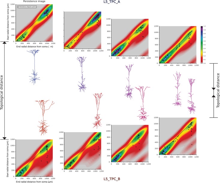
“Kanari et al. [2018] refer to this as the persistence image.”
BackgroundMethodInfluentialPredicting Synaptic Connectivity for Large-Scale Microcircuit Simulations Using Snudda
Hjorth, J. J. J., Kotaleski, J. H., & Kozlov, A. (2021) · Neuroinformatics · doi:10.1007/s12021-021-09531-w · Cited by 10
Quantifying neuronal structural changes over time using dynamic morphometrics
Hogg, P. W., Coleman, P., Toth, T. D., & Haas, K. Z. (2021) · Trends in Neurosciences · doi:10.1016/j.tins.2021.10.011 · Cited by 10
Higher-Order Description of Brain Function
Expert, P., & Petri, G. (2022) · Understanding complex systems · doi:10.1007/978-3-030-91374-8_17 · Cited by 9
Measurements of neuronal morphological variation across the rat neocortex
Friedman, R. (2020) · Neuroscience Letters · doi:10.1016/j.neulet.2020.135077 · Cited by 9
Barcodes distinguishing morphology of neuronal tauopathy
Beers, D., Goniotaki, D., Hanger, D. P., Goriely, A., & Harrington, H. A. (2023) · Physical Review Research · doi:10.1103/physrevresearch.5.043006 · Cited by 9
Kanari, L., Schmidt, S., Casalegno, F., Delattre, É., Lukic, J. B., Negrello, T., Shi, Y., Meystre, J., Defferrard, M., Schürmann, F., et al. (2026) · Scientific Reports · doi:10.1038/s41598-026-71217-7 · Cited by 8
Self-citationReproducibility of predictive networks for mouse visual cortex
Turishcheva, P., Burg, M. F., Sinz, F. H., & Ecker, A. S. (2024) · neural information processing systems · doi:10.52202/079017-0255 · Cited by 7
“As we do not know the true underlying number of clusters, we explore a range of k = 5 up to k = 100 clusters, which covers the typical amount of cell types suggested in various works and modalities [41, 42, 45, 59].”
Augmenting learning in neuro-embodied systems through neurobiological first principlesPreprint
Rodriguez-Garcia, A., Ghosh, A., Mei, J., & Ramaswamy, S. (2024) · arXiv (Cornell University) · doi:10.48550/arxiv.2407.04525 · Cited by 7
Self-citationMulti-level feature fusion network for neuronal morphology classification
Sun, C., & Zhao, F. (2024) · Frontiers in Neuroscience · doi:10.3389/fnins.2024.1465642 · Cited by 6
Modeling and simulation of neocortical micro- and mesocircuitry (Part I, anatomy)
Reimann, M., Bolaños‐Puchet, S., Courcol, J.-D., Santander, D. E., Arnaudon, A., Coste, B., Delalondre, F., Delemontex, T., Devresse, A., Dictus, H., et al. (2026) · eLife · doi:10.7554/elife.99688.3 · Cited by 6
Self-citationThe fiber of persistent homology for trees
Beers, D., & Leygonie, J. (2025) · Journal of Applied and Computational Topology · doi:10.1007/s41468-025-00213-z · Cited by 6
“for analyzing neuronal morphologies [13, 15] and brain functionalities [1].”
Effect of Phosphorylated Tau on Cortical Pyramidal Neuron Morphology during Hibernation
Regalado-Reyes, M., Benavides‐Piccione, R., Fernaud-Espinosa, I., DeFelipe, J., & León‐Espinosa, G. (2020) · Cerebral Cortex Communications · doi:10.1093/texcom/tgaa018 · Cited by 6
“Furthermore, they constitute the major source of cortical excitatory synapses, and their dendritic spines are the principal cortical postsynaptic targets of excitatory synapses (DeFelipe and Fariñas 1992; Kanari et al. 2019).”
BackgroundTopological Adventures in Neuroscience
Hess, K. P. (2020) · Abel symposia · doi:10.1007/978-3-030-43408-3_11 · Cited by 6
Self-citationA systematic evaluation of interneuron morphology representations for cell type discriminationPreprint
Laturnus, S., Kobak, D., & Berens, P. (2019) · bioRxiv (Cold Spring Harbor Laboratory) · doi:10.1101/591370 · Cited by 6
Ecker, A., Santander, D. E., Abdellah, M., Alonso, J. B., Bolaños‐Puchet, S., Chindemi, G., Mariyappan, D. P. G., Isbister, J. B., King, J. G., Kumbhar, P., et al. (2024) · eLife · doi:10.7554/elife.101850.1 · Cited by 6
Self-citationDiscovering optimal features for neuron-type identification from extracellular recordings
Haynes, V., Zhou, Y., & Crook, S. M. (2024) · Frontiers in Neuroinformatics · doi:10.3389/fninf.2024.1303993 · Cited by 5
“Cortical pyramidal cells primarily differ in their dendritic properties (Spruston, 2008; Kanari et al., 2019) as opposed to cortical interneurons which differ based on axonal properties (Markram et al., 2004; Jiang et al., 2015).”
BackgroundEcker, A., Santander, D. E., Abdellah, M., Alonso, J. B., Bolaños‐Puchet, S., Chindemi, G., Mariyappan, D. P. G., Isbister, J. B., King, J. G., Kumbhar, P., et al. (2025) · eLife · doi:10.1101/2023.08.07.552264 · Cited by 4
“In line with the biological variability, excitatory cells are modeled as a di-110 verse set of morphologies (Reimann et al., 2024a; Kanari et al., 2019) (Figure 1B) equipped with conductances distributed across all electrical compartments (Reva et al., 2023) (Supplementary Figure S1A).”
Self-citationStability of topological descriptors for neuronal morphologyPreprint
Beers, D., Harrington, H. A., & Goriely, A. (2022) · arXiv (Cornell University) · doi:10.48550/arxiv.2211.09058 · Cited by 4
“have already used unweighted analogues of reflected persistence images without the transformation (x, y) 7→ (x, y−x) in their previous work on the TMD [8, 9].”
MethodManjunatha, K. K. H., Bruzzone, M., Nicoletti, G., Suweis, S. S., & Maschio, M. D. (2023) · bioRxiv (Cold Spring Harbor Laboratory) · doi:10.1101/2023.05.21.541471 · Cited by 4
“Similar to Kanari et al 67 , we preserve essential network features, but with the added advantage of incorporating spatial constraints that better maintain known anatomical relationships.”
Moon, J. H., Seong, K., Kim, H. G., Mun, W., Min, S. H., Park, S., Jeong, J., Koh, C. S., Jung, H. H., & Eom, K. (2025) · Advanced Healthcare Materials · doi:10.1002/adhm.202502443 · Cited by 3
Generating brain-wide connectome using synthetic axonal morphologies
Petkantchin, R., Berchet, A., Peng, H., Markram, H., & Kanari, L. (2025) · Nature Communications · doi:10.1038/s41467-025-62030-3 · Cited by 3
“Furthermore, the computationally synthesized axons generate large-scale inter-regional connectivity, de fi ning the projectome and the connectome of the brain, thereby enabling the in silico experimentation of large brain regions.”
Self-citationDensity Visualization Pipeline: A Tool for Cellular and Network Density Visualization and Analysis
Grein, S., Qi, G., & Queisser, G. (2020) · Frontiers in Computational Neuroscience · doi:10.3389/fncom.2020.00042 · Cited by 4
Bitanihirwe, B. K. Y., & Woo, T.-U. W. (2021) · Elsevier eBooks · doi:10.1016/b978-0-12-817986-4.00037-7 · Cited by 3
Sun, C., Guo, Q., Yang, G., & Zhao, F. (2023) · Lecture notes in computer science · doi:10.1007/978-3-031-46005-0_12 · Cited by 3
Wang, W. X., & Lefebvre, J. L. (2020) · doi:10.1101/2020.02.29.971366
Efficient and Lightweight Neuron Morphology Classification Using Gabor Convolutional Networks
He, F., Wei, Y., Feng, F., & Qian, Y. (2023) · Procedia Computer Science · doi:10.1016/j.procs.2023.08.161 · Cited by 2
Shi, Y., & Wang, G. (2024) · STAR Protocols · doi:10.1016/j.xpro.2024.102917 · Cited by 2
Self-citationBoges, D. J., Calì, C., Magistretti, P. J., Hadwiger, M., Sicat, R., & Agus, M. (2019) · ACM Symposium on Virtual Reality Software and Technology (VRST) · doi:10.1145/3359996.3365042 · Cited by 2
Advances in approaches to study cell-type specific cortical circuits throughout development
Hanson, M. A., & Wester, J. C. (2022) · Frontiers in Cellular Neuroscience · doi:10.3389/fncel.2022.1031389 · Cited by 2
“However, each major neuronal class contains several distinct cell types that can be parsed according to additional morphological, electrophysiological, and molecular features (Kubota, 2014; Pelkey et al., 2017; Nigro et al., 2018; Huang and Paul, 2019; Kanari et al., 2019; Winnubst et al., 2019; Que et al., 2021).”
BackgroundTemporal Topology Provides an Interpretable Framework for Neuronal Morphogenesis
Rigaux, K., Castro, A. F., & Kanari, L. (2026) · Neuroinformatics · doi:10.1007/s12021-026-09795-0 · Cited by 2
“Specifically, we modify the traditional definition of vineyards to establish correspondences between persistence diagrams extracted at different time points using the Topological Morphology Descriptor (TMD) [35, 34].”
InfluentialSelf-citationComputational characteristics and hardware implications of brain tissue simulations
Cremonesi, F. (2019) · Infoscience (Ecole Polytechnique Fédérale de Lausanne) · doi:10.5075/epfl-thesis-9767 · Cited by 2
“, 2007), tens of different cell types in rat’s neocortex alone (Kanari et al., 2019; Narayanan et al., 2017) and in the order of a hundred different cell types in the whole mouse brain (Hodge et al.”
BackgroundEasley, T. O., Freese, K., Munch, E., & Bijsterbosch, J. D. (2023) · PubMed Central · doi:10.48550/arxiv.2306.13802 · Cited by 2
Novel definition and quantitative analysis of branch structure with topological data analysisPreprint
Oda, H., Kida, M., Nakata, Y., & Kurihara, H. (2024) · arXiv (Cornell University) · doi:10.48550/arxiv.2402.07436 · Cited by 2
“When we focus on branch structures, Reimann et al. (2017)[19] and Kanari et al. (2018, 2019)[20, 21] deal with neuronal structures.”
BackgroundSpike sorting biases and information loss in a detailed cortical modelPreprint
Laquitaine, S., Imbeni, M., Tharayil, J., Isbister, J., & Reimann, M. (2025) · Research Square · doi:10.21203/rs.3.rs-6234254/v1 · Cited by 2
A Detailed Model for Understanding the Human NeocortexPreprint
Barros-Zulaica, N., Kanari, L., Sood, V., Rai, P., Arnaudon, A., Shi, Y., Mandge, D., Geit, W. A. H. V., Zbili, M., Reva, M., et al. (2026) · bioRxiv (Cold Spring Harbor Laboratory) · doi:10.64898/2026.01.29.702592 · Cited by 2
“…from all possible perspectives: its anatomical structure [1, 4]; its cell classification from different angles, such as: morphological features [5–7], molecular [8], transcriptomics [9] and single cell physiology [6]; its connectivity [10]; synaptic physiology between pairs of connected cells…”
Self-citationShah, U., Agus, M., Gamal, M., Alzubaidi, M. S., Calì, C., Magistretti, P. J., Bouzerdoum, A., & Househ, M. (2026) · arXiv (Cornell University) · doi:10.48550/arxiv.2603.20970 · Cited by 2
Properties of Cultured Cells and Selection of Culture Media
Mani, S. (2023) · Techniques in life science and biomedicine for the non-expert · doi:10.1007/978-3-031-19485-6_6 · Cited by 1
Liao, M., Wan, G., Hu, W., & Du, B. (2025) · Neural Networks · doi:10.1016/j.neunet.2025.107603 · Cited by 1
In silico neuronal morphology classification: A systematic review
Lobato, F. M. F., Leite, J. B. P., Jacob, A. C., & Santana, R. (2026) · Neuroscience · doi:10.1016/j.neuroscience.2026.02.044 · Cited by 1
Application of the mirror technique for block-face scanning electron microscopy
Talapka, P., Bába, B. B., Mészár, Z., Kisvárday, R. E., Kocsis, Z., Srivastava, M., & Kisvárday, Z. F. (2022) · Brain Structure and Function · doi:10.1007/s00429-022-02506-w · Cited by 1
“In this regard, most dendrites run quasi-perpendicular to the brain surface which is quite evident for the apical dendrite of pyramidal cells (Kanari et al. 2019) but also for nonpyramidal cells (Jin et al.”
BackgroundVerasztó, C., Roussel, Y., Kanari, L., Piluso, S., Markram, H., & Keller, D. X. (2026) · PLoS Computational Biology · doi:10.1371/journal.pcbi.1014106 · Cited by 1
Self-citationRecut: a Concurrent Framework for Sparse Reconstruction of Neuronal MorphologyPreprint
Marrett, K., Zhu, M., Chi, Y., Choi, C., Chen, Z., Dong, H., Park, C. S., Yang, X. W., & Cong, J. (2021) · bioRxiv (Cold Spring Harbor Laboratory) · doi:10.1101/2021.12.07.471686 · Cited by 1
“Persistence images have been used to classify neuronal types with subtle morphological differences that are difficult for even human experts to discriminate [25].”
MethodAuthor response: Modeling and Simulation of Neocortical Micro- and Mesocircuitry. Part I: Anatomy
Reimann, M., Bolaños‐Puchet, S., Courcol, J.-D., Santander, D. E., Arnaudon, A., Coste, B., Delalondre, F., Delemontex, T., Devresse, A., Dictus, H., et al. (2024) · eLife · doi:10.7554/elife.99688.1.sa4 · Cited by 1
Self-citationZhang, J., & Goetz, S. M. (2025) · arXiv (Cornell University) · doi:10.48550/arxiv.2509.00291 · Cited by 1
“However, the brain contains a number of different neuron types and shapes, which differ also in their nonlinear activation dynamics [3].”
Topology for trees and curves: theory and applications
Beers, D. (2023) · Oxford University Research Archive (ORA) (University of Oxford) · doi:10.5287/ora-7jbz87djb · Cited by 1
Uncovering the Basis of Human Connectome Complexity: The Role of Neuronal MorphologyPreprint
Barros-Zulaica, N., Santander, D. E., Kanari, L., Shi, Y., Perin, R., Pezzoli, M., Benavides‐Piccione, R., DeFelipe, J., de Kock, C. P. J., Segev, I., et al. (2026) · bioRxiv (Cold Spring Harbor Laboratory) · doi:10.64898/2026.02.12.705493 · Cited by 1
“This image-based topological summary [50] has been shown to be a suitable format for quantitative analysis and has been used in several applications, including neuronal morphology classification [31, 51] and glia classification [52].”
Self-citationThe development of an idiographic method of measurement for use in dynamic assessment
Hurley, E. (2019) · Arrow@dit (Dublin Institute of Technology)
Vroman, R., & McKay, L. S. (2021) · bioRxiv (Cold Spring Harbor Laboratory) · doi:10.1101/2021.11.17.468122
Zhao, S., Qian, P., & Liu, L. (2023) · Research Square · doi:10.1101/2023.09.26.559442
Etude de l'inhibition corticale au cours du cycle veille-sommeil
Brécier, A. (2020) · theses.fr (ABES) · doi:10.70675/6e744687zf366z48fdz9ab1z44fdc38a56a7
Abdellah, M., Foni, A., Cantero, J. J. G., Guerrero, N. R., Boci, E., Fleury, A. C., Coggan, J. S., Keller, D. X., Planas, J., Courcol, J.-D., et al. (2024) · Briefings in Bioinformatics · doi:10.1093/bib/bbae393
Self-citation
Metadata
- Authors
- Kanari, L., Ramaswamy, S., Shi, Y., Morand, S., Meystre, J., Perin, R., Abdellah, M., Wang, Y., Hess, K., & Markram, H.
- Affiliations
- École Polytechnique Fédérale de Lausanne (CH)
- Allen Institute for Brain Science (US)
- Corresponding author
- Kanari, Lida
- Markram, Henry
- ORCID
- Venue
- Cerebral Cortex
- Publisher
- Oxford University Press (OUP)
- Published
- 2019-01-31
- Volume
- 29
- Issue
- 4
- Pages
- 1719-1735
- ISSN
- 1047-3211 (ISSN-L)
- 1460-2199
- PMC
- PMC6418396
- OpenAlex
- W2911934184
- Cited by
- 94
- References
- 88
- Citations by year
- 2026: 9 · 2025: 12 · 2024: 18 · 2023: 11 · 2022: 14 · 2021: 9 · 2020: 16 · 2019: 5
- FWCI
- 5.33
- Type
- journal article
- Language
- en
- Open access
- hybrid
- doi.org
- Licence
- Locations
- Cerebral Cortex (publishedVersion, cc-by-nc)
- bioRxiv (Cold Spring Harbor Laboratory) (acceptedVersion)
- Europe PMC (PubMed Central) (publishedVersion, cc-by-nc)
- PubMed Central (publishedVersion, cc-by-nc)
- PubMed (publishedVersion)
- Infoscience (Ecole Polytechnique Fédérale de Lausanne) (submittedVersion, cc-by-nc-nd)
- Funders
- European Union Seventh (604102)
- HBP (720270)
- HBP SGA1
- National Natural Science Foundation (No. 31070951)
- Topics
- Life Sciences › Biochemistry, Genetics and Molecular Biology › Biophysics › Cell Image Analysis Techniques
- Physical Sciences › Computer Science › Computational Theory and Mathematics › Topological and Geometric Data Analysis
- Life Sciences › Neuroscience › Cognitive Neuroscience › Neural dynamics and brain function
- Keywords
- neocortical pyramidal cells, dendritic branching, algebraic topology, topological classification, somatosensory cortex, rats
- MeSH terms
- Supervised Machine Learning, Animals, Lysine, Pattern Recognition, Automated, Somatosensory Cortex, Pyramidal Cells, Neocortex, Imaging, Three-Dimensional, Rats
About these checks
Registry, retraction, Cites and statistics results are computed, not judged, with one exception: a printed reference that no index lists is read once by a model before it counts as a citation error. Claims are extracted by a model and kept only when their quote appears verbatim in the text; each cited source is then checked, with a model’s help, for whether it says what the claim relies on. Those verdicts can be wrong — read the claim’s quote and the cited source before relying on one.
Statistics and arithmetic are recomputed, not judged.
Confidence bands are shown only for fields calibrated on our annotated test set. None is calibrated yet, so no band is shown.
What counts as a claim ›How we measure confidence ›How we check this ›
Are you an author, or do you see a problem with this page? Request a review
Data sources
Bibliographic data: Semantic Scholar, OpenAlex, Crossref.
Contains information from the Semantic Scholar Academic Graph Datasets, made available under the ODC Attribution License (ODC-BY 1.0).
Semantic Scholar’s data is described in: Kinney, R., et al. (2023). The Semantic Scholar Open Data Platform. arXiv:2301.10140.
