

Development and Validation of a Deep Learning–based Automatic Detection Algorithm for Active Pulmonary Tuberculosis on Chest Radiographs
Open accessThe DOI matches its Crossref record; 31 of the 31 references it cites were found in a registry or an index.
Abstract
Background: Detection of active pulmonary tuberculosis on chest radiographs (CRs) is critical for the diagnosis and screening of tuberculosis. An automated system may help streamline the tuberculosis screening process and improve diagnostic performance. Methods: We developed a deep learning–based automatic detection (DLAD) algorithm using 54c221 normal CRs and 6768 CRs with active pulmonary tuberculosis that were labeled and annotated by 13 board-certified radiologists. The performance of DLAD was validated using 6 external multicenter, multinational datasets. To compare the performances of DLAD with physicians, an observer performance test was conducted by 15 physicians including nonradiology physicians, board-certified radiologists, and thoracic radiologists. Image-wise classification and lesion-wise localization performances were measured using area under the receiver operating characteristic (ROC) curves and area under the alternative free-response ROC curves, respectively. Sensitivities and specificities of DLAD were calculated using 2 cutoffs (high sensitivity [98%] and high specificity [98%]) obtained through in-house validation. Results: DLAD demonstrated classification performance of 0.977–1.000 and localization performance of 0.973–1.000. Sensitivities and specificities for classification were 94.3%–100% and 91.1%–100% using the high-sensitivity cutoff and 84.1%–99.0% and 99.1%–100% using the high-specificity cutoff. DLAD showed significantly higher performance in both classification (0.993 vs 0.746–0.971) and localization (0.993 vs 0.664–0.925) compared to all groups of physicians. Conclusions: Our DLAD demonstrated excellent and consistent performance in the detection of active pulmonary tuberculosis on CR, outperforming physicians, including thoracic radiologists.
Overview
The claims and statistics of this paper haven’t been checked yet.
Free: it reads the open-access text and checks the first 5 claims that cite a source we can check. Create a free account to check every claim.
Integrity signals
Registry
Cites
Statistics
Figures
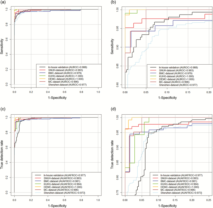
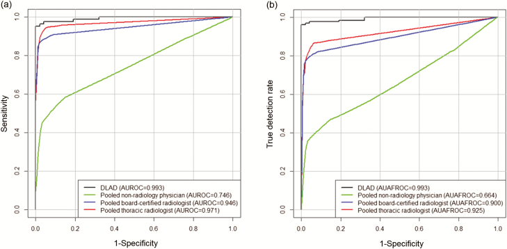
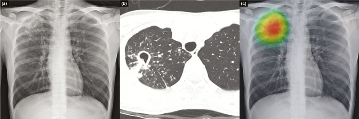
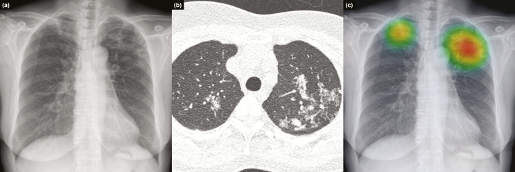
Verification
Claims the paper makes
The claims and statistics of this paper haven’t been checked yet.
Free: it reads the open-access text and checks the first 5 claims that cite a source we can check. Create a free account to check every claim.
Claims other papers make about this one
Each is checked against this paper’s own text: does it say what the citing paper says it does?
The cited source says what the claim attributes to it.
In the citing paper
“13 (refs. 63–75) studies validated algorithms on external data”
From Diagnostic accuracy of deep learning in medical imaging: a systematic review and meta-analysis (2021) · cites this paper as [67]
The cited source says what the claim attributes to it.
In the citing paper
“Twenty-one54,63–67,70,72,76–88 studies compared algorithm performance against healthcare professionals.”
From Diagnostic accuracy of deep learning in medical imaging: a systematic review and meta-analysis (2021) · cites this paper as [67]
Self-check
Percentages, sums and reported differences re-computed from the paper’s own numbers.
The self-check needs the paper’s full text.
Citations
Cites
R: A Language and Environment for Statistical Computing
R Core Team (2025) · doi:10.32614/r.manuals · Cited by 352,181
ImageNet classification with deep convolutional neural networks
Krizhevsky, A., Sutskever, I., & Hinton, G. E. (2017) · Communications of the ACM · doi:10.1145/3065386 · Cited by 112,155
A Simple Sequentially Rejective Multiple Test Procedure
(1979) · Scandinavian Journal of Statistics · doi:10.2307/4615733 · Cited by 21,526 · cited 1×
“Holm–Bonferroni methods were used to correct multiple comparisons [24].”
MethodDermatologist-level classification of skin cancer with deep neural networks
Esteva, A., Kuprel, B., Novoa, R. A., Ko, J., Swetter, S. M., Blau, H. M., & Thrun, S. (2017) · Nature · doi:10.1038/nature21056 · Cited by 14,362
Advances in Neural Information Processing Systems 25
Zhang, Y., Sutton, C. A., Storkey, A., & Zoubin Ghahramani, A. (2012) · Cited by 12,653
Gulshan, V., Peng, L., Coram, M., Stumpe, M. C., Wu, D., Narayanaswamy, A., Venugopalan, S., Widner, K., Madams, T., Cuadros, J., et al. (2016) · JAMA · doi:10.1001/jama.2016.17216 · Cited by 7,697 · cited 1×
“Meanwhile, after overwhelming success in the ImageNet Large Scale Visual Recognition Competition in 2012 [12], the deep learning technique demonstrated successful results even at medical image classifications [13, 14].”
MethodLakhani, P., & Sundaram, B. (2017) · Radiology · doi:10.1148/radiol.2017162326 · Cited by 1,775
Samek, W., Wiegand, T., & Müller, K.-R. (2017) · arXiv · doi:10.48550/arxiv.1708.08296 · Cited by 1,080
Two public chest X-ray datasets for computer-aided screening of pulmonary diseases.
(2014) · PubMed Central · doi:10.3978/j.issn.2223-4292.2014.11.20 · Cited by 918
Dorfman, D. D., Berbaum, K. S., & Metz, C. E. (1992) · PubMed · Cited by 724
Research works printed but not linked: 3
- 2017., Global tuberculosis report 2017
- 2013., Systematic screening for active tuberculosis: principles and recommendations
- Zhai, RJafroc: analysis of data acquired using the receiver operating characteristic paradigm and its extensions
Cited by
Items 101–200 of 266
Hwang, E. J., Park, J., Hong, W., Lee, H., Choi, H., Kim, H., Nam, J. G., Goo, J. M., Yoon, S. H., Lee, C. H., et al. (2022) · European Radiology · doi:10.1007/s00330-022-08593-x · Cited by 23
Self-citationHan, D., Chen, Y., Li, X., Li, W., Zhang, X., He, T., Yu, Y., Dou, Y., Duan, H., & Yu, N. (2022) · La radiologia medica · doi:10.1007/s11547-022-01580-8 · Cited by 23
The application of artificial intelligence to chest medical image analysis
Liu, F., Tang, J., Ma, J., Wang, C., Ha, Q., Yu, Y., & Zhou, Z. (2021) · Intelligent Medicine · doi:10.1016/j.imed.2021.06.004 · Cited by 22
Choi, S. Y., Choi, A., Baek, S., Ahn, J. Y., Roh, Y. H., & Kim, J. H. (2023) · Scientific Reports · doi:10.1038/s41598-023-47146-0 · Cited by 21
Self-citationZhang, Y., Liu, M., Zhang, L., Wang, L., Zhao, K., Hu, S., Chen, X., & Xie, X. (2023) · JAMA Network Open · doi:10.1001/jamanetworkopen.2022.55113 · Cited by 20
Han, D., He, T., Yu, Y., Guo, Y., Chen, Y., Duan, H., & Yu, N. (2022) · Academic Radiology · doi:10.1016/j.acra.2021.12.025 · Cited by 20
Nojomi, M., Babaee, E., Rampisheh, Z., Benis, M. R., Soheyli, M., & Raz, N. R. (2025) · Medical Journal of the Islamic Republic of Iran · doi:10.47176/mjiri.39.81 · Cited by 19
Yang, Y., Xia, L., Liu, P., Yang, F., Wu, Y., Pan, H., Hou, D., Liu, N., & Lu, S. (2023) · Frontiers in Medicine · doi:10.3389/fmed.2023.1195451 · Cited by 19
Kim, H., Park, C. M., & Goo, J. M. (2020) · European Radiology · doi:10.1007/s00330-019-06589-8 · Cited by 19
Self-citationKim, H., Kwak, N., Yoon, S. H., Park, N., Kim, Y. R., Lee, J. H., Lee, J. Y., Park, Y., Kang, Y. A., Kim, S., et al. (2024) · Scientific Reports · doi:10.1038/s41598-024-63885-0 · Cited by 19
Self-citationWang, X., Yang, T., Zhang, Y., Liu, X., Zhang, Y., Sun, L., Gu, X., Chen, Z., Guo, Y., Xue, C., et al. (2022) · Prenatal Diagnosis · doi:10.1002/pd.6220 · Cited by 17
Early pleural effusion detection from respiratory diseases including COVID-19 via deep learning
Serte, S., & Serener, A. (2020) · Medical Technologies Congress (TIPTEKNO) · doi:10.1109/tiptekno50054.2020.9299300 · Cited by 17
Lee, S., Lee, H. W., Kim, H., Kim, D. K., Yim, J., Yoon, S. H., & Kwak, N. (2022) · CHEST Journal · doi:10.1016/j.chest.2022.06.018 · Cited by 16
Self-citationEvolving and Novel Applications of Artificial Intelligence in Thoracic Imaging
Chang, J. Y., & Makary, M. S. (2024) · Diagnostics · doi:10.3390/diagnostics14131456 · Cited by 15
Hwang, E. J., Kim, H., Lee, J. H., Goo, J. M., & Park, C. M. (2020) · European Radiology · doi:10.1007/s00330-020-07062-7 · Cited by 15
Self-citationKim, H. W., Myong, J., & Kim, J. S. (2020) · The Korean Journal of Internal Medicine · doi:10.3904/kjim.2020.144 · Cited by 15
Self-citationArtificial Intelligence in Chest Radiography—A Comparative Review of Human and Veterinary Medicine
Rubini, A., Via, R. D., Pastore, V. P., Signore, F. D., Rosto, M., Bonis, A. D., Odone, F., & Vignoli, M. (2025) · Veterinary Sciences · doi:10.3390/vetsci12050404 · Cited by 13
Jiang, W., Zhang, H., Li, Z., Jiang, X., Shao, J., Yang, X., Xiong, J., Zhou, P., Zhang, H., Wang, H., et al. (2025) · Journal of Medical Internet Research · doi:10.2196/83041 · Cited by 13
Chest Radiography of Tuberculosis: Determination of Activity Using Deep Learning Algorithm
Choi, Y. R., Yoon, S. H., Kim, J., Yoo, J. Y., Kim, H., & Jin, K. N. (2023) · Tuberculosis & respiratory diseases · doi:10.4046/trd.2023.0020 · Cited by 13
Self-citationHong, J. H., Yoon, S. H., Goo, J. M., Yim, J., & Jeon, Y. K. (2020) · PLoS ONE · doi:10.1371/journal.pone.0231537 · Cited by 13
Self-citationTuberculosis detection using deep and hybrid learning techniques using X-ray images
Shilpa, N., & Banu, W. A. (2025) · Neural Computing and Applications · doi:10.1007/s00521-025-11173-x · Cited by 12
Wang, S., Karaca, Y., Zhang, X., & Zhang, Y. (2021) · Fractals · doi:10.1142/s0218348x22400473 · Cited by 12
An Efficient, Lightweight, Tiny 2D-CNN Ensemble Model to Detect Cardiomegaly in Heart CT Images
Doppala, B. P., Bataineh, A. A., & Vamsi, B. (2023) · Journal of Personalized Medicine · doi:10.3390/jpm13091338 · Cited by 12
Blood RNA signatures predict recent tuberculosis exposure in mice, macaques and humans
Ault, R., Headley, C. A., Hare, A. E., Carruthers, B., Mejías, A., & Turner, J. (2020) · Scientific Reports · doi:10.1038/s41598-020-73942-z · Cited by 12
A Framework for Assurance of Medication Safety using Machine LearningPreprint
Jia, Y., Lawton, T., McDermid, J. A., Rojas, E., & Habli, I. (2021) · arXiv (Cornell University) · doi:10.48550/arxiv.2101.05620 · Cited by 12
Zhu, X., Zhu, L., Song, D., Wang, D., Wu, F., & Wu, J. (2022) · Clinical Radiology · doi:10.1016/j.crad.2022.09.114 · Cited by 11
Emerging Trends and Innovations in Radiologic Diagnosis of Thoracic Diseases
Song, J., Hwang, E. J., Yoon, S. H., Park, C. M., & Goo, J. M. (2025) · Investigative Radiology · doi:10.1097/rli.0000000000001179 · Cited by 10
Self-citationHong, E. K., Kim, H. W., Song, O. K., Lee, K.-C., Kim, D. K., Cho, J.-B., Kim, J., Lee, S., Bae, W., & Roh, B. (2025) · American Journal of Roentgenology · doi:10.2214/ajr.25.33059 · Cited by 10
Self-citationLopes, S., Mascarenhas, M., Fonseca, J., Fernandes, M. G. O., & Leite-Moreira, A. F. (2025) · Diagnostics · doi:10.3390/diagnostics15141734 · Cited by 10
Ensemble Learning With Attention-Based Multiple Instance Pooling for Classification of SPT
Zhou, Q., Zhang, X., & Zhang, Y. (2021) · IEEE Transactions on Circuits & Systems II Express Briefs · doi:10.1109/tcsii.2021.3124165 · Cited by 10
Huang, M., Chen, C., Horng, M., Li, C.-I., Hsu, I.-L., Su, C.-M., Sun, Y., & Lai, C. (2022) · Anesthesiology · doi:10.1097/aln.0000000000004378 · Cited by 10
Shetty, S., Ananthanarayana, V. S., & Mahale, A. R. (2022) · Mathematics · doi:10.3390/math10193646 · Cited by 10
Chung, W. Y., Yoon, J., Yoon, D., Kim, S., Kim, Y., Park, J. E., & Kang, Y. A. (2024) · Journal of Medical Internet Research · doi:10.2196/58413 · Cited by 9
the MultiomIcs claSSIfier for pulmOnary Nodules (MISSION) Collaborative Group, Deng, J., Zhao, M., Li, Q., Zhang, Y., Ma, M., Li, C., Wang, J., She, Y., Jiang, Y., et al. (2021) · Translational Lung Cancer Research · doi:10.21037/tlcr-21-971 · Cited by 9
Cai, J., Guo, L., Zhu, L., Li, X., Qian, L., Lure, Y. M. F., & Yin, X.-P. (2023) · Frontiers in Oncology · doi:10.3389/fonc.2023.1140635 · Cited by 9
An Analysis of Adaptable Intelligent Models for Pulmonary Tuberculosis Detection and Classification
Siddiqui, A. K., & Garg, V. K. (2021) · SN Computer Science · doi:10.1007/s42979-021-00890-4 · Cited by 9
Hanis, T. M., Ruhaiyem, N. I. R., Arifin, W. N., Haron, J., Rahman, W. F. W. A., Abdullah, R., & Musa, K. I. (2022) · Diagnostics · doi:10.3390/diagnostics12112826 · Cited by 9
Khan, S. H., Sohail, A., Khan, A., & Lee, Y. S. (2022) · SSRN Electronic Journal · doi:10.2139/ssrn.4029649 · Cited by 9
Kazemzadeh, S., Yu, J., Jamshy, S., Pilgrim, R., Nabulsi, Z., Chen, C., Beladia, N., Lau, C. T., McKinney, S. M., Hughes, T., et al. (2021) · arXiv (Cornell University) · doi:10.48550/arxiv.2105.07540 · Cited by 9
Anichini, G., & Kotras, B. (2024) · Science Technology & Human Values · doi:10.1177/01622439241262854 · Cited by 8
Distinguishing infectivity in patients with pulmonary tuberculosis using deep learning
Gao, Y., Zhang, Y., Hu, C., He, P., Fu, J. Y., Lin, F., Liu, K., Fu, X., Liu, R., Sun, J., et al. (2023) · Frontiers in Public Health · doi:10.3389/fpubh.2023.1247141 · Cited by 8
Zhang, G., Shang, L., Cao, Y., Zhang, J., Li, S., Qian, R., Liu, H., Zhang, Z., Pu, H., Man, Q., et al. (2024) · Quantitative Imaging in Medicine and Surgery · doi:10.21037/qims-24-33 · Cited by 8
Park, J., Hwang, E. J., Lee, J. H., Hong, W., Nam, J. G., Lim, W. H., Kim, J. H., Goo, J. M., & Park, C. M. (2023) · Journal of Thoracic Imaging · doi:10.1097/rti.0000000000000691 · Cited by 8
Self-citationKhan, S. H., Sohail, A., Khan, A., & Lee, Y. S. (2022) · SSRN Electronic Journal · doi:10.2139/ssrn.4060772 · Cited by 8
Yoon, M. S., Kwon, G., Oh, J., Ryu, J., Lim, J., Kang, B.-K., Lee, J., & Han, D.-K. (2023) · Journal of Imaging Informatics in Medicine · doi:10.1007/s10278-022-00772-y · Cited by 7
Self-citationSmith, E. F., Thomas, M., Kutukcu, E. C., Torres‐Sánchez, I., Santiago, M. G., Quijano-Campos, J. C., Sylvester, K. P., Burtin, C., Šajnić, A., Brandt, J. D., et al. (2020) · ERJ Open Research · doi:10.1183/23120541.00808-2020 · Cited by 7
Deep Learning in Medicine. Are We Ready?
Ting, D. S. W., Rim, T. H., Choi, Y. S., & Ledsam, J. R. (2019) · Annals of the Academy of Medicine Singapore · doi:10.47102/annals-acadmedsg.v48n1p1 · Cited by 6
Classification of pulmonary diseases from chest radiographs using deep transfer learning
Shamas, M., Tauseef, H., Ahmad, A., Raza, A., Ghadi, Y. Y., Mamyrbayev, O. J., Momynzhanova, K., & Alahmadi, T. J. (2025) · PLoS ONE · doi:10.1371/journal.pone.0316929 · Cited by 5
Use of Artificial Intelligence in Diagnosing Vertical Root Fractures—A Systematic Review
Alshahrani, A. S., Alelyani, A. A., Jabali, A. H., Malwi, A. A. A., Alroomy, R., Shaiban, A. S., Almnea, R. A., Mehta, V., & Moaleem, M. M. A. (2026) · Diagnostics · doi:10.3390/diagnostics16030406 · Cited by 5
Abhishek, A., Chalga, M. S., Yadav, R. M., Agarwal, K., Vohra, V., Tayade, A., Kumar, A., Basil, M. V., Mrigpuri, P., Manek, H., et al. (2026) · The Indian Journal of Medical Research · doi:10.25259/ijmr_1854_2025 · Cited by 5
AI-Aided Disease Prediction in Visualized Medicine
Du, J., Huang, M., & Liu, L. (2023) · Advances in experimental medicine and biology · doi:10.1007/978-981-32-9902-3_6 · Cited by 5
A systematic survey on deep learning techniques for chest disease detection using chest radiographs
Soni, A., & Rai, A. (2023) · Journal of Current Science and Technology · doi:10.59796/jcst.v13n2.2023.1744 · Cited by 5
Liu, M., Zhou, Y., Ding, J., Wei, F., Wang, F., Nie, S., Chen, X., Jiang, Y., Huang, M., & Hu, L. (2025) · Frontiers in Medicine · doi:10.3389/fmed.2025.1508736 · Cited by 5
Sudarshan, V. K., Ramachandra, R. A., Tan, N. S. M., Ojha, S., & Tan, R. (2022) · International Journal of Imaging Systems and Technology · doi:10.1002/ima.22715 · Cited by 5
Hong, W., Hwang, E. J., Park, C. M., & Goo, J. M. (2023) · Korean Journal of Radiology · doi:10.3348/kjr.2023.0255 · Cited by 5
Self-citationA Tuberculosis Detection Method Using Attention and Sparse R-CNN
Xu, X., Zhang, J., Cheng, X., Lu, L., Zhao, Y., Xu, Z., & Gu, Z. (2022) · KSII Transactions on Internet and Information Systems · doi:10.3837/tiis.2022.07.001 · Cited by 5
Inteligencia artificial en radiología torácica. ¿Un reto en tiempos de la COVID-19?
Corbacho-Abelaira, D., Ruano‐Raviña, A., & Fernández‐Villar, A. (2020) · Archivos de Bronconeumología · doi:10.1016/j.arbres.2020.10.008 · Cited by 5
Pathology-Aware Generative Adversarial Networks for Medical Image AugmentationPreprint
Han, C. (2021) · arXiv (Cornell University) · doi:10.48550/arxiv.2106.01915 · Cited by 5
Chiu, W.-C., Chang, S., Lin, C., Chen, T.-W., & Fang, W.-H. (2025) · Bioengineering · doi:10.3390/bioengineering12121377 · Cited by 4
Application of Artificial Intelligence in Thoracic Radiology: A Narrative Review
Lim, W. H., & Kim, H. (2024) · Tuberculosis & respiratory diseases · doi:10.4046/trd.2024.0062 · Cited by 4
Revolutionizing Tuberculosis Detection: Advanced TBNet Classifier for Chest X-Rays
Prova, N. N. I., Keshetti, S., Jamili, L. K., Hassan, A., Punjabi, V. N., & Ravi, V. (2025) · International Conference on Advances in Computer Science, Electrical, Electronics, and Communication Technologies (CE2CT) · doi:10.1109/ce2ct64011.2025.10939749 · Cited by 4
Rhee, C., Hong, K. J., Kim, K. H., Goo, J. M., & Hwang, E. J. (2025) · Korean Journal of Radiology · doi:10.3348/kjr.2025.0237 · Cited by 4
Self-citationDeep learning-based anomaly detection in orthopedic medical imaging: A systematic literature review
Ounasser, N., Rhanoui, M., Mikram, M., & Asri, B. E. (2025) · Journal of Orthopaedics · doi:10.1016/j.jor.2025.07.015 · Cited by 4
Nkouanga, H. Y., & Vajda, S. (2021) · Proceedings - International Conference on Pattern Recognition/Proceedings/International Conference on Pattern Recognition · doi:10.1109/icpr48806.2021.9412430 · Cited by 4
Artificial Intelligence for Cardiothoracic Imaging: Overview of Current and Emerging Applications
Hochhegger, B., Pasini, R., Carvalho, A. R., Rodrigues, R. S., Altmayer, S., Bittencourt, L. K., Marchiori, E. D. S., & Forghani, R. (2023) · Seminars in Roentgenology · doi:10.1053/j.ro.2023.02.001 · Cited by 4
Artificial Intelligence in Diagnostic Imaging
Sollini, M., Loiacono, D., Volpe, D., Levra, A. G., Lomeo, E., Giacomello, E., Kirienko, M., Chiti, A., & Lanzi, P. L. (2023) · Wiley eBooks · doi:10.1002/9781119603627.ch31 · Cited by 4
Classification Of Sleep-Wake State In A Ballistocardiogram System Based On Deep LearningPreprint
Ahmed, N., Singh, A., Srivyshnav, K. S., Kumar, G., Parchani, G., & Saran, V. (2020) · arXiv (Cornell University) · doi:10.48550/arxiv.2011.08977 · Cited by 4
Kvak, D., Chromcová, A., Ovesná, P., Dandár, J., Biros, M., Hrubý, R., Dufek, D., & Pajdaković, M. (2023) · arXiv (Cornell University) · doi:10.48550/arxiv.2305.10116 · Cited by 4
Cho, H. S., Hwang, E. J., Yi, J., Choi, B., & Park, C. M. (2024) · Diagnostic and Interventional Radiology · doi:10.4274/dir.2024.242835 · Cited by 3
Self-citationGuo, L., Li, X., Zheng, Q., Zheng, B., Jaeger, S., Giger, M. L., Fuhrman, J. D., Li, H., Lure, F. Y. M., Li, H., et al. (2024) · Journal of X-Ray Science and Technology · doi:10.3233/xst-240051 · Cited by 3
HEGAZY, N. Y., & Sawah, M. S. (2026) · Scientific Reports · doi:10.1038/s41598-026-42492-1 · Cited by 3
Effect of an artificial-intelligent chest radiographs reporting system in an emergency department
Yoon, D. H., Heo, S., Yu, J. H., Lee, S. U., Hwang, S. Y., Yoon, H. J., Shin, T. G., Lee, G. T., Park, J. S., Chang, H., et al. (2023) · Signa Vitae · doi:10.22514/sv.2023.108 · Cited by 3
Zanetti, C., George, A. E., Stiegmann, R. A., & Phelan, D. (2019) · Advances in medical education, research, and ethics (AMERE) book series · doi:10.4018/978-1-7998-1468-9.ch021 · Cited by 3
Brand LOGO Image Recognition and Inquiry Creative Teaching Design Based on Deep Learning
Gao, X., & Chen, W. (2022) · Wireless Communications and Mobile Computing · doi:10.1155/2022/5396468 · Cited by 3
Dikici, E., Nguyen, X. V., Takacs, N., & Prevedello, L. M. S. (2023) · Computers in Biology and Medicine · doi:10.1016/j.compbiomed.2023.106901 · Cited by 3
Tuberculosis Detection Using a Deep Neural Network
Patil, D. H., & Gadekar, A. (2023) · Proceedings in adaptation, learning and optimization · doi:10.1007/978-3-031-31164-2_51 · Cited by 3
Sorace, L., Raju, N., O'Shaughnessy, J., Kachel, S., Jansz, K., Yang, N., & Lim, R. (2023) · Radiography · doi:10.1016/j.radi.2023.10.014 · Cited by 3
Hwang, E. J. (2024) · Journal of the Korean Society of Radiology · doi:10.3348/jksr.2024.0052 · Cited by 3
Self-citationBedoya, O. F., Aristizábal, H. S. G., & Agudelo, J. (2022) · Revista EIA · doi:10.24050/reia.v20i39.1617 · Cited by 3
Kim, E. Y., Kim, Y. J., Choi, W.-J., Lee, G. P., Choi, Y. R., Jin, K. N., & Cho, Y. J. (2021) · PLoS ONE · doi:10.1371/journal.pone.0251045 · Cited by 3
Self-citationLi, H., Nijiati, M., Zhou, R., Damaola, M., Li, L., Qian, B., Abulizi, A., & Zou, X. (2022) · Research Square · doi:10.21203/rs.3.rs-1266973/v1 · Cited by 3
Institutional Tuberculosis Control and Elimination Program
Yu, S. N., Kim, T. H., Han, S. H., & Kim, Y. (2023) · Korean Journal of Healthcare-Associated Infection Control and Prevention · doi:10.14192/kjicp.2023.28.1.22 · Cited by 2
Wu, S., Liu, S., Zhong, M., de Loos, E. R., Hartert, M., Fuentes-Martín, Á., Lenzini, A., Wang, D., & Qing, Q. (2024) · Journal of Thoracic Disease · doi:10.21037/jtd-24-679 · Cited by 2
Oh, S. N., Yang, H., Lee, C. K., Park, S. H., Han, C. H., Heo, H., & Kim, Y. S. (2024) · Journal of the American College of Radiology · doi:10.1016/j.jacr.2024.11.009 · Cited by 2
Self-citationHashmi, M. F., Patil, R. B., Kene, J. D., & Waje, M. G. (2024) · Traitement du signal · doi:10.18280/ts.410616 · Cited by 2
YOLOv11-MFF: A multi-scale frequency-adaptive fusion network for enhanced CXR anomaly detection
Guan, L., Zhang, R., & Zhao, Y. M. (2025) · PLoS ONE · doi:10.1371/journal.pone.0334283 · Cited by 2
Matsuo, H., Nishio, M., Fujimoto, K., Deperrois, N., Matsunaga, T., Nooralahzadeh, F., Krauthammer, M., & Murakami, T. (2026) · npj Health Systems · doi:10.1038/s44401-026-00087-y · Cited by 3
Spatial lung imaging in clinical and translational settings
Persson, I. M., Božović, G., Westergren‐Thorsson, G., & Enes, S. R. (2024) · Breathe · doi:10.1183/20734735.0224-2023 · Cited by 2
Prabu, S., Chandra, N. S., Bhavani, M., Varma, V. K. C., & Soni, M. (2025) · International Conference on Integrated Circuits and Communication Systems (ICICACS) · doi:10.1109/icicacs65178.2025.10968948 · Cited by 2
Artificial Intelligence in the Analysis of PET Scans of the Human Brain
Mouridsen, K., & Borra, R. (2020) · Springer eBooks · doi:10.1007/978-3-030-53168-3_5 · Cited by 2
Revolution in Detecting Tuberculosis using Radiology with Application of Deep Learning Algorithm
Satya, P. G. A. N., & Parikesit, A. A. (2021) · Cermin Dunia Kedokteran · doi:10.55175/cdk.v48i4.1475 · Cited by 2
Ibrahim, S. J. A., Kumar, V., Suchitra, S., Sathya, M., Sahini, V., Chakravarthy, N. S. K., & Saruchi, S. (2022) · Current Materials Science · doi:10.2174/2666145415666220816163634 · Cited by 2
Lim, C. Y., Ki, Y., Chung, M. J., Park, S., Park, S., Woo, J. H., & Kim, J. H. (2023) · Diagnostics · doi:10.3390/diagnostics13122060 · Cited by 2
Self-citationAbubakar, M. Z., Kaya, M., Eriş, M., Abubakar, M. M., Karakuş, S., & Sani, K. J. (2024) · Turkish Journal of Science and Technology · doi:10.55525/tjst.1222836 · Cited by 2
Systematic Analysis of Tuberculosis prediction using Deep Learning Technique
Riaz, U., Abdullah, A., Aslam, H., Nawaz, H., & Rasool, S. (2022) · Machines and Algorithms · doi:10.66108/mna.v1i3.32 · Cited by 2
Dašić, L., & Filipović, N. (2024) · Springer eBooks · doi:10.1007/978-3-031-60044-9_14 · Cited by 2
Survey of the Detection and Classification of Pulmonary Lesions via CT and X-RayPreprint
Sun, Y., Li, C., Zhang, Q., Zhou, A., & Zhang, G. (2020) · arXiv (Cornell University) · doi:10.48550/arxiv.2012.15442 · Cited by 2
Jiang, F., Wang, Y., Xu, C., & Xu, Q. (2022) · Radiology of Infectious Diseases · doi:10.4103/rid.rid_39_22 · Cited by 1
Advances in imaging studies of active pulmonary tuberculosis
Zhang, Y., Li, H., Ma, Z., Wang, J., & Yin, X. (2023) · Radiology of Infectious Diseases · doi:10.4103/rid.rid-d-23-00012 · Cited by 1
Ghimire, S., Sitaula, J., Karki, S., Khatri, D., & Jha, S. (2024) · International Conference on Advancements in Computing (ICAC) · doi:10.1109/icac64487.2024.10851094 · Cited by 1
Metadata
- Authors
- Hwang, E. J., Park, S., Jin, K.-N., Kim, J. I., Choi, S. Y., Lee, J. H., Goo, J. M., Aum, J., Yim, J.-J., Park, C. M., Deep Learning-Based Automatic Detection Algorithm Development and Evaluation Group, Kim, D. H., Woo, W., Choi, C., Hwang, I. P., Song, Y. S., Lim, L., Kim, K., Wi, J. Y., Oh, S. S., & Kang, M.-J.
- Affiliations
- Seoul National University (KR)
- Kyung Hee University Hospital at Gangdong (KR)
- Daejeon Eulji Medical Center, Eulji University (KR)
- Corresponding author
- Park, Chang Min
- ORCID
- Venue
- Clinical Infectious Diseases
- Publisher
- Oxford University Press (OUP)
- Published
- 2018-11-08
- Volume
- 69
- Issue
- 5
- Pages
- 739-747
- ISSN
- 1058-4838 (ISSN-L)
- 1537-6591
- PMC
- PMC6695514
- OpenAlex
- W2900683726
- Cited by
- 241
- References
- 31
- Citations by year
- 2026: 23 · 2025: 26 · 2024: 28 · 2023: 38 · 2022: 45 · 2021: 40 · 2020: 27 · 2019: 14
- FWCI
- 13.97
- Type
- journal article
- Language
- en
- Open access
- hybrid
- doi.org
- Licence
- Locations
- Clinical Infectious Diseases (publishedVersion, cc-by)
- Europe PMC (PubMed Central) (publishedVersion, cc-by)
- PubMed Central (publishedVersion, cc-by)
- PubMed (publishedVersion)
- Seoul National University Open Repository (Seoul National University) (submittedVersion)
- Funders
- Seoul National University Hospital Research (04-2016-3000)
- Seoul Research & Business Development Program (FI170002)
- Topics
- Health Sciences › Medicine › Radiology, Nuclear Medicine and Imaging › COVID-19 diagnosis using AI
- Physical Sciences › Computer Science › Artificial Intelligence › AI in cancer detection
- Health Sciences › Medicine › Radiology, Nuclear Medicine and Imaging › Radiomics and Machine Learning in Medical Imaging
- Keywords
- active pulmonary tuberculosis, chest radiograph, deep learning, lesion localization, observer performance comparison, tuberculosis screening, diagnostic accuracy
- MeSH terms
- Deep Learning, Adult, Aged, Algorithms, Automation, Female, Humans, Male, Middle Aged, Radiography, ROC Curve, Sensitivity and Specificity, Thorax, Tuberculosis, Pulmonary
About these checks
Registry, retraction, Cites and statistics results are computed, not judged, with one exception: a printed reference that no index lists is read once by a model before it counts as a citation error. Claims are extracted by a model and kept only when their quote appears verbatim in the text; each cited source is then checked, with a model’s help, for whether it says what the claim relies on. Those verdicts can be wrong — read the claim’s quote and the cited source before relying on one.
Statistics and arithmetic are recomputed, not judged.
Confidence bands are shown only for fields calibrated on our annotated test set. None is calibrated yet, so no band is shown.
What counts as a claim ›How we measure confidence ›How we check this ›
Are you an author, or do you see a problem with this page? Request a review
Data sources
Bibliographic data: Semantic Scholar, OpenAlex, Crossref.
Contains information from the Semantic Scholar Academic Graph Datasets, made available under the ODC Attribution License (ODC-BY 1.0).
Semantic Scholar’s data is described in: Kinney, R., et al. (2023). The Semantic Scholar Open Data Platform. arXiv:2301.10140.
