

Development and Validation of a Deep Learning–based Automatic Detection Algorithm for Active Pulmonary Tuberculosis on Chest Radiographs
Open accessThe DOI matches its Crossref record; 31 of the 31 references it cites were found in a registry or an index.
Abstract
Background: Detection of active pulmonary tuberculosis on chest radiographs (CRs) is critical for the diagnosis and screening of tuberculosis. An automated system may help streamline the tuberculosis screening process and improve diagnostic performance. Methods: We developed a deep learning–based automatic detection (DLAD) algorithm using 54c221 normal CRs and 6768 CRs with active pulmonary tuberculosis that were labeled and annotated by 13 board-certified radiologists. The performance of DLAD was validated using 6 external multicenter, multinational datasets. To compare the performances of DLAD with physicians, an observer performance test was conducted by 15 physicians including nonradiology physicians, board-certified radiologists, and thoracic radiologists. Image-wise classification and lesion-wise localization performances were measured using area under the receiver operating characteristic (ROC) curves and area under the alternative free-response ROC curves, respectively. Sensitivities and specificities of DLAD were calculated using 2 cutoffs (high sensitivity [98%] and high specificity [98%]) obtained through in-house validation. Results: DLAD demonstrated classification performance of 0.977–1.000 and localization performance of 0.973–1.000. Sensitivities and specificities for classification were 94.3%–100% and 91.1%–100% using the high-sensitivity cutoff and 84.1%–99.0% and 99.1%–100% using the high-specificity cutoff. DLAD showed significantly higher performance in both classification (0.993 vs 0.746–0.971) and localization (0.993 vs 0.664–0.925) compared to all groups of physicians. Conclusions: Our DLAD demonstrated excellent and consistent performance in the detection of active pulmonary tuberculosis on CR, outperforming physicians, including thoracic radiologists.
Overview
The claims and statistics of this paper haven’t been checked yet.
Free: it reads the open-access text and checks the first 5 claims that cite a source we can check. Create a free account to check every claim.
Integrity signals
Registry
Cites
Statistics
Figures
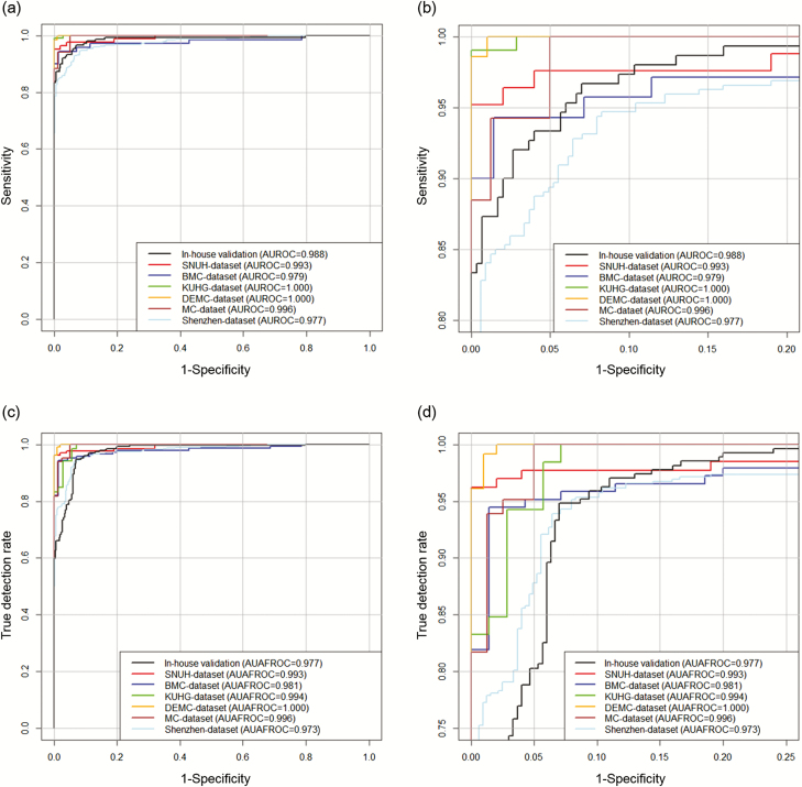
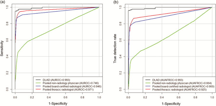
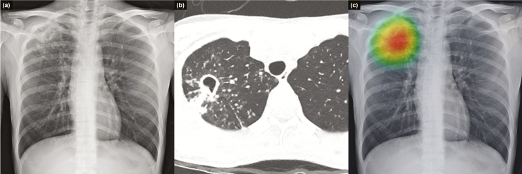
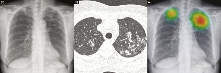
Verification
Claims the paper makes
The claims and statistics of this paper haven’t been checked yet.
Free: it reads the open-access text and checks the first 5 claims that cite a source we can check. Create a free account to check every claim.
Claims other papers make about this one
Each is checked against this paper’s own text: does it say what the citing paper says it does?
The cited source says what the claim attributes to it.
In the citing paper
“13 (refs. 63–75) studies validated algorithms on external data”
From Diagnostic accuracy of deep learning in medical imaging: a systematic review and meta-analysis (2021) · cites this paper as [67]
The cited source says what the claim attributes to it.
In the citing paper
“Twenty-one54,63–67,70,72,76–88 studies compared algorithm performance against healthcare professionals.”
From Diagnostic accuracy of deep learning in medical imaging: a systematic review and meta-analysis (2021) · cites this paper as [67]
Self-check
Percentages, sums and reported differences re-computed from the paper’s own numbers.
The self-check needs the paper’s full text.
Citations
Cites
R: A Language and Environment for Statistical Computing
R Core Team (2025) · doi:10.32614/r.manuals · Cited by 352,181
ImageNet classification with deep convolutional neural networks
Krizhevsky, A., Sutskever, I., & Hinton, G. E. (2017) · Communications of the ACM · doi:10.1145/3065386 · Cited by 112,155
A Simple Sequentially Rejective Multiple Test Procedure
(1979) · Scandinavian Journal of Statistics · doi:10.2307/4615733 · Cited by 21,526 · cited 1×
“Holm–Bonferroni methods were used to correct multiple comparisons [24].”
MethodDermatologist-level classification of skin cancer with deep neural networks
Esteva, A., Kuprel, B., Novoa, R. A., Ko, J., Swetter, S. M., Blau, H. M., & Thrun, S. (2017) · Nature · doi:10.1038/nature21056 · Cited by 14,362
Advances in Neural Information Processing Systems 25
Zhang, Y., Sutton, C. A., Storkey, A., & Zoubin Ghahramani, A. (2012) · Cited by 12,653
Gulshan, V., Peng, L., Coram, M., Stumpe, M. C., Wu, D., Narayanaswamy, A., Venugopalan, S., Widner, K., Madams, T., Cuadros, J., et al. (2016) · JAMA · doi:10.1001/jama.2016.17216 · Cited by 7,697 · cited 1×
“Meanwhile, after overwhelming success in the ImageNet Large Scale Visual Recognition Competition in 2012 [12], the deep learning technique demonstrated successful results even at medical image classifications [13, 14].”
MethodLakhani, P., & Sundaram, B. (2017) · Radiology · doi:10.1148/radiol.2017162326 · Cited by 1,775
Samek, W., Wiegand, T., & Müller, K.-R. (2017) · arXiv · doi:10.48550/arxiv.1708.08296 · Cited by 1,080
Two public chest X-ray datasets for computer-aided screening of pulmonary diseases.
(2014) · PubMed Central · doi:10.3978/j.issn.2223-4292.2014.11.20 · Cited by 918
Dorfman, D. D., Berbaum, K. S., & Metz, C. E. (1992) · PubMed · Cited by 724
Research works printed but not linked: 3
- 2017., Global tuberculosis report 2017
- 2013., Systematic screening for active tuberculosis: principles and recommendations
- Zhai, RJafroc: analysis of data acquired using the receiver operating characteristic paradigm and its extensions
Cited by
Items 201–266 of 266
Shwetha, V., Banerjee, B., Laxmi, V., & Kamath, P. R. (2025) · International Journal of Biomedical Imaging · doi:10.1155/ijbi/3559598 · Cited by 1
Chiang, L.-F. (2026) · Cureus · doi:10.7759/cureus.103134 · Cited by 1
Bruce, A. T. I., de Faria, M. G. B. F., Andrade, R. L. D. P., Arcêncio, R. A., Pinto, I. C., Gomes, D. M. D. O., Rocha, E. S. C., Tavares, M. E. D. A., & Monroe, A. A. (2026) · Revista Brasileira de Epidemiologia · doi:10.1590/1980-549720260012 · Cited by 1
Xu, S., Guo, C., Xu, Q., Zuo, J., Yang, M., Chen, B., Li, S., Xiao, J., Tang, X., & Qin, Y. (2026) · Journal of Advanced Research · doi:10.1016/j.jare.2026.04.013 · Cited by 1
Rajaraman, S., Marini, N., Liang, Z., Xue, Z., & Antani, S. (2026) · Journal of Medical Systems · doi:10.1007/s10916-026-02368-3 · Cited by 2
Agbeko, E. (2026) · American Journal of Medicine and Health Studies · doi:10.32674/ex08x042 · Cited by 1
Jin, Y. Y., Ding, J., Ouyang, J., Yao, Z., Wang, L., Xu, R., & Jin, X. (2026) · Frontiers in Medicine · doi:10.3389/fmed.2026.1858126 · Cited by 1
PREDICTION OF DRUG-RESISTANT TUBERCULOSIS USING MACHINE LEARNING
Nurgozhaeva, D. (2026) · doi:10.70286/eoss-06.07.2026.011.186-191 · Cited by 1
Irmak, Ç., Süner, A. F., Akbulut, İ., & Atalay, S. (2026) · Infection · doi:10.1007/s15010-026-02934-4 · Cited by 1
A Study on the Clinical Effectiveness of Deep Learning CAD Technology
Ju-Hyuck, H., Oh, H., & Kim, W.-S. (2022) · International Conference on Artificial Intelligence in Information and Communication (ICAIIC) · doi:10.1109/icaiic54071.2022.9722681 · Cited by 1
External Validation of Deep Learning Algorithm for Tuberculosis Detection in Thai Population
Rajak, A., Chaisangmongkon, W., Chamveha, I., Promwiset, T., Rungsinaporn, K., Saiviroonporn, P., & Tongdee, T. (2022) · doi:10.1109/incit56086.2022.10067327 · Cited by 1
Literature Review of Deep Learning for Tuberculosis Based on Chest Imaging
Sara, L., & Ziti, S. (2023) · Lecture notes in networks and systems · doi:10.1007/978-3-031-35248-5_46 · Cited by 1
Lancaster, A. C., Cardin, M., Nguyen, J. A., Mehta, T. I., Öncel, D., Bai, H. X., Cohen, K. A., & Lin, C. T. (2023) · Journal of Thoracic Imaging · doi:10.1097/rti.0000000000000745 · Cited by 1
Convolutional neural network for IT lung diagnostics
Vishniakou, U. А., & He, T. (2024) · «System analysis and applied information science» · doi:10.21122/2309-4923-2024-1-59-64 · Cited by 1
Mirza, I. R. (2024) · Journal of Innovative Computing and Emerging Technologies · doi:10.56536/jicet.v4i2.121 · Cited by 1
Anmol, A., Ilyas, M., Bilal, M., Khan, H. U., & Ramzan, M. (2024) · University of Sindh Journal of Information and Communication Technology · Cited by 1
Detection and Classification of Lung Diseases for Pneumonia: A Survey
Anita, G. (2024) · International Journal of Intelligent Systems and Applications in Engineering · Cited by 1
A distillation based approach for the diagnosis of diseasesPreprint
Bandyopadhyay, H., Dastidar, S. G., Mondal, B., Banerjee, B., & Das, N. (2021) · arXiv (Cornell University) · doi:10.48550/arxiv.2108.03470 · Cited by 1
AI-driven Detection, Characterization and Classification of Chronic Lung Diseases
Chassagnon, G. (2019) · theses.fr (ABES) · doi:10.70675/0abbd9a6zf9c6z47e0z96fbz3c544a648d65 · Cited by 1
Manan, M. R., & Manan, H. (2021) · TURKISH MEDICAL STUDENT JOURNAL · doi:10.4274/tmsj.galenos.2021.08.01.03
Karthik, T., Kasiraman, V., Paski, B., Gurram, K., Talwar, A., & Donkena, M. (2021) · doi:10.36227/techrxiv.16725727.v1
Karthik, T., Kasiraman, V., Paski, B., Gurram, K., Talwar, A., & Donkena, M. (2021) · doi:10.36227/techrxiv.16725727
Kim, J. H., Han, S. G., Cho, A., Shin, H. J., & Baek, S. (2021) · doi:10.2196/preprints.29612
Self-citationOtte, A. (2022) · Springer eBooks · doi:10.1007/978-3-030-91255-0_2
MetaEfficientNet: A Few-Shot Learning Approach for Lung Disease Classification
Nimbolkar, S., Thakare, A., Mitra, S., Biranje, O., & Sutar, A. (2022) · Algorithms for intelligent systems · doi:10.1007/978-981-19-0863-7_19
Guo, C. (2022) · Research Square · doi:10.21203/rs.3.rs-2232950/v1
A Comparative Study Based on Lung Cancer with Deep Learning and Machine Learning Models
Afreen, Y. N., & Reddy, P. V. B. (2023) · Algorithms for intelligent systems · doi:10.1007/978-981-19-7892-0_4
Koç, H., Hızıroğlu, A., & Erbaycu, A. E. (2023) · DergiPark
- Retracted
A Deep Learning-Based Approach of Detecting Tuberculosis Lesion of Tuberculous MeningoencephalitisPreprint
Li, W., Peng, H., Zhu, Z., Ma, W., Liang, W., Li, J., & Yang, H. (2023) · SSRN Electronic Journal · doi:10.2139/ssrn.4568782
Tuberculosis and chest radiology
Kang, E.-Y. (2023) · Journal of Korean Medical Association · doi:10.5124/jkma.2023.66.11.667
Detecting Pulmonary Lesions in Low-Prevalence Real-World Settings Using Deep Learning
Kvak, D., Chromcová, A., Ovesná, P., Dandár, J., Biros, M., Hrubý, R., Dufek, D., & Pajdaković, M. (2024) · Lecture notes in electrical engineering · doi:10.1007/978-981-97-1335-6_1
AI for Lawyers: A Gentle Introduction
McDermid, J. A., Jia, Y., & Habli, I. (2024) · Cambridge University Press eBooks · doi:10.1017/9781108980197.002
Deep Learning for Clinical Practice.
Sogancioglu, E. (2024) · Radboud University Press eBooks · doi:10.54195/9789493296770
KI-unterstützte Krankheitsvorhersage in der visualisierten Medizin
Du, J., Huang, M., & Liu, L. (2025) · Springer eBooks · doi:10.1007/978-981-97-9693-9_6
A semi-supervised TB detection model based on improved YOLOv5
Dong, H., Liu, Q., & Yang, F. (2025) · International Conference on Signal Processing Systems (ICSPS) · doi:10.1117/12.3061504
Dandár, J., Gerold, J., Křístek, J., Straka, Z., Kvak, D., Biros, M., & Hrubý, R. (2025) · Lecture notes in electrical engineering · doi:10.1007/978-981-96-3863-5_33
Scaling laws for Haralick texture features of linear gradients
Oprisan, S. A., & Oprisan, A. (2025) · PeerJ Computer Science · doi:10.7717/peerj-cs.2856
Aranay, O. M. (2022) · Scholars Archive - University at Albany (University at Albany, State University of New York) · doi:10.54014/22c3-k94e
Innovative Imaging Solutions for Tuberculosis Discovery by Deep Learning
Rao, B. S. K., & Maheshwary, P. (2025) · Lecture notes in networks and systems · doi:10.1007/978-981-96-1264-2_15
Wang, Q., Zhang, Z., Li, X., Guo, X., Xiao, Q., Guo, L., & Geng, H. (2025) · Scientific Reports · doi:10.1038/s41598-025-23172-y
Specific fine-tuned GPT-enhanced medical imaging diagnosis recommendations
Wang, Z., Li, J., Sun, J., Wang, X., Ren, P., Cai, L., Wang, Z., & Lv, H. (2025) · Intelligent Medicine · doi:10.1016/j.imed.2025.03.005 · Cited by 1
Ahmad, C. W. I. C. W., Lukman, K. A., Jeffree, M. S., Omar, R. M. R., Avoi, R., Hijazi, M. H. A., Muien, M. Z. A., Kieu, S. T. H., & Ibrahim, M. Y. (2025) · IEEE International Conference on Artificial Intelligence in Engineering and Technology (IICAIET) · doi:10.1109/iicaiet67254.2025.11265054
Mohammad, Q., & Justus Liebig University Giessen (2020) · University writing server of the Justus Liebig University Giessen (Justus-Liebig-Universität Gießen) · doi:10.22029/jlupub-14829
Park, M. W., Lee, Y., Kim, S., Kim, Y.-J., Kim, S. Y., Kim, Y., & Kim, H.-M. (2023) · Figshare · doi:10.6084/m9.figshare.22603226
AI in Cancer Screening and Early Detection
Sachdeva, K., & Sachdeva, R. (2026) · Wiley eBooks · doi:10.1002/9781394389131.ch35
Girma, A., Abnew, J., & Kelkay, B. (2026) · The Journal of Engineering · doi:10.1049/tje2.70171
Wei, Y., Cui, X., Chong, L., Wang, C., Liu, M., Chen, X., & Zhong, L. (2026) · PLoS ONE · doi:10.1371/journal.pone.0338810
Jeon, W., Jang, H., Lee, H., Park, C., Lyu, J., & Choi, S. (2026) · Sensors · doi:10.3390/s26072100
Self-citationSharma, A., Chaudhary, V. K., Sharma, C., Sharma, A., Sharma, S., Sharma, S., & Verma, N. (2026) · Quality & Quantity · doi:10.1007/s11135-026-02897-6
METABOLIC SYNDROME: GASTROENTERECOLOGICAL ASPECTS, PATHOGENESIS AND MODERN THERAPY
Zhexenbek, N., Makhanbet, N., & Berdimurat, O. (2026) · doi:10.70286/eoss-06.07.2026.012.191-197
Mishra, I., Kondabala, R., Suresh, K., N, K. S. B. V., V, & Arasi, P. R. E. (2026) · International Journal of Drug Delivery Technology · doi:10.25258/ijddt.16.66s.39
Parate, J., Borkar, A., Umate, L., Kadadi, S., & Parate, J. (2026) · IEEE International Conference on Intelligent Technologies for a Sustainable and Inclusive Future (ICITSIF) · doi:10.1109/icitsif69060.2026.11608375
A Multi-Metric Evaluation of K-Means Clustering for Tuberculosis Detection in Healthcare domain
Bendarkar, A. P., & Lahande, P. V. (2026) · International Conference on Intelligent Technologies (CONIT) · doi:10.1109/conit69683.2026.11620642
Detecting Tuberculosis Disease from Chest X-Ray Using Deep Learning
Boujelb, Z., Benba, A., Idrissi, A., Achenani, Y., & Chakir, E. M. (2026) · Lecture notes in networks and systems · doi:10.1007/978-3-032-28097-8_44
Deep learning–based tuberculosis detection with an image fusion strategy
Yang, T.-C., Lee, C.-C., Hwang, J.-I., Chang, Y., Chen, P.-J., Lin, S.-H., & Chu, Y.-W. (2026) · Chinese Journal of Physics · doi:10.1016/j.cjph.2026.09.001
YOLOv10-FMW: A Multiscale Convolutional Detection Method for Pulmonary Tuberculosis
Lu, M., Wang, M., Liu, J., Sun, Q., Xu, B., Cong, J., Shi, J., & Wu, Y. (2026) · Journal of Imaging Informatics in Medicine · doi:10.1007/s10278-026-02218-1
Kadam, S., & Soni, U. (2026) · International Journal of Advances in Signal and Image Sciences · doi:10.29284/payy6a84
Yusoffdeen, N. (2023) · International Journal on Advanced Computer Engineering and Communication Technology · doi:10.65521/ijacect.v12i2.3760
کاربرد هوش مصنوعی در تشخیص بیماریهای ریوی
قنبری, م., محبی, س., & زاده, س. ع. (2024) · Nafas Journal
Nair, N., Bahl, S., Menon, J. C., Narayanan, S., & Nedungadi, P. (2026) · Lecture notes in computer science · doi:10.1007/978-981-92-6513-8_35
Bilfaqih, S. M., & Maram, B. (2026) · IGI Global eBooks · doi:10.4018/979-8-2600-4881-8.ch010
Haider, A., Ullah, I., Hakeem, S., & Ali, K. (2026) · Pakistan Journal of Medical & Cardiological Review · doi:10.66021/pakmcr1834
Nijiati, M., Zhou, R., Damaola, M., Hu, C., Li, L., Qian, B., Abulizi, A., Kaisaier, A., Cai, C., Li, H., et al. (2022) · doi:10.3389/fmolb.2022.1086047.s001
Chien, S.-C., Huang, C.-C., & Huang, C.-C. (2026) · Journal of Medical Systems · doi:10.1007/s10916-026-02488-w
Metadata
- Authors
- Hwang, E. J., Park, S., Jin, K.-N., Kim, J. I., Choi, S. Y., Lee, J. H., Goo, J. M., Aum, J., Yim, J.-J., Park, C. M., Deep Learning-Based Automatic Detection Algorithm Development and Evaluation Group, Kim, D. H., Woo, W., Choi, C., Hwang, I. P., Song, Y. S., Lim, L., Kim, K., Wi, J. Y., Oh, S. S., & Kang, M.-J.
- Affiliations
- Seoul National University (KR)
- Kyung Hee University Hospital at Gangdong (KR)
- Daejeon Eulji Medical Center, Eulji University (KR)
- Corresponding author
- Park, Chang Min
- ORCID
- Venue
- Clinical Infectious Diseases
- Publisher
- Oxford University Press (OUP)
- Published
- 2018-11-08
- Volume
- 69
- Issue
- 5
- Pages
- 739-747
- ISSN
- 1058-4838 (ISSN-L)
- 1537-6591
- PMC
- PMC6695514
- OpenAlex
- W2900683726
- Cited by
- 241
- References
- 31
- Citations by year
- 2026: 23 · 2025: 26 · 2024: 28 · 2023: 38 · 2022: 45 · 2021: 40 · 2020: 27 · 2019: 14
- FWCI
- 13.97
- Type
- journal article
- Language
- en
- Open access
- hybrid
- doi.org
- Licence
- Locations
- Clinical Infectious Diseases (publishedVersion, cc-by)
- Europe PMC (PubMed Central) (publishedVersion, cc-by)
- PubMed Central (publishedVersion, cc-by)
- PubMed (publishedVersion)
- Seoul National University Open Repository (Seoul National University) (submittedVersion)
- Funders
- Seoul National University Hospital Research (04-2016-3000)
- Seoul Research & Business Development Program (FI170002)
- Topics
- Health Sciences › Medicine › Radiology, Nuclear Medicine and Imaging › COVID-19 diagnosis using AI
- Physical Sciences › Computer Science › Artificial Intelligence › AI in cancer detection
- Health Sciences › Medicine › Radiology, Nuclear Medicine and Imaging › Radiomics and Machine Learning in Medical Imaging
- Keywords
- active pulmonary tuberculosis, chest radiograph, deep learning, lesion localization, observer performance comparison, tuberculosis screening, diagnostic accuracy
- MeSH terms
- Deep Learning, Adult, Aged, Algorithms, Automation, Female, Humans, Male, Middle Aged, Radiography, ROC Curve, Sensitivity and Specificity, Thorax, Tuberculosis, Pulmonary
About these checks
Registry, retraction, Cites and statistics results are computed, not judged, with one exception: a printed reference that no index lists is read once by a model before it counts as a citation error. Claims are extracted by a model and kept only when their quote appears verbatim in the text; each cited source is then checked, with a model’s help, for whether it says what the claim relies on. Those verdicts can be wrong — read the claim’s quote and the cited source before relying on one.
Statistics and arithmetic are recomputed, not judged.
Confidence bands are shown only for fields calibrated on our annotated test set. None is calibrated yet, so no band is shown.
What counts as a claim ›How we measure confidence ›How we check this ›
Are you an author, or do you see a problem with this page? Request a review
Data sources
Bibliographic data: Semantic Scholar, OpenAlex, Crossref.
Contains information from the Semantic Scholar Academic Graph Datasets, made available under the ODC Attribution License (ODC-BY 1.0).
Semantic Scholar’s data is described in: Kinney, R., et al. (2023). The Semantic Scholar Open Data Platform. arXiv:2301.10140.
