

Development and Validation of a Deep Learning–based Automatic Detection Algorithm for Active Pulmonary Tuberculosis on Chest Radiographs
Open accessThe DOI matches its Crossref record; 31 of the 31 references it cites were found in a registry or an index.
Abstract
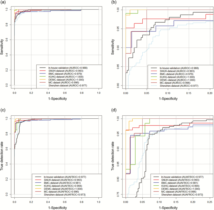
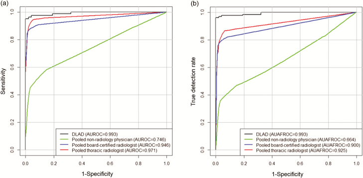
Background: Detection of active pulmonary tuberculosis on chest radiographs (CRs) is critical for the diagnosis and screening of tuberculosis. An automated system may help streamline the tuberculosis screening process and improve diagnostic performance. Methods: We developed a deep learning–based automatic detection (DLAD) algorithm using 54c221 normal CRs and 6768 CRs with active pulmonary tuberculosis that were labeled and annotated by 13 board-certified radiologists. The performance of DLAD was validated using 6 external multicenter, multinational datasets. To compare the performances of DLAD with physicians, an observer performance test was conducted by 15 physicians including nonradiology physicians, board-certified radiologists, and thoracic radiologists. Image-wise classification and lesion-wise localization performances were measured using area under the receiver operating characteristic (ROC) curves and area under the alternative free-response ROC curves, respectively. Sensitivities and specificities of DLAD were calculated using 2 cutoffs (high sensitivity [98%] and high specificity [98%]) obtained through in-house validation. Results: DLAD demonstrated classification performance of 0.977–1.000 and localization performance of 0.973–1.000. Sensitivities and specificities for classification were 94.3%–100% and 91.1%–100% using the high-sensitivity cutoff and 84.1%–99.0% and 99.1%–100% using the high-specificity cutoff. DLAD showed significantly higher performance in both classification (0.993 vs 0.746–0.971) and localization (0.993 vs 0.664–0.925) compared to all groups of physicians. Conclusions: Our DLAD demonstrated excellent and consistent performance in the detection of active pulmonary tuberculosis on CR, outperforming physicians, including thoracic radiologists.
Overview
The claims and statistics of this paper haven’t been checked yet.
Free: it reads the open-access text and checks the first 5 claims that cite a source we can check. Create a free account to check every claim.
Integrity signals
Registry
Cites
Statistics
Figures
Verification
Claims the paper makes
The claims and statistics of this paper haven’t been checked yet.
Free: it reads the open-access text and checks the first 5 claims that cite a source we can check. Create a free account to check every claim.
Claims other papers make about this one
Each is checked against this paper’s own text: does it say what the citing paper says it does?
The cited source says what the claim attributes to it.
In the citing paper
“13 (refs. 63–75) studies validated algorithms on external data”
From Diagnostic accuracy of deep learning in medical imaging: a systematic review and meta-analysis (2021) · cites this paper as [67]
The cited source says what the claim attributes to it.
In the citing paper
“Twenty-one54,63–67,70,72,76–88 studies compared algorithm performance against healthcare professionals.”
From Diagnostic accuracy of deep learning in medical imaging: a systematic review and meta-analysis (2021) · cites this paper as [67]
Self-check
Percentages, sums and reported differences re-computed from the paper’s own numbers.
The self-check needs the paper’s full text.
Citations
Cites
R: A Language and Environment for Statistical Computing
R Core Team (2025) · doi:10.32614/r.manuals · Cited by 352,181
ImageNet classification with deep convolutional neural networks
Krizhevsky, A., Sutskever, I., & Hinton, G. E. (2017) · Communications of the ACM · doi:10.1145/3065386 · Cited by 112,155
A Simple Sequentially Rejective Multiple Test Procedure
(1979) · Scandinavian Journal of Statistics · doi:10.2307/4615733 · Cited by 21,526 · cited 1×
“Holm–Bonferroni methods were used to correct multiple comparisons [24].”
MethodDermatologist-level classification of skin cancer with deep neural networks
Esteva, A., Kuprel, B., Novoa, R. A., Ko, J., Swetter, S. M., Blau, H. M., & Thrun, S. (2017) · Nature · doi:10.1038/nature21056 · Cited by 14,362
Advances in Neural Information Processing Systems 25
Zhang, Y., Sutton, C. A., Storkey, A., & Zoubin Ghahramani, A. (2012) · Cited by 12,653
Gulshan, V., Peng, L., Coram, M., Stumpe, M. C., Wu, D., Narayanaswamy, A., Venugopalan, S., Widner, K., Madams, T., Cuadros, J., et al. (2016) · JAMA · doi:10.1001/jama.2016.17216 · Cited by 7,697 · cited 1×
“Meanwhile, after overwhelming success in the ImageNet Large Scale Visual Recognition Competition in 2012 [12], the deep learning technique demonstrated successful results even at medical image classifications [13, 14].”
MethodLakhani, P., & Sundaram, B. (2017) · Radiology · doi:10.1148/radiol.2017162326 · Cited by 1,775
Samek, W., Wiegand, T., & Müller, K.-R. (2017) · arXiv · doi:10.48550/arxiv.1708.08296 · Cited by 1,080
Two public chest X-ray datasets for computer-aided screening of pulmonary diseases.
(2014) · PubMed Central · doi:10.3978/j.issn.2223-4292.2014.11.20 · Cited by 918
Dorfman, D. D., Berbaum, K. S., & Metz, C. E. (1992) · PubMed · Cited by 724
Research works printed but not linked: 3
- 2017., Global tuberculosis report 2017
- 2013., Systematic screening for active tuberculosis: principles and recommendations
- Zhai, RJafroc: analysis of data acquired using the receiver operating characteristic paradigm and its extensions
Cited by
Liu, X., Faes, L., Kale, A. U., Wagner, S. K., Fu, D. J., Bruynseels, A., Mahendiran, T., Moraes, G., Shamdas, M., Kern, C., et al. (2019) · The Lancet Digital Health · doi:10.1016/s2589-7500(19)30123-2 · Cited by 1,883
Diagnostic accuracy of deep learning in medical imaging: a systematic review and meta-analysis
Aggarwal, R., Sounderajah, V., Martin, G., Ting, D. S. W., Karthikesalingam, A., King, D., Ashrafian, H., & Darzi, A. (2021) · npj Digital Medicine · doi:10.1038/s41746-021-00438-z · Cited by 946
Deep learning in ophthalmology: The technical and clinical considerations
Ting, D. S. W., Peng, L., Varadarajan, A. V., Keane, P. A., Burlina, P. M., Chiang, M. F., Schmetterer, L., Pasquale, L. R., Bressler, N. M., Webster, D. R., et al. (2019) · Progress in Retinal and Eye Research · doi:10.1016/j.preteyeres.2019.04.003 · Cited by 534
Deep learning for chest X-ray analysis: A survey
Çallı, E., Sogancioglu, E., van Ginneken, B., van Leeuwen, K. G., & Murphy, K. (2021) · Medical Image Analysis · doi:10.1016/j.media.2021.102125 · Cited by 506
Hwang, E. J., Park, S., Jin, K.-N., Kim, J. I., Choi, S. Y., Lee, J. H., Goo, J. M., Aum, J., Yim, J.-J., Cohen, J. G., et al. (2019) · JAMA Network Open · doi:10.1001/jamanetworkopen.2019.1095 · Cited by 447
Self-citationExternal Validation of Deep Learning Algorithms for Radiologic Diagnosis: A Systematic Review
Yu, A. C., Mohajer, B., & Eng, J. (2022) · Radiology: Artificial Intelligence · doi:10.1148/ryai.210064 · Cited by 408
Medical image analysis based on deep learning approach
Puttagunta, M., & Ravi, S. (2021) · Multimedia Tools and Applications · doi:10.1007/s11042-021-10707-4 · Cited by 321
Qin, Z. Z., Sander, M. S., Rai, B., Titahong, C. N., Sudrungrot, S., Laah, S. N., Adhikari, L. M., Carter, E. J., Puri, L., Codlin, A. J., et al. (2019) · Scientific Reports · doi:10.1038/s41598-019-51503-3 · Cited by 289
“The greater the abnormality score, the higher probability of having TB. Current versions of qXR 16 and Lunit 17 detect several discrete pulmonary abnormalities, such as calcification, cavitation, opacities etc.”
BackgroundKaplan, A., Cao, H., FitzGerald, J. M., Iannotti, N., Yang, E., Kocks, J. W. H., Kostikas, K., Price, D., Reddel, H. K., Tsiligianni, I., et al. (2021) · The Journal of Allergy and Clinical Immunology: In Practice · doi:10.1016/j.jaip.2021.02.014 · Cited by 219
Deep Learning Techniques to Diagnose Lung Cancer
Wang, L. (2022) · Cancers · doi:10.3390/cancers14225569 · Cited by 209
Artificial intelligence applications for thoracic imaging
Chassagnon, G., Vakalopoulou, M., Paragios, N., & Revel, M.-P. (2020) · European Journal of Radiology · doi:10.1016/j.ejrad.2019.108774 · Cited by 203
Nafisah, S. I., & Muhammad, G. (2022) · Neural Computing and Applications · doi:10.1007/s00521-022-07258-6 · Cited by 194
Where Is the Artificial Intelligence Applied in Dentistry? Systematic Review and Literature Analysis
Thurzo, A., Urbanová, W., Novák, B., Czako, L., Siebert, T., Stano, P., Mareková, S., Fountoulaki, G., Kosnáčová, H., & Varga, I. (2022) · Healthcare · doi:10.3390/healthcare10071269 · Cited by 189
Kieu, S. T. H., Bade, A., Hijazi, M. H. A., & Kolivand, H. (2020) · Journal of Imaging · doi:10.3390/jimaging6120131 · Cited by 179
Harris, M., Qi, A., Jeagal, L., Torabi, N., Menzies, D., Korobitsyn, A., Pai, M., Nathavitharana, R. R., & Ahmad Khan, F. (2019) · PLOS ONE · doi:10.1371/journal.pone.0221339 · Cited by 177
Artificial Intelligence Screening for Diabetic Retinopathy: the Real-World Emerging Application
Bellemo, V., Lim, G., Rim, T. H., Tan, G. S. W., Cheung, C. Y., Sadda, S., He, M.-G., Tufail, A., Lee, M. L., Hsu, W., et al. (2019) · Current Diabetes Reports · doi:10.1007/s11892-019-1189-3 · Cited by 176
Image Enhancement for Tuberculosis Detection Using Deep Learning
Munadi, K., Muchtar, K., Maulina, N., & Pradhan, B. (2020) · IEEE Access · doi:10.1109/access.2020.3041867 · Cited by 172
Deep Learning for Chest Radiograph Diagnosis in the Emergency Department
Hwang, E. J., Nam, J. G., Lim, W. H., Park, S. J., Jeong, Y. S., Kang, J. H., Hong, E. K., Kim, T. M., Goo, J. M., Park, S., et al. (2019) · Radiology · doi:10.1148/radiol.2019191225 · Cited by 164
Self-citationHabuza, T., Navaz, A. N., Hashim, F., Alnajjar, F., Zaki, N., Serhani, M. A., & Statsenko, Y. (2021) · Informatics in Medicine Unlocked · doi:10.1016/j.imu.2021.100596 · Cited by 160
Machine and Deep Learning for Tuberculosis Detection on Chest X-Rays: Systematic Literature Review
Hansun, S., Argha, A., Liaw, S., Celler, B. G., & Marks, G. B. (2023) · Journal of Medical Internet Research · doi:10.2196/43154 · Cited by 154
Rajpurkar, P., O’Connell, C., Schechter, A., Asnani, N., Li, J., Kiani, A., Ball, R. L., Mendelson, M., Maartens, G., van Hoving, D. J., et al. (2020) · npj Digital Medicine · doi:10.1038/s41746-020-00322-2 · Cited by 137
Khan, F. A., Majidulla, A., Tavaziva, G., Nazish, A., Abidi, S. K., Benedetti, A., Menzies, D., Johnston, J. C., Khan, A. J., & Saeed, S. (2020) · The Lancet Digital Health · doi:10.1016/s2589-7500(20)30221-1 · Cited by 134
Advances in Deep Learning for Tuberculosis Screening using Chest X-rays: The Last 5 Years Review
Santosh, K., Allu, S., Rajaraman, S., & Antani, S. (2022) · Journal of Medical Systems · doi:10.1007/s10916-022-01870-8 · Cited by 127
Applications of artificial intelligence and machine learning in respiratory medicine
Gonem, S., Janssens, W., Das, N., & Topalovic, M. (2020) · Thorax · doi:10.1136/thoraxjnl-2020-214556 · Cited by 99
Nam, J. G., Kim, M., Park, J., Hwang, E. J., Lee, J. H., Hong, J. H., Goo, J. M., & Park, C. M. (2021) · European Respiratory Journal · doi:10.1183/13993003.03061-2020 · Cited by 103
Self-citationUnsupervised Deep Anomaly Detection in Chest Radiographs
Nakao, T., Hanaoka, S., Nomura, Y., Murata, M., Takenaga, T., Miki, S., Watadani, T., Yoshikawa, T., Hayashi, N., & Abe, O. (2021) · Journal of Imaging Informatics in Medicine · doi:10.1007/s10278-020-00413-2 · Cited by 101
Ting, D. S. W., Cheung, C. Y., Nguyen, Q., Sabanayagam, C., Lim, G., Lim, Z. W., Tan, G. S. W., Soh, Y. Q., Schmetterer, L., Wang, Y. X., et al. (2019) · npj Digital Medicine · doi:10.1038/s41746-019-0097-x · Cited by 82
Ensemble deep learning for tuberculosis detection using chest X-ray and canny edge detected images
Hijazi, M. H. A., Hwa, S. K. T., Bade, A., Yaakob, R., & Jeffree, M. S. (2019) · IAES International Journal of Artificial Intelligence · doi:10.11591/ijai.v8.i4.pp429-435 · Cited by 90
COVID-19 Detection in Chest X-ray Images Using a New Channel Boosted CNN
Khan, S. H., Sohail, A., Khan, A., & Lee, Y.-S. (2022) · Diagnostics · doi:10.3390/diagnostics12020267 · Cited by 87
Hwang, E. J., & Park, C. M. (2020) · Korean Journal of Radiology · doi:10.3348/kjr.2019.0821 · Cited by 71
Self-citationRetinal disease identification using upgraded CLAHE filter and transfer convolution neural network
Sheet, S. S. M., Swee, T. T., As’ari, M. A., Hitam, W. H. W., & Sia, J. S. Y. (2021) · ICT Express · doi:10.1016/j.icte.2021.05.002 · Cited by 80
Current and emerging artificial intelligence applications in chest imaging: a pediatric perspective
Schalekamp, S., Klein, W. M., & van Leeuwen, K. G. (2021) · Pediatric Radiology · doi:10.1007/s00247-021-05146-0 · Cited by 77
Shao, J., Ma, J., Yu, Y., Zhang, S., Wang, W., Li, W., & Wang, C. (2024) · The Innovation · doi:10.1016/j.xinn.2024.100648 · Cited by 78
Sung, J., Park, S., Lee, S. M., Bae, W., Park, B., Jung, E., Seo, J. B., & Jung, K.-H. (2021) · Radiology · doi:10.1148/radiol.2021202818 · Cited by 71
Self-citationLiang, S., Ma, J., Wang, G., Shao, J., Li, J., Deng, H., Wang, C., & Li, W. (2022) · Frontiers in Medicine · doi:10.3389/fmed.2022.935080 · Cited by 74
Artificial intelligence, fetal echocardiography, and congenital heart disease
Day, T. G., Kainz, B., Hajnal, J. V., Razavi, R., & Simpson, J. M. (2021) · Prenatal Diagnosis · doi:10.1002/pd.5892 · Cited by 74
Deep learning applied to automatic disease detection using chest X‐rays
Moses, D. A. (2021) · Journal of Medical Imaging and Radiation Oncology · doi:10.1111/1754-9485.13273 · Cited by 74
A Systematic Review of Deep Learning Techniques for Tuberculosis Detection From Chest Radiograph
Oloko-Oba, M., & Viriri, S. (2022) · Frontiers in Medicine · doi:10.3389/fmed.2022.830515 · Cited by 71
Wang, L., Ding, W., Mo, Y., Shi, D., Zhang, S., Zhong, L., Wang, K., Wang, J., Huang, C., Zhang, S., et al. (2021) · European Journal of Nuclear Medicine and Molecular Imaging · doi:10.1007/s00259-021-05432-x · Cited by 70
Kotei, E., & Thirunavukarasu, R. (2023) · Archives of Computational Methods in Engineering · doi:10.1007/s11831-023-09987-w · Cited by 68
Identifying tuberculous pleural effusion using artificial intelligence machine learning algorithms
Ren, Z., Hu, Y., & Xu, L. (2019) · Respiratory Research · doi:10.1186/s12931-019-1197-5 · Cited by 64
Han, C., Rundo, L., Murao, K., Nemoto, T., & Nakayama, H. (2020) · IFIP Advances in Information and Communication Technology · doi:10.1007/978-3-030-49186-4_27 · Cited by 45
Fan, W., Yang, Y., Qi, J., Zhang, Q., Liao, C., Li, W., Wang, S., Wang, G., Yu, X., Wu, Q., et al. (2024) · Nature Communications · doi:10.1038/s41467-024-45599-z · Cited by 64
Hansun, S., Argha, A., Bakhshayeshi, I., Wicaksana, A., Alinejad‐Rokny, H., Fox, G. J., Liaw, S., Celler, B. G., & Marks, G. B. (2025) · Journal of Medical Internet Research · doi:10.2196/69068 · Cited by 59
Zhan, Y., Wang, Y., Zhang, W., Ying, B., & Wang, C. (2022) · Journal of Clinical Medicine · doi:10.3390/jcm12010303 · Cited by 58
A deep learning-based algorithm for pulmonary tuberculosis detection in chest radiography
Chen, C.-F., Hsu, C.-H., Jiang, Y.-C., Lin, W.-R., Hong, W.-C., Chen, I.-Y., Lin, M.-H., Chu, K., Lee, C.-H., Lee, D. L., et al. (2024) · Scientific Reports · doi:10.1038/s41598-024-65703-z · Cited by 59
Urooj, S., Suchitra, S., Krishnasamy, L., Sharma, N., & Pathak, N. (2022) · IEEE Access · doi:10.1109/access.2022.3208882 · Cited by 56
Kotei, E., & Thirunavukarasu, R. (2022) · Healthcare · doi:10.3390/healthcare10112335 · Cited by 52
Machine learning in the loop for tuberculosis diagnosis support
Orjuela-Cañón, Á. D., Jutinico, A. L., Awad, C., Vergara, E., & Palencia, M. A. (2022) · Frontiers in Public Health · doi:10.3389/fpubh.2022.876949 · Cited by 51
Cao, X., Li, Y., Xin, H., Zhang, H., Pai, M., & Gao, L. (2021) · Chronic Diseases and Translational Medicine · doi:10.1016/j.cdtm.2021.02.001 · Cited by 51
Hwang, E. J., Goo, J. M., Yoon, S. H., Beck, K. S., Seo, J. B., Choi, B. W., Chung, M. J., Park, C. M., Jin, K. N., & Lee, S. M. (2021) · Korean Journal of Radiology · doi:10.3348/kjr.2021.0544 · Cited by 43
Self-citationAI for Detection of Tuberculosis: Implications for Global Health
Hwang, E. J., Jeong, W. G., David, P., Arentz, M., Rühwald, M., & Yoon, S. H. (2024) · Radiology Artificial Intelligence · doi:10.1148/ryai.230327 · Cited by 49
Self-citationTuberculosis detection using deep learning and contrastenhanced canny edge detected X-Ray images
Hwa, S. K. T., Bade, A., Hijazi, M. H. A., & Jeffree, M. S. (2020) · IAES International Journal of Artificial Intelligence · doi:10.11591/ijai.v9.i4.pp713-720 · Cited by 49
Hong, W., Hwang, E. J., Lee, J. H., Park, J., Goo, J. M., & Park, C. M. (2022) · Radiology · doi:10.1148/radiol.211706 · Cited by 49
Self-citationTuberculosis Diagnosis Using Deep Transferred EfficientNet
Huang, C., Wang, W., Zhang, X., Wang, S., & Zhang, Y. (2022) · IEEE Transactions on Computational Biology and Bioinformatics · doi:10.1109/tcbb.2022.3199572 · Cited by 49
Han, Z.-L., Zhang, Y.-Y., Li, J., Gao, S., Liu, W., Yang, W., & Xing, Z. (2025) · Journal of Thoracic Disease · doi:10.21037/jtd-2025-604 · Cited by 48
Guidance for Studies Evaluating the Accuracy of Tuberculosis Triage Tests
Nathavitharana, R. R., Yoon, C., Macpherson, P., Dowdy, D. W., Cattamanchi, A., Somoskovi, A., Broger, T., Ottenhoff, T. H. M., Arinaminpathy, N., Lonnroth, K., et al. (2019) · The Journal of Infectious Diseases · doi:10.1093/infdis/jiz243 · Cited by 45
Lee, J. H., Park, S., Hwang, E. J., Goo, J. M., Lee, W. Y., Lee, S., Kim, H., Andrews, J. R., & Park, C. M. (2021) · European Radiology · doi:10.1007/s00330-020-07219-4 · Cited by 47
Self-citationHwang, E. J., Hong, J. H., Lee, K. H., Kim, J. I., Nam, J. G., Kim, D., Choi, H., Yoo, S., Goo, J. M., & Park, C. M. (2020) · European Radiology · doi:10.1007/s00330-020-06771-3 · Cited by 47
Self-citationDeep Learning to Determine the Activity of Pulmonary Tuberculosis on Chest Radiographs
Lee, S., Yim, J., Kwak, N., Lee, Y. J., Lee, J., Lee, J. Y., Kim, J. S., Kang, Y. A., Jeon, D., Jang, M., et al. (2021) · Radiology · doi:10.1148/radiol.2021210063 · Cited by 47
Self-citationDoherty, G., McLaughlin, L., Hughes, C., McConnell, J., Bond, R., & McFadden, S. (2024) · Radiography · doi:10.1016/j.radi.2023.12.019 · Cited by 38
Zhou, W., Cheng, G., Zhang, Z., Zhu, L., Jaeger, S., Lure, F. Y. M., & Guo, L. (2022) · Quantitative Imaging in Medicine and Surgery · doi:10.21037/qims-21-676 · Cited by 46
A descriptive framework for the field of deep learning applications in medical images
Tian, Y., & Fu, S. (2020) · Knowledge-Based Systems · doi:10.1016/j.knosys.2020.106445 · Cited by 45
Liu, C.-J., Tsai, C. C., Kuo, L.-C., Kuo, P., Lee, M., Wang, J., Ko, J., Shih, J.-Y., Wang, H., & Yu, C. (2023) · Insights into Imaging · doi:10.1186/s13244-023-01395-9 · Cited by 45
Ma, L., Wang, Y., Guo, L., Zhang, Y., Wang, P., Pei, X., Qian, L., Jaeger, S., Ke, X., Yin, X.-P., et al. (2020) · Journal of X-Ray Science and Technology · doi:10.3233/xst-200662 · Cited by 43
Kim, J. H., Han, S. G., Cho, A., Shin, H. J., & Baek, S.-E. (2021) · BMC Medical Informatics and Decision Making · doi:10.1186/s12911-021-01679-4 · Cited by 38
Self-citationEnsemble of EfficientNets for the Diagnosis of Tuberculosis
Oloko-Oba, M., & Viriri, S. (2021) · Computational Intelligence and Neuroscience · doi:10.1155/2021/9790894 · Cited by 42
Deep learning assistance for tuberculosis diagnosis with chest radiography in low-resource settings
Nijiati, M., Zhang, Z., Abulizi, A., Miao, H., Tuluhong, A., Quan, S., Guo, L., Xu, T., & Zou, X. (2021) · Journal of X-Ray Science and Technology · doi:10.3233/xst-210894 · Cited by 40
Kim, C., Yang, Z., Park, S. H., Hwang, S. H., Oh, Y., Kang, E.-Y., & Yong, H. S. (2023) · European Radiology · doi:10.1007/s00330-022-09315-z · Cited by 38
Self-citationTBNet: a context-aware graph network for tuberculosis diagnosis
Lu, S., Wang, S., Zhang, X., & Zhang, Y. (2021) · Computer Methods and Programs in Biomedicine · doi:10.1016/j.cmpb.2021.106587 · Cited by 38
Engle, E., Gabrielian, A., Long, A., Hurt, D. E., & Rosenthal, A. (2020) · PLOS ONE · doi:10.1371/journal.pone.0224445 · Cited by 37
Lung image segmentation with improved U-Net, V-Net and Seg-Net techniques
Turk, F. J., & Kılıçaslan, M. (2025) · PeerJ Computer Science · doi:10.7717/peerj-cs.2700 · Cited by 37
AI Applications for Thoracic Imaging: Considerations for Best Practice
Hwang, E. J., Goo, J. M., & Park, C. M. (2025) · Radiology · doi:10.1148/radiol.240650 · Cited by 37
Self-citationZhang, Y., Nayak, D. R., Zhang, X., & Wang, S. (2020) · Journal of Ambient Intelligence and Humanized Computing · doi:10.1007/s12652-020-02612-9 · Cited by 37
Wang, S., Govindaraj, V., Górriz, J. M., Zhang, X., & Zhang, Y. (2021) · Journal of Ambient Intelligence and Humanized Computing · doi:10.1007/s12652-021-02998-0 · Cited by 37
Diagnostic performance of artificial intelligence model for pneumonia from chest radiography
Kwon, T., Lee, S. P., Kim, D., Jang, J., Lee, M., Kang, S. U., Kim, H. J., Oh, K.-Y., On, J., Kim, Y. J., et al. (2021) · PLoS ONE · doi:10.1371/journal.pone.0249399 · Cited by 37
Self-citationJin, K. N., Kim, E. Y., Kim, Y. J., Lee, G. P., Kim, H., Oh, S., Kim, Y., Han, J., & Cho, Y. J. (2022) · European Radiology · doi:10.1007/s00330-021-08397-5 · Cited by 37
Self-citationTavolara, T. E., Niazi, M. K. K., Ginese, M. L., Piedra-Mora, C., Gatti, D. M., Beamer, G., & Gürcan, M. N. (2020) · EBioMedicine · doi:10.1016/j.ebiom.2020.103094 · Cited by 36
Ng, C. K. C. (2023) · Children · doi:10.3390/children10081372 · Cited by 37
Sufian, M. A., Hamzi, W., Sharifi, T., Zaman, S., Alsadder, L., Lee, E., Hakim, A., & Hamzi, B. (2024) · Journal of Personalized Medicine · doi:10.3390/jpm14080856 · Cited by 35
Kim, E. Y., Kim, Y. J., Choi, W.-J., Jeon, J. S., Kim, M. Y., Oh, D. H., Jin, K. N., & Cho, Y. J. (2022) · PLoS ONE · doi:10.1371/journal.pone.0264383 · Cited by 35
Self-citationDeep Learning for Detection of Pulmonary Metastasis on Chest Radiographs
Hwang, E. J., Lee, J. S., Lee, J. H., Lim, W. H., Kim, J. H., Choi, K. S., Choi, T. W., Kim, T., Goo, J. M., & Park, C. M. (2021) · Radiology · doi:10.1148/radiol.2021210578 · Cited by 34
Self-citationGovindarajan, A., Govindarajan, A., Tanamala, S., Chattoraj, S., Reddy, B., Agrawal, R., Iyer, D., Srivastava, A., Kumar, P., & Putha, P. (2022) · Diagnostics · doi:10.3390/diagnostics12112724 · Cited by 34
Integration of AI and ML in Tuberculosis (TB) Management: From Diagnosis to Drug Discovery
Memon, S., Bibi, S., & He, G. (2025) · Diseases · doi:10.3390/diseases13060184 · Cited by 33
ViT-TB: Ensemble Learning Based ViT Model for Tuberculosis Recognition
Ammar, L. B., Gasmi, K., & Ltaifa, I. B. (2022) · Cybernetics & Systems · doi:10.1080/01969722.2022.2162736 · Cited by 33
Artificial Intelligence in Paediatric Tuberculosis
Naidoo, J., Shelmerdine, S. C., Ugas-Charcape, C. F., & Sodhi, A. S. (2023) · Pediatric Radiology · doi:10.1007/s00247-023-05606-9 · Cited by 33
Park, M., Lee, Y., Kim, S., Kim, Y.-J., Kim, Y., Kim, Y., & Kim, H. (2023) · BMC Infectious Diseases · doi:10.1186/s12879-023-07996-5 · Cited by 32
Kim, Y., Park, J. Y., Hwang, E. J., Lee, S. M., & Park, C. M. (2021) · Journal of Thoracic Disease · doi:10.21037/jtd-21-1342 · Cited by 31
“(20) developed a 27-layer deep CNN model and validated it with six external multicenter, multinational datasets.”
BackgroundSelf-citationWang, S., Satapathy, S. C., Zhou, Q., Zhang, X., & Zhang, Y. (2021) · Journal of Grid Computing · doi:10.1007/s10723-021-09596-6 · Cited by 31
Ruan, J., Yu, M., Zhao, F., Gu, H., He, L., & Gong, X. (2022) · Academic Radiology · doi:10.1016/j.acra.2021.12.001 · Cited by 31
Nijiati, M., Zhou, R., Damaola, M., Hu, C., Li, L., Qian, B., Abulizi, A., Kaisaier, A., Cai, C., Li, H., et al. (2022) · Frontiers in Molecular Biosciences · doi:10.3389/fmolb.2022.1086047 · Cited by 29
Liang, S., Xu, X., Yang, Z., Du, Q., Zhou, L., Shao, J., Guo, J., Ying, B., Li, W., & Wang, C. (2024) · MedComm · doi:10.1002/mco2.487 · Cited by 29
Hwang, E. J., Goo, J. M., Nam, J. G., Park, C. M., Hong, K. J., & Kim, K. H. (2023) · Korean Journal of Radiology · doi:10.3348/kjr.2022.0651 · Cited by 28
Self-citationKim, E. Y., Kim, Y. J., Choi, W.-J., Lee, G. P., Choi, Y. R., Jin, K. N., & Cho, Y. J. (2021) · PLoS ONE · doi:10.1371/journal.pone.0246472 · Cited by 27
Self-citationChoi, S. Y., Park, S., Kim, M., Park, J., Choi, Y. R., & Jin, K. N. (2021) · Medicine · doi:10.1097/md.0000000000025663 · Cited by 27
Self-citationShin, H. J., Lee, S., Kim, S., Son, N., & Kim, E. (2023) · PLoS ONE · doi:10.1371/journal.pone.0282123 · Cited by 27
Okada, K., Yamada, N., Takayanagi, K., Hiasa, Y., Kitamura, Y., Hoshino, Y., Hirao, S., Yoshiyama, T., Onozaki, I., & Kato, S. (2024) · Tropical Medicine and Health · doi:10.1186/s41182-023-00560-6 · Cited by 27
Artificial Intelligence in Lung Imaging
Choe, J., Lee, S. M., Hwang, H. J., Yun, J., Kim, N., & Seo, J. B. (2022) · Seminars in Respiratory and Critical Care Medicine · doi:10.1055/s-0042-1755571 · Cited by 25
Healthcare’s new Frontier: AI-driven early cancer detection for improved well-being
Deepa, R., Arunkumar, S., Jayaraj, V., & Sivasamy, A. (2023) · AIP Advances · doi:10.1063/5.0177640 · Cited by 25
Wu, J., Li, R., Gan, J., Zheng, Q., Wang, G., Tao, W., Yang, M. S., Li, W., Ji, G., & Li, W. (2024) · Thoracic Cancer · doi:10.1111/1759-7714.15428 · Cited by 24
Metadata
- Authors
- Hwang, E. J., Park, S., Jin, K.-N., Kim, J. I., Choi, S. Y., Lee, J. H., Goo, J. M., Aum, J., Yim, J.-J., Park, C. M., Deep Learning-Based Automatic Detection Algorithm Development and Evaluation Group, Kim, D. H., Woo, W., Choi, C., Hwang, I. P., Song, Y. S., Lim, L., Kim, K., Wi, J. Y., Oh, S. S., & Kang, M.-J.
- Affiliations
- Seoul National University (KR)
- Kyung Hee University Hospital at Gangdong (KR)
- Daejeon Eulji Medical Center, Eulji University (KR)
- Corresponding author
- Park, Chang Min
- ORCID
- Venue
- Clinical Infectious Diseases
- Publisher
- Oxford University Press (OUP)
- Published
- 2018-11-08
- Volume
- 69
- Issue
- 5
- Pages
- 739-747
- ISSN
- 1058-4838 (ISSN-L)
- 1537-6591
- PMC
- PMC6695514
- OpenAlex
- W2900683726
- Cited by
- 241
- References
- 31
- Citations by year
- 2026: 23 · 2025: 26 · 2024: 28 · 2023: 38 · 2022: 45 · 2021: 40 · 2020: 27 · 2019: 14
- FWCI
- 13.97
- Type
- journal article
- Language
- en
- Open access
- hybrid
- doi.org
- Licence
- Locations
- Clinical Infectious Diseases (publishedVersion, cc-by)
- Europe PMC (PubMed Central) (publishedVersion, cc-by)
- PubMed Central (publishedVersion, cc-by)
- PubMed (publishedVersion)
- Seoul National University Open Repository (Seoul National University) (submittedVersion)
- Funders
- Seoul National University Hospital Research (04-2016-3000)
- Seoul Research & Business Development Program (FI170002)
- Topics
- Health Sciences › Medicine › Radiology, Nuclear Medicine and Imaging › COVID-19 diagnosis using AI
- Physical Sciences › Computer Science › Artificial Intelligence › AI in cancer detection
- Health Sciences › Medicine › Radiology, Nuclear Medicine and Imaging › Radiomics and Machine Learning in Medical Imaging
- Keywords
- active pulmonary tuberculosis, chest radiograph, deep learning, lesion localization, observer performance comparison, tuberculosis screening, diagnostic accuracy
- MeSH terms
- Deep Learning, Adult, Aged, Algorithms, Automation, Female, Humans, Male, Middle Aged, Radiography, ROC Curve, Sensitivity and Specificity, Thorax, Tuberculosis, Pulmonary
About these checks
Registry, retraction, Cites and statistics results are computed, not judged, with one exception: a printed reference that no index lists is read once by a model before it counts as a citation error. Claims are extracted by a model and kept only when their quote appears verbatim in the text; each cited source is then checked, with a model’s help, for whether it says what the claim relies on. Those verdicts can be wrong — read the claim’s quote and the cited source before relying on one.
Statistics and arithmetic are recomputed, not judged.
Confidence bands are shown only for fields calibrated on our annotated test set. None is calibrated yet, so no band is shown.
What counts as a claim ›How we measure confidence ›How we check this ›
Are you an author, or do you see a problem with this page? Request a review
Data sources
Bibliographic data: Semantic Scholar, OpenAlex, Crossref.
Contains information from the Semantic Scholar Academic Graph Datasets, made available under the ODC Attribution License (ODC-BY 1.0).
Semantic Scholar’s data is described in: Kinney, R., et al. (2023). The Semantic Scholar Open Data Platform. arXiv:2301.10140.
