

The DOI matches its Crossref record; 44 of the 44 references it cites were found in a registry or an index.
The role of the FPR2/ALX receptor in atherosclerosis development and plaque stability
Open accessAbstract
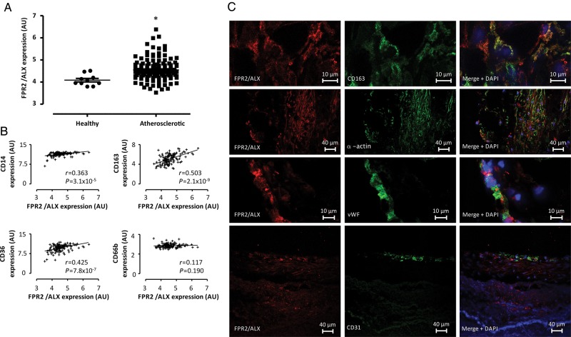
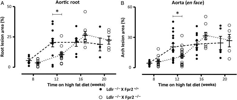
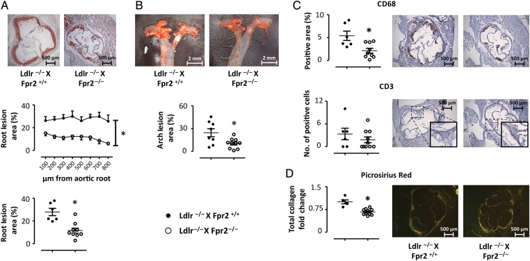
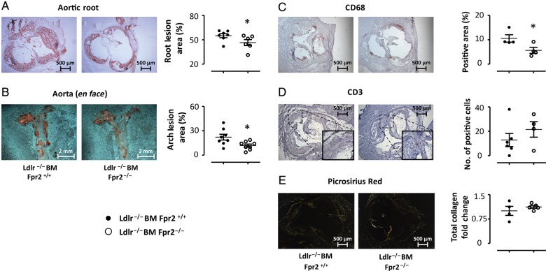
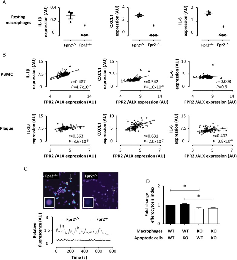
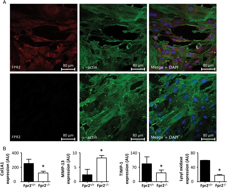
AIMS: The formyl peptide receptor (FPR) subtype FPR2/ALX transduces pro-inflammatory responses and participates in the resolution of inflammation depending on activation. The aim of the present study was to unravel the role of FPR2/ALX signalling in atherosclerosis. METHODS AND RESULTS: Expression of FPR2/ALX was analysed in 127 human carotid atherosclerotic lesions and revealed that this receptor was expressed on macrophages, smooth muscle cells (SMCs), and endothelial cells. Furthermore, FPR2/ALX mRNA levels were significantly up-regulated in atherosclerotic lesions compared with healthy vessels. In multiple regression, age, creatinine, and clinical signs of increased cerebral ischaemia were independent predictors of FPR2/ALX expression. To provide mechanistic insights into these observations, we generated Ldlr(-/-)xFpr2(-/-) mice, which exhibited delayed atherosclerosis development and less macrophage infiltration compared with Ldlr(-/-)xFpr2(+/+) mice. These findings were reproduced by transplantation of Fpr2(-/-) bone marrow into Ldlr(-/-) mice and further extended by in vitro experiments, demonstrating a lower inflammatory state in Fpr2(-/-) macrophages. FPR2/ALX expression correlated with chemo- and cytokines in human atherosclerotic lesions and leucocytes. Finally, atherosclerotic lesions in Ldlr(-/-)xFpr2(-/-) mice exhibited decreased collagen content, and Fpr2(-/-) SMCs exhibited a profile of increased collagenase and decreased collagen production pathways. CONCLUSION: FPR2/ALX is proatherogenic due to effects on bone marrow-derived cells, but promoted a more stable plaque phenotype through effects on SMCs. Taken together, these results suggest a dual role of FPR2/ALX signalling in atherosclerosis by way of promoting disease progression and but increasing plaque stability.
Overview
The claims and statistics of this paper haven’t been checked yet.
Run the check — free, no account needed. It reads the open-access text and checks the sources it cites.
Integrity signals
Registry
Cites
Statistics
Figures
Verification
Claims the paper makes
The claims and statistics of this paper haven’t been checked yet.
Run the check — free, no account needed. It reads the open-access text and checks the sources it cites.
Claims other papers make about this one
Each is checked against this paper’s own text: does it say what the citing paper says it does?
The source backs part of the claim, not all of it — or backs it for a different population, condition or comparison.
In the citing paper
“These receptors are expressed in human aortic, coronary and carotid atherosclerotic lesions104–106, indicating that their pro-resolving ligands exert local effects within the vascular wall (TABLE 1).”
From Inflammation and its resolution in atherosclerosis: mediators and therapeutic opportunities (2019) · cites this paper as [104]
Experimental — accuracy not yet measuredIn the citing paper
“A study on genetic targeting of the murine ALX/FPR2 homologue in hyperlipidaemic mice provided arguments in favour of this hypothesis by showing that ALX/FPR2 deficiency reduced atherosclerosis consistently in different mouse models99,104.”
From Inflammation and its resolution in atherosclerosis: mediators and therapeutic opportunities (2019) · cites this paper as [104]
Self-check
Percentages, sums and reported differences re-computed from the paper’s own numbers.
The self-check needs the paper’s full text.
Citations
Cites
Ridker, P. M., Hennekens, C. H., Buring, J. E., & Rifai, N. (2000) · New England Journal of Medicine · doi:10.1056/nejm200003233421202 · Cited by 5,850
Serhan, C. N., Hong, S., Gronert, K., Colgan, S. P., Devchand, P. R., Mirick, G., & Moussignac, R.-L. (2002) · The Journal of Experimental Medicine · doi:10.1084/jem.20020760 · Cited by 1,650
Macrophage death and defective inflammation resolution in atherosclerosis
Tabas, I. (2010) · Nature Reviews Immunology · doi:10.1038/nri2675 · Cited by 1,120
Ye, R. D., Boulay, F., Wang, J. M., Dahlgren, C., Gerard, C. J., Parmentier, M., Serhan, C. N., & Murphy, P. M. (2009) · Pharmacological Reviews · doi:10.1124/pr.109.001578 · Cited by 778
Resolvin D1 binds human phagocytes with evidence for proresolving receptors
Krishnamoorthy, S., Recchiuti, A., Chiang, N., Yacoubian, S., Lee, C., Yang, R., Petasis, N. A., & Serhan, C. N. (2010) · Proceedings of the National Academy of Sciences · doi:10.1073/pnas.0907342107 · Cited by 752
Immune Effector Mechanisms Implicated in Atherosclerosis: From Mice to Humans
Libby, P., Lichtman, A. H., & Hansson, G. K. (2013) · Immunity · doi:10.1016/j.immuni.2013.06.009 · Cited by 685
Self-citationDisruption of the 12/15-lipoxygenase gene diminishes atherosclerosis in apo E–deficient mice
Cyrus, T., Witztum, J. L., Rader, D. J., Tangirala, R., Fazio, S., Linton, M. F., & Funk, C. D. (1999) · Journal of Clinical Investigation · doi:10.1172/jci5897 · Cited by 523
The Lipoxin Receptor ALX: Potent Ligand-Specific and Stereoselective Actions in Vivo
Chiang, N., Serhan, C. N., Dahlén, S., Drazen, J. M., Hay, D. W. P., Rovati, G. E., Shimizu, T., Yokomizo, T., & Brink, C. (2006) · Pharmacological Reviews · doi:10.1124/pr.58.3.4 · Cited by 464
Elssner, A., Duncan, M., Gavrilin, M. A., & Wewers, M. D. (2004) · The Journal of Immunology · doi:10.4049/jimmunol.172.8.4987 · Cited by 425
Merched, A. J., Ko, K., Gotlinger, K. H., Serhan, C. N., & Chan, L. (2008) · The FASEB Journal · doi:10.1096/fj.08-112201 · Cited by 414
Cited by
Sorted by citations
Inflammation and its resolution in atherosclerosis: mediators and therapeutic opportunities
Bäck, M., Yurdagul, A., Tabas, I., Öörni, K., & Kovanen, P. T. (2019) · Nature Reviews Cardiology · doi:10.1038/s41569-019-0169-2 · Cited by 1,341
Self-citationTargeting inflammation in atherosclerosis — from experimental insights to the clinic
Soehnlein, O., & Libby, P. (2021) · Nature Reviews Drug Discovery · doi:10.1038/s41573-021-00198-1 · Cited by 1,085
Inflammation and plaque vulnerability
Hansson, G. K., Libby, P., & Tabas, I. A. (2015) · Journal of Internal Medicine · doi:10.1111/joim.12406 · Cited by 809
Self-citationMore friend than foe: the emerging role of neutrophils in tissue repair
Peiseler, M., & Kubes, P. (2019) · Journal of Clinical Investigation · doi:10.1172/jci124616 · Cited by 423
The role of non-resolving inflammation in atherosclerosis
Kasikara, C., Doran, A. C., Cai, B., & Tabas, I. (2018) · Journal of Clinical Investigation · doi:10.1172/jci97950 · Cited by 266
Pro-resolving lipid mediators: regulators of inflammation, metabolism and kidney function
Brennan, E. P., Kantharidis, P., Cooper, M. E., & Godson, C. (2021) · Nature Reviews Nephrology · doi:10.1038/s41581-021-00454-y · Cited by 284
New pro-resolving n-3 mediators bridge resolution of infectious inflammation to tissue regeneration
Serhan, C. N., Chiang, N., & Dalli, J. (2017) · Molecular Aspects of Medicine · doi:10.1016/j.mam.2017.08.002 · Cited by 253
Sansbury, B. E., & Spite, M. R. (2016) · Circulation Research · doi:10.1161/circresaha.116.307308 · Cited by 238
Inflammation Resolution: Implications for Atherosclerosis
Doran, A. C. (2022) · Circulation Research · doi:10.1161/circresaha.121.319822 · Cited by 181
Lipoxin and Resolvin Receptors Transducing the Resolution of Inflammation in Cardiovascular Disease
Pirault, J., & Bäck, M. (2018) · Frontiers in Pharmacology · doi:10.3389/fphar.2018.01273 · Cited by 174
Self-citationPotential of anti-inflammatory agents for treatment of atherosclerosis
Chistiakov, D. A., Melnichenko, A. А., Grechko, A. V., Myasoedova, V. A., & Orekhov, A. N. (2018) · Experimental and Molecular Pathology · doi:10.1016/j.yexmp.2018.01.008 · Cited by 169
Fredman, G., & Serhan, C. N. (2024) · Nature Reviews Cardiology · doi:10.1038/s41569-023-00984-x · Cited by 176
Dahlgren, C., Gabl, M., Holdfeldt, A., Winther, M., & Forsman, H. F. (2016) · Biochemical Pharmacology · doi:10.1016/j.bcp.2016.04.014 · Cited by 141
Laguna-Fernandez, A., Checa, A., Carracedo, M., Artiach, G., Petri, M. H., Baumgartner, R., Forteza, M. J., Jiang, X., Andonova, T., Walker, M. E., et al. (2018) · Circulation · doi:10.1161/circulationaha.117.032801 · Cited by 141
Self-citationAspirin‐triggered lipoxin A4 inhibits atherosclerosis progression in apolipoprotein E−/− mice
Petri, M. H., Laguna‐Fernandez, A., Arnardottir, H., Wheelock, C. E., Perretti, M., Hansson, G. K., & Bäck, M. (2017) · British Journal of Pharmacology · doi:10.1111/bph.13707 · Cited by 120
Self-citationZhuang, Y., Liu, H., Zhou, X. E., Verma, R. K., de Waal, P. W., Jang, W., Xu, T.-H., Wang, L., Meng, X. M., Zhao, G., et al. (2020) · Nature Communications · doi:10.1038/s41467-020-14728-9 · Cited by 112
Annexin-A1: Therapeutic Potential in Microvascular Disease
Purvis, G. S. D., Solito, E., & Thiemermann, C. (2019) · Frontiers in Immunology · doi:10.3389/fimmu.2019.00938 · Cited by 107
Regulation of atherosclerotic plaque inflammation
Bäck, M., Weber, C., & Lutgens, E. (2015) · Journal of Internal Medicine · doi:10.1111/joim.12367 · Cited by 98
Self-citationTargeting immune cell recruitment in atherosclerosis
Döring, Y., van der Vorst, E. P. C., & Weber, C. (2024) · Nature Reviews Cardiology · doi:10.1038/s41569-024-01023-z · Cited by 99
Carracedo, M., Artiach, G., Arnardottir, H. H., & Bäck, M. (2019) · Seminars in Immunopathology · doi:10.1007/s00281-019-00767-y · Cited by 100
Self-citationThe resolvin D1 receptor GPR32 transduces inflammation resolution and atheroprotection
Arnardottir, H., Thul, S., Pawelzik, S., Karadimou, G., Artiach, G., Gallina, A. L., Mysdotter, V., Carracedo, M., Tarnawski, L., Caravaca, A. S., et al. (2021) · Journal of Clinical Investigation · doi:10.1172/jci142883 · Cited by 87
Self-citationKain, V., Liu, F., Kozlovskaya, V., Ingle, K. A., Bolisetty, S., Agarwal, A., Khedkar, S. A., Prabhu, S. D., Kharlampieva, E., & Halade, G. V. (2017) · Scientific Reports · doi:10.1038/s41598-017-10441-8 · Cited by 81
Qin, C. X., Norling, L. V., Vecchio, E. A., Brennan, E. P., May, L. T., Wootten, D. L., Godson, C., Perretti, M., & Ritchie, R. H. (2022) · British Journal of Pharmacology · doi:10.1111/bph.15919 · Cited by 76
den Hartigh, L. J., May, K. S., Zhang, X., Chait, A., & Blaser, M. J. (2023) · Frontiers in Cardiovascular Medicine · doi:10.3389/fcvm.2023.1197432 · Cited by 81
Serum amyloid A and atherosclerosis
Getz, G. S., Krishack, P. A., & Reardon, C. A. (2016) · Current Opinion in Lipidology · doi:10.1097/mol.0000000000000331 · Cited by 68
Tourki, B., Kain, V., Pullen, A. B., Norris, P. C., Patel, N., Arora, P. K., Leroy, X., Serhan, C. N., & Halade, G. V. (2019) · Molecular Metabolism · doi:10.1016/j.molmet.2019.10.008 · Cited by 68
Wu, B., Mottola, G., Schaller, M. S., Upchurch, G. R., & Conte, M. S. (2017) · Molecular Aspects of Medicine · doi:10.1016/j.mam.2017.07.005 · Cited by 66
Maciuszek, M., Cacace, A., Brennan, E. P., Godson, C., & Chapman, T. M. (2021) · European Journal of Medicinal Chemistry · doi:10.1016/j.ejmech.2021.113167 · Cited by 63
Petri, M. H., Laguna-Fernandez, A., Tseng, C.-N., Hedin, U., Perretti, M., & Bäck, M. (2015) · International Journal of Cardiology · doi:10.1016/j.ijcard.2014.11.010 · Cited by 60
Self-citationBeyond host defense and tissue injury: the emerging role of neutrophils in tissue repair
Rizo-Téllez, S. A., & Filep, J. G. (2024) · American Journal of Physiology-Cell Physiology · doi:10.1152/ajpcell.00652.2023 · Cited by 65
Petri, M. H., Thul, S., Andonova, T., Liljeqvist, M. L., Jin, H. H., Skenteris, N.-T., Arnardottir, H. H., Mäegdefessel, L., Caidahl, K., Perretti, M., et al. (2018) · JACC Basic to Translational Science · doi:10.1016/j.jacbts.2018.08.005 · Cited by 52
Self-citationTorp, M., Vaage, J., & Stensløkken, K. (2023) · Acta Physiologica · doi:10.1111/apha.13920 · Cited by 54
Formyl peptide receptor 2 regulates monocyte recruitment to promote intestinal mucosal wound repair
Birkl, D., O’Leary, M. N., Quirós, M., Azcutia, V., Schaller, M. A., Reed, M., Nishio, H., Keeney, J. N., Neish, A. S., Lukacs, N. W., et al. (2019) · The FASEB Journal · doi:10.1096/fj.201901163r · Cited by 49
Formyl peptide receptor 2 is an emerging modulator of inflammation in the liver
Lee, C., Han, J., & Jung, Y. (2023) · Experimental & Molecular Medicine · doi:10.1038/s12276-023-00941-1 · Cited by 52
Formyl Peptide Receptors in Cellular Differentiation and Inflammatory Diseases
Lee, H. Y., Lee, M., & Bae, Y. (2017) · Journal of Cellular Biochemistry · doi:10.1002/jcb.25877 · Cited by 48
Personalized Nutrition and -Omics
Chaudhary, N., Kumar, V., Sangwan, P., Pant, N. C., Saxena, A., Joshi, S., & Yadav, A. N. (2020) · Elsevier eBooks · doi:10.1016/b978-0-08-100596-5.22880-1 · Cited by 55
Li, X., Yang, Y., Wang, Z., Jiang, S., Meng, Y., Song, X., Zhao, L., Zou, L., Li, M., & Yu, T. (2021) · International Journal of Biological Sciences · doi:10.7150/ijbs.62506 · Cited by 49
Caso, V. M., Manzo, V., Cimmino, T. P., Conti, V., Caso, P., Esposito, G., Russo, V., Filippelli, A., Ammendola, R., & Cattaneo, F. (2021) · Life · doi:10.3390/life11030243 · Cited by 45
Perivascular delivery of resolvin D1 inhibits neointimal hyperplasia in a rabbit vein graft model
Wu, B., Werlin, E. C., Chen, M., Mottola, G., Chatterjee, A., Lance, K. D., Bernards, D. A., Sansbury, B. E., Spite, M. R., Desai, T. A., et al. (2018) · Journal of Vascular Surgery · doi:10.1016/j.jvs.2018.05.206 · Cited by 43
Carracedo, M., Artiach, G., Witasp, A., Clària, J., Carlström, M., Laguna-Fernández, A., Stenvinkel, P., & Bäck, M. (2018) · Cardiovascular Research · doi:10.1093/cvr/cvy316 · Cited by 44
Self-citationBardin, M., Pawelzik, S., Lagrange, J., Mahdi, A., Arnardottir, H. H., Regnault, V., Fève, B., Lacolley, P. J., Michel, J., Mercier, N., et al. (2022) · Biochemical Pharmacology · doi:10.1016/j.bcp.2022.115075 · Cited by 43
Self-citationSingle-Cell Analyses Identify Dysfunctional CD16+ CD8 T Cells in Smokers
Martos, S. N., Campbell, M. R., Lozoya, O. A., Wang, X., Bennett, B. D., Thompson, I. J. B., Wan, M., Pittman, G. S., & Bell, D. A. (2020) · Cell Reports Medicine · doi:10.1016/j.xcrm.2020.100054 · Cited by 45
Specialized pro-resolving lipid mediators in cardiovascular disease, diagnosis, and therapy
Kim, A. S., & Conte, M. S. (2020) · Advanced Drug Delivery Reviews · doi:10.1016/j.addr.2020.07.011 · Cited by 42
Formyl peptide receptor 2 and heart disease
Lupisella, J. A., Shirude, P. S., Wurtz, N. R., & Garcia, R. A. (2022) · Seminars in Immunology · doi:10.1016/j.smim.2022.101602 · Cited by 39
Lipoxin-mediated signaling: ALX/FPR2 interaction and beyond
Sánchez‐García, S., Jaén, R. I., Fernández‐Velasco, M., Delgado, C., Boscá, L., & Prieto, P. (2023) · Pharmacological Research · doi:10.1016/j.phrs.2023.106982 · Cited by 40
Annexin A protein family in atherosclerosis
Li, Y., Wang, Y., Huang, L., Zhao, Y.-Y., Chen, L.-H., & Zhang, C. (2022) · Clinica Chimica Acta · doi:10.1016/j.cca.2022.05.009 · Cited by 37
Mai, J., Liu, W., Fang, Y., Zhang, S., Qiu, Q., Yang, Y., Wang, X., Huang, T., Zhang, H., Xie, Y., et al. (2018) · Atherosclerosis · doi:10.1016/j.atherosclerosis.2018.09.025 · Cited by 34
Targeting formyl peptide receptors to facilitate the resolution of inflammation
Filep, J. G., Sekheri, M., & Kebir, D. E. (2018) · European Journal of Pharmacology · doi:10.1016/j.ejphar.2018.06.025 · Cited by 35
Specialized Pro-Resolving Lipid Mediators: New Therapeutic Approaches for Vascular Remodeling
del Campo, L. S. D., Rodrigues‐Díez, R., Salaíces, M., Briones, A. M., & García‐Redondo, A. B. (2022) · International Journal of Molecular Sciences · doi:10.3390/ijms23073592 · Cited by 36
Apoproteins E, A-I, and SAA in Macrophage Pathobiology Related to Atherogenesis
Getz, G. S., & Reardon, C. A. (2019) · Frontiers in Pharmacology · doi:10.3389/fphar.2019.00536 · Cited by 36
Interferon-β regulates proresolving lipids to promote the resolution of acute airway inflammation
Sekheri, M., Rizo-Téllez, S. A., Othman, A., Kebir, D. E., & Filep, J. G. (2022) · Proceedings of the National Academy of Sciences · doi:10.1073/pnas.2201146119 · Cited by 32
Cammalleri, M., Monte, M. D., Locri, F., Marsili, S., Lista, L., ROSA, M. D., Pavone, V., Rusciano, D., & Bagnoli, P. (2017) · Journal of Diabetes Research · doi:10.1155/2017/2904150 · Cited by 29
Artiach, G., Carracedo, M., Clària, J., Laguna-Fernández, A., & Bäck, M. (2018) · Frontiers in Pharmacology · doi:10.3389/fphar.2018.01327 · Cited by 29
Self-citationG-Protein Coupled Receptor Targeting on Myeloid Cells in Atherosclerosis
van der Vorst, E. P. C., Peters, L. J. F., Müller, M., Gencer, S., Yan, Y., Weber, C., & Döring, Y. (2019) · Frontiers in Pharmacology · doi:10.3389/fphar.2019.00531 · Cited by 29
Kraft, J. D., Blomgran, R., Bergström, I., Soták, M., Clark, M., Rani, A., Rajan, M. R., Dalli, J., Nyström, S., Quiding‐Järbrink, M., et al. (2022) · The FASEB Journal · doi:10.1096/fj.202101219rr · Cited by 29
Chait, A., Wang, S., Goodspeed, L., Gomes, D. J., Turk, K. E., Wietecha, T., Tang, J., Storey, C., O’Brien, K. D., Rubinow, K. B., et al. (2021) · Arteriosclerosis Thrombosis and Vascular Biology · doi:10.1161/atvbaha.121.316066 · Cited by 28
Molecular Pharmacology of Inflammation Resolution in Atherosclerosis
Kotlyarov, S. N., & Котлярова, А. А. (2022) · International Journal of Molecular Sciences · doi:10.3390/ijms23094808 · Cited by 31
Su, C., Zhang, J., Zhao, F., Xu, D., Pan, J., & Liu, T. (2023) · Frontiers in Immunology · doi:10.3389/fimmu.2023.1091753 · Cited by 27
Extrinsic and intrinsic modulators of inflammation-resolution signaling in heart failure
Rao, A., Gupta, A., Kain, V., & Halade, G. V. (2023) · American Journal of Physiology-Heart and Circulatory Physiology · doi:10.1152/ajpheart.00276.2023 · Cited by 27
Specialized Proresolving Lipid Mediators: A Potential Therapeutic Target for Atherosclerosis
Salazar, J. J., Pírela, D., Nava, M., Castro, A. V., Angarita, L., Parra, H., Durán‐Agüero, S., Rojas-Gómez, D. M., Galbán, N., Áñez, R., et al. (2022) · International Journal of Molecular Sciences · doi:10.3390/ijms23063133 · Cited by 26
Ganekal, P., Vastrad, B. M., Kavatagimath, S., Vastrad, C. M., & Kotrashetti, S. (2023) · Medicina · doi:10.3390/medicina59020309 · Cited by 22
Lipoxin A4 improves cardiac remodeling and function in diabetes-associated cardiac dysfunction
Fu, T., Mohan, M., Bose, M., Brennan, E. P., Kiriazis, H., Deo, M., Nowell, C. J., Godson, C., Cooper, M. E., Zhao, P., et al. (2024) · Cardiovascular Diabetology · doi:10.1186/s12933-024-02501-x · Cited by 27
Acute-serum amyloid A and A-SAA-derived peptides as formyl peptide receptor (FPR) 2 ligands
Salama, S. A., Gouwy, M., Damme, J. V., & Struyf, S. (2023) · Frontiers in Endocrinology · doi:10.3389/fendo.2023.1119227 · Cited by 22
Barua, J. D., Omit, S. B. S., Rana, H. K., Podder, N. K., Chowdhury, U. N., & Rahman, M. H. (2022) · Cardiovascular Therapeutics · doi:10.1155/2022/9034996 · Cited by 20
Karayiğit, O., Nurkoç, S. G., Başyiğit, F., & Kızıltunç, E. (2022) · Medical Principles and Practice · doi:10.1159/000527064 · Cited by 20
DAMP signaling networks: from receptors to diverse pathophysiological functions
Chen, R., Zou, J., Zhong, X. Y., Liu, J., Kang, R., & Tang, D. (2025) · Journal of Advanced Research · doi:10.1016/j.jare.2025.07.047 · Cited by 26
Chen, R., Li, J., Zhou, J., Wang, Y., Zhao, X., Li, N., Liu, W., Liu, C., Zhou, P., Chen, Y., et al. (2022) · Pharmacological Research · doi:10.1016/j.phrs.2022.106618 · Cited by 18
Pro-resolving lipid mediators in diseases: exploring the molecular basis and clinical implication
Li, C., Wang, Z., Yang, Y., Jiang, Q., Jiang, Y., Xiao, J., Shen, L., Wu, W., & Li, C. (2026) · Molecular Biomedicine · doi:10.1186/s43556-025-00400-5 · Cited by 22
Fukuishi, N., Takahama, K., Kurosaki, H., Ono, S., & Asai, H. (2025) · International Journal of Molecular Sciences · doi:10.3390/ijms26041491 · Cited by 18
Chen, Y., Zheng, Y., Xin, L., Zhong, S., Liu, A., Lai, W., Liu, L., Lin, C., Liao, C., Zeng, J., et al. (2019) · Biomedicine & Pharmacotherapy · doi:10.1016/j.biopha.2019.109099 · Cited by 16
Liu, L., Yu, Y., Hu, L., Dong, Q., Hu, F., Zhu, L., Liang, Q., Yu, L., Bao, H.-H., & Cheng, X. (2021) · Medical Science Monitor · doi:10.12659/msm.928366 · Cited by 14
Lipoxins as Modulators of Diseases
Saqib, U., Pandey, M., Vyas, A., Patidar, P., Hajela, S., Ali, A., Tiwari, M., Sarkar, S., Yadav, N., Patel, S. S., et al. (2025) · Cells · doi:10.3390/cells14161244 · Cited by 14
Differential regulation of monocytic expression of leukotriene and lipoxin receptors
Petri, M. H., Thul, S., Овчинникова, О. А., & Bäck, M. (2015) · Prostaglandins & Other Lipid Mediators · doi:10.1016/j.prostaglandins.2015.07.005 · Cited by 13
Self-citationExploiting oxidized lipids and the lipid‐binding GPCRs against cardiometabolic diseases
Guimarães, R. C., Gonçalves, T. T., & Leiria, L. O. (2020) · British Journal of Pharmacology · doi:10.1111/bph.15321 · Cited by 15
Val‐Blasco, A., Prieto, P., Jaén, R. I., Fernández, M. G., Pajares, M., Doménech, N., Terrón, V., Tamayo, M., Jorge, I., Vázquez, J., et al. (2022) · JACC Basic to Translational Science · doi:10.1016/j.jacbts.2022.01.009 · Cited by 13
DHA, RvD1, RvD5, and MaR1 reduce human coronary arteries contractions induced by PGE2
Bouhadoun, A., Manikpurage, H. D., Deschildre, C., Zalghout, S., Dubourdeau, M., Urbach, V., Ho‐Tin‐Noé, B., Deschamps, L., Michel, J., Longrois, D., et al. (2022) · Prostaglandins & Other Lipid Mediators · doi:10.1016/j.prostaglandins.2022.106700 · Cited by 13
Spite, M. R., & Fredman, G. (2023) · Advances in pharmacology · doi:10.1016/bs.apha.2022.12.005 · Cited by 13
Hughes, F. M., Harper, S. N., Nosé, B. D., Allkanjari, A., Zheng, M., Jin, H., & Purves, J. T. (2021) · American Journal of Physiology-Renal Physiology · doi:10.1152/ajprenal.00205.2021 · Cited by 14
wRAPping Up Early Monocyte and Neutrophil Recruitment in Atherogenesis via AnnexinA1/FPR2 Signaling
Butcher, M. J., & Galkina, E. (2015) · Circulation Research · doi:10.1161/circresaha.115.305920 · Cited by 13
Thakur, M., & Tupe, R. S. (2022) · Prostaglandins & Other Lipid Mediators · doi:10.1016/j.prostaglandins.2022.106698 · Cited by 13
Cheng, W., Zhang, J., Peng, S., Liu, J., Xu, Y. Q., Zhao, M., Xu, S., Pan, W. D., Yin, Z., Zheng, Z., et al. (2023) · Journal of Hypertension · doi:10.1097/hjh.0000000000003610 · Cited by 10
Basic Mechanisms of Atherosclerosis
Bäck, M., & Hansson, G. K. (2017) · Elsevier eBooks · doi:10.1016/b978-0-323-42880-4.00004-2 · Cited by 12
Self-citationZhao, A., Yang, Y., Yang, Y., Chi, Z., & Sun, Y. (2024) · American Journal of Reproductive Immunology · doi:10.1111/aji.70017 · Cited by 10
Taylor, L. C., Arthur, G., Cruz, M. D. C., Stec, D. E., & Badmus, O. O. (2024) · International Journal of Translational Medicine · doi:10.3390/ijtm4040052 · Cited by 12
Increased atherogenic index in the general hearing loss population
Zhang, H., Wang, D., Ma, H., Ren, Y., Li, C., Zheng, Y.-H., Dai, X., Yang, L., & Xu, L. (2020) · Open Medicine · doi:10.1515/med-2020-0003 · Cited by 9
Inflammation-resolution signalling in cardiac repair, remodelling, and heart failure
Halade, G. V., Bäck, M., & Kain, V. (2025) · European Heart Journal Open · doi:10.1093/ehjopen/oeaf157 · Cited by 9
Self-citationUnleashing the power of formyl peptide receptor 2 in cardiovascular disease
Prevete, N., Poto, R., Marone, G., & Varricchi, G. (2023) · Cytokine · doi:10.1016/j.cyto.2023.156298 · Cited by 8
Proresolving receptor tames inflammation in atherosclerosis
Mena, H. A., & Spite, M. R. (2021) · Journal of Clinical Investigation · doi:10.1172/jci155240 · Cited by 8
Millar, B., & de Gaetano, M. (2023) · Frontiers in Pharmacology · doi:10.3389/fphar.2023.1125858 · Cited by 7
Maciuszek, M., Ortega‐Gómez, A., Maas, S. L., Garrido‐Mesa, J., Ferraro, B., Perretti, M., Merritt, A. T., Nicolaes, G. A. F., Soehnlein, O., & Chapman, T. M. (2021) · European Journal of Medicinal Chemistry · doi:10.1016/j.ejmech.2021.113805 · Cited by 6
Immune-Related Genes in the Pathogenesis of Atherosclerosis: Based on Sex Differences
Zhang, P., Lin, H., Guo, Y. P., Peng, F., & Meng, L. (2023) · Journal of Inflammation Research · doi:10.2147/jir.s429247 · Cited by 5
Differential gene expression and miRNA regulatory network in coronary slow flow
Sun, L., Wang, J., Lei, J., Zhang, Y., Zhang, Y., Zhang, Y., & Xing, S. (2024) · Scientific Reports · doi:10.1038/s41598-024-58745-w · Cited by 5
ALX-chemy: adding spice to the inflammatory soup of atherosclerosis
Libby, P. (2014) · Cardiovascular Research · doi:10.1093/cvr/cvu245 · Cited by 5
Formyl peptide receptor 1 and its antagonist T0080 in atherosclerosis
Li, Y.-J., Zhao, X., Wu, S., Yao, N., Zhang, X., Liu, Y., Tian, X., Li, Y., Gao, B., Johnston, S. C., et al. (2025) · Cell Death and Differentiation · doi:10.1038/s41418-025-01506-7 · Cited by 4
Kim, Y., Ahn, S. Y., Sung, D. K., Chang, Y. S., & Park, W. S. (2022) · International Journal of Molecular Sciences · doi:10.3390/ijms231810604 · Cited by 4
Formyl peptide receptor 2: a potential therapeutic target for inflammation-related diseases
Wang, J., Miao, Z., Gao, Y., Xie, Z., Liu, M., & Zou, W. (2025) · Pharmacological Reports · doi:10.1007/s43440-025-00704-x · Cited by 7
Fine-tuning inflammation-resolution programs
Fredman, G., Sadhu, S., & Rymut, N. (2016) · Current Opinion in Clinical Nutrition & Metabolic Care · doi:10.1097/mco.0000000000000351 · Cited by 4
Kagaya, H., Kim, A. S., Chen, M., Lin, P., Yin, X., Spite, M. R., & Conte, M. S. (2024) · Physiological Reports · doi:10.14814/phy2.16178 · Cited by 3
Xiao, S., & Kuang, C. (2022) · PubMed · Cited by 5
Vojinović, D., Kavousi, M., Ghanbari, M., Brouwer, R. W. W., van Rooij, J., van den Hout, M. C. G. N., Kraaij, R., van IJcken, W. F. J., Uitterlinden, A. G., van Duijn, C. M., et al. (2018) · Frontiers in Genetics · doi:10.3389/fgene.2018.00420 · Cited by 4
Metadata
- Authors
- Petri, M. H., Laguna-Fernández, A., Gonzalez-Diez, M., Paulsson-Berne, G., Hansson, G. K., & Bäck, M.
- Affiliations
- Karolinska University Hospital (SE)
- Corresponding author
- Bäck, Magnus
- Venue
- Cardiovascular Research
- Publisher
- Oxford University Press (OUP)
- Published
- 2014-10-23
- Volume
- 105
- Issue
- 1
- Pages
- 65-74
- ISSN
- 0008-6363 (ISSN-L)
- 1755-3245
- PMC
- PMC4277257
- OpenAlex
- W2000987582
- Cited by
- 125
- References
- 44
- Citations by year
- 2026: 3 · 2025: 11 · 2024: 9 · 2023: 13 · 2022: 20 · 2021: 14 · 2020: 7 · 2019: 10 · 2018: 13 · 2017: 10 · 2016: 7 · 2015: 6 · 2014: 2
- FWCI
- 3.81
- Type
- journal article
- Language
- en
- Open access
- hybrid
- doi.org
- Licence
- cc-by-nc
- Locations
- Cardiovascular Research (publishedVersion, cc-by-nc)
- PubMed Central (publishedVersion, cc-by-nc)
- PubMed (publishedVersion)
- Funders
- Stockholms Läns Landsting (20120297)
- Vetenskapsrådet (2011-02988, 20120827, 2012-02577, 20120474)
- Topics
- Life Sciences › Immunology and Microbiology › Immunology › Atherosclerosis and Cardiovascular Diseases
- Health Sciences › Medicine › Cardiology and Cardiovascular Medicine › Cerebrovascular and Carotid Artery Diseases
- Life Sciences › Biochemistry, Genetics and Molecular Biology › Biochemistry › Cholesterol and Lipid Metabolism
- Keywords
- FPR2/ALX, formyl peptide receptor, atherosclerosis, plaque stability, macrophage infiltration, smooth muscle cells, collagen synthesis, collagenase, inflammation, bone marrow-derived cells, Ldlr knockout mice, cerebral ischemia
- MeSH terms
- Aged, Aged, 80 and over, Animals, Carotid Artery Diseases, Collagen, Female, Humans, Macrophages, Male, Middle Aged, Receptors, LDL, RNA, Messenger, Signal Transduction, Up-Regulation, Bone Marrow Transplantation, Carotid Stenosis, Mice, Knockout, Myocytes, Smooth Muscle, Receptors, Formyl Peptide, Receptors, Lipoxin, Atherosclerosis, Mice, Plaque, Atherosclerotic
About these checks
Registry, retraction, Cites and statistics results are computed, not judged, with one exception: a printed reference that no index lists is read once by a model before it counts as a citation error. Claims are extracted by a model and kept only when their quote appears verbatim in the text; each cited source is then checked, with a model’s help, for whether it says what the claim relies on. Those verdicts can be wrong — read the claim’s quote and the cited source before relying on one.
Statistics and arithmetic are recomputed, not judged.
Confidence bands are shown only for fields calibrated on our annotated test set. None is calibrated yet, so no band is shown.
What counts as a claim ›How we measure confidence ›How we check this ›
Are you an author, or do you see a problem with this page? Request a review
