

The DOI matches its Crossref record; 4 of the 4 references it cites were found in a registry or an index.
In-frame deletion in a novel centrosomal/ciliary protein CEP290/NPHP6 perturbs its interaction with RPGR and results in early-onset retinal degeneration in the rd16 mouse
Overview
The claims and statistics of this paper haven’t been checked yet.
Run the check — free, no account needed. It reads the open-access text and checks the sources it cites.
Integrity signals
Registry
Cites
Statistics
Figures
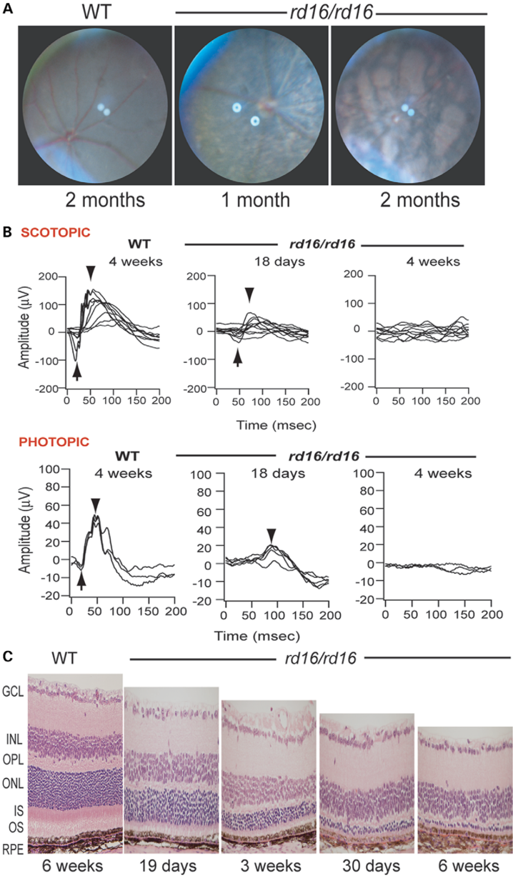
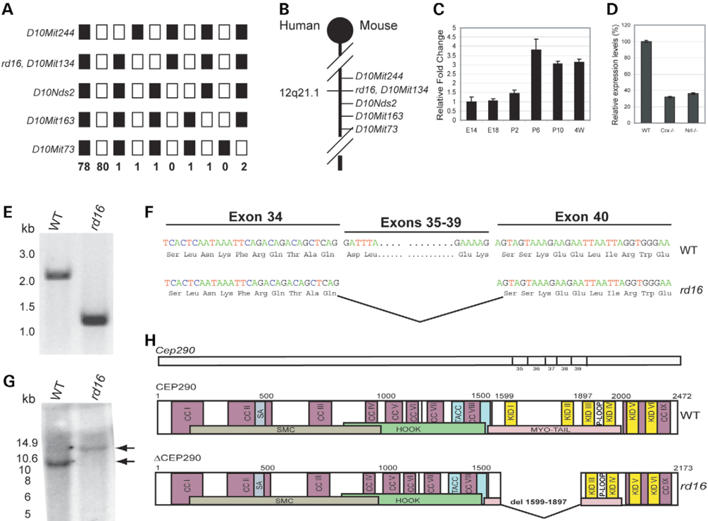
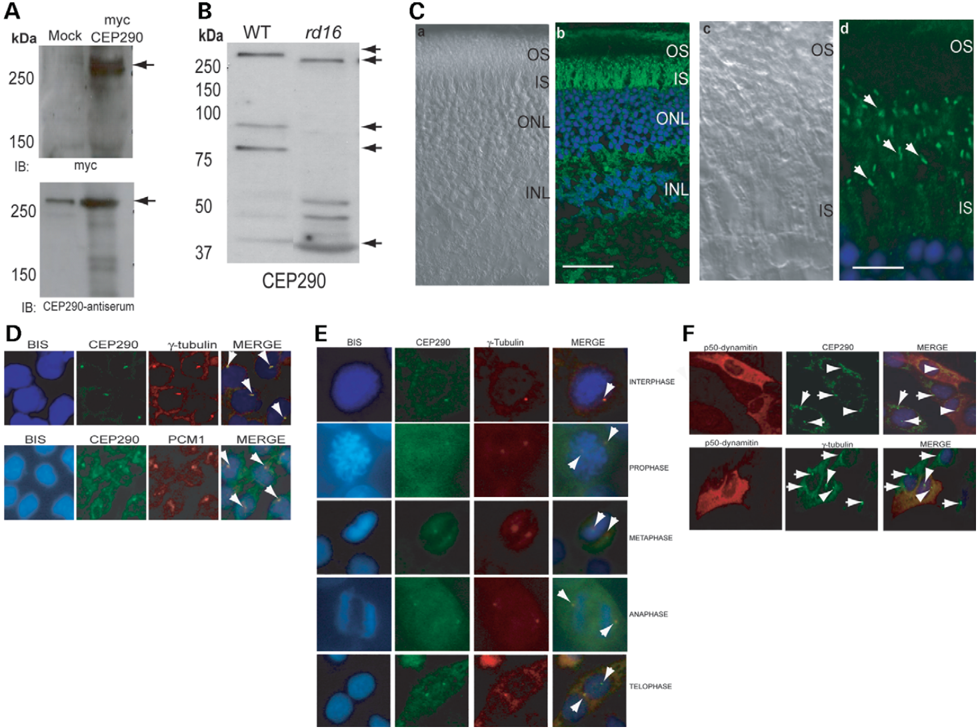
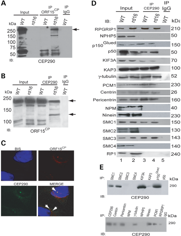
Verification
Claims the paper makes
The claims and statistics of this paper haven’t been checked yet.
Run the check — free, no account needed. It reads the open-access text and checks the sources it cites.
Claims other papers make about this one
Each is checked against this paper’s own text: does it say what the citing paper says it does?
The source backs part of the claim, not all of it — or backs it for a different population, condition or comparison.
In the citing paper
“CEP290 is a ubiquitously expressed, 2,480 amino-acid protein that plays roles in ciliary transport and centrosomal function.11”
From Effect of genome size on AAV vector packaging (2010) · cites this paper as [11]
Experimental — accuracy not yet measuredThe cited source says what the claim attributes to it.
In the citing paper
“The rd16 mouse carries an in-frame deletion in the CEP290 gene and exhibits an early-onset retinal degeneration that is similar to the human disease.11”
From Effect of genome size on AAV vector packaging (2010) · cites this paper as [11]
Self-check
Percentages, sums and reported differences re-computed from the paper’s own numbers.
The self-check needs the paper’s full text.
Citations
Cites
Proteomic characterization of the human centrosome by protein correlation profiling
Andersen, J. S., Wilkinson, C. J., Mayor, T., Mortensen, P., Nigg, E. A., & Mann, M. (2003) · Nature · doi:10.1038/nature02166 · Cited by 1,319
Nrl is required for rod photoreceptor development
Mears, A. J., Kondo, M., Swain, P. K., Takada, Y., Bush, R. A., Saunders, T. L., Sieving, P. A., & Swaroop, A. (2001) · Nature Genetics · doi:10.1038/ng774 · Cited by 912
Self-citationCole, D. G., Diener, D. R., Himelblau, A. L., Beech, P. L., Fuster, J. C., & Rosenbaum, J. L. (1998) · The Journal of Cell Biology · doi:10.1083/jcb.141.4.993 · Cited by 898
Akhmedov, N. B., Piriev, N. I., Chang, B., Rapoport, A. L., Hawes, N. L., Nishina, P. M., Nusinowitz, S., Heckenlively, J. R., Roderick, T. H., Kozak, C. A., et al. (2000) · Proceedings of the National Academy of Sciences · doi:10.1073/pnas.97.10.5551 · Cited by 221
Self-citation
Research works printed but not linked: 43
- Nigg, E.A. (2004) Centrosomes in Development and Disease, Wiley-VCH Verlag GmbH & Co. KGaA. · DOI
- Doxsey, S. (2001) Re-evaluating centrosome function. Nat. Rev. Mol. Cell. Biol., 2, 688–698. · DOI
- Beisson, J. and Wright, M. (2003) Basal body/centriole assembly and continuity. Curr. Opin. Cell. Biol., 15, 96–104. · DOI
- Badano, J.L., Teslovich, T.M. and Katsanis, N. (2005) The centrosome in human genetic disease. Nat. Rev. Genet., 6, 194–205. · DOI
- Young, R.W. (1968) Passage of newly formed protein through the connecting cilium of retina rods in the frog. J. Ultrastruct. Res., 23, 462–473. · DOI
- Marszalek, J.R., Liu, X., Roberts, E.A., Chui, D., Marth, J.D., Williams, D.S. and Goldstein, L.S. (2000) Genetic evidence for selective transport of opsin and arrestin by kinesin-II in mammalian photoreceptors. Cell, 102, 175–187. · DOI
- Besharse, J.C., Baker, S.A., Luby-Phelps, K. and Pazour, G.J. (2003) Photoreceptor intersegmental transport and retinal degeneration: a conserved pathway common to motile and sensory cilia. Adv. Exp. Med. Biol., 533, 157–164. · DOI
- Bowes, C., Li, T., Danciger, M., Baxter, L.C., Applebury, M.L. and Farber, D.B. (1990) Retinal degeneration in the rd mouse is caused by a defect in the beta subunit of rod cGMP-phosphodiesterase. Nature, 347, 677–680. · DOI
- Travis, G.H., Brennan, M.B., Danielson, P.E., Kozak, C.A. and Sutcliffe, J.G. (1989) Identification of a photoreceptor-specific mRNA encoded by the gene responsible for retinal degeneration slow (rds). Nature, 338, 70–73. · DOI
- McLaughlin, M.E., Sandberg, M.A., Berson, E.L. and Dryja, T.P. (1993) Recessive mutations in the gene encoding the beta-subunit of rod phosphodiesterase in patients with retinitis pigmentosa. Nat. Genet., 4, 130–134. · DOI
- Haider, N.B., Jacobson, S.G., Cideciyan, A.V., Swiderski, R., Streb, L.M., Searby, C., Beck, G., Hockey, R., Hanna, D.B., Gorman, S. et al. (2000) Mutation of a nuclear receptor gene, NR2E3, causes enhanced S cone syndrome, a disorder of retinal cell fate. Nat. Genet., 24, 127–131. · DOI · Paper page
- Farrar, G.J., Kenna, P., Jordan, S.A., Kumar-Singh, R., Humphries, M.M., Sharp, E.M., Sheils, D.M. and Humphries, P. (1991) A three-base-pair deletion in the peripherin-RDS gene in one form of retinitis pigmentosa. Nature, 354, 478–480. · DOI
- Chang, B., Hawes, N.L., Hurd, R.E., Davisson, M.T., Nusinowitz, S. and Heckenlively, J.R. (2002) Retinal degeneration mutants in the mouse. Vision Res., 42, 517–525. · DOI · Paper page
- Akimoto, M., Cheng, H., Zhu, D., Brzezinski, J.A., Khanna, R., Filippova, E., Oh, E.C., Jing, Y., Linares, J.-L., Brooks, M. et al. (2006) Targeting of green fluorescent protein to new-born rods by Nrl promoter and temporal expression profiling of flow-sorted photoreceptors. Proc. Natl Acad. Sci. USA, 103, 3890–3895. · DOI
- Blackshaw, S., Harpavat, S., Trimarchi, J., Cai, L., Huang, H., Kuo, W.P., Weber, G., Lee, K., Fraioli, R.E., Cho, S.H. et al. (2004) Genomic analysis of mouse retinal development. PLoS. Biol., 2, E247. · DOI
- Furukawa, T., Morrow, E.M., Li, T., Davis, F.C. and Cepko, C.L. (1999) Retinopathy and attenuated circadian entrainment in Crx-deficient mice. Nat. Genet., 23, 466–470. · DOI
- Nasmyth, K. and Haering, C.H. (2005) The structure and function of SMC and kleisin complexes. Annu. Rev. Biochem., 74, 595–648. · DOI
- Guo, J., Jin, G., Meng, L., Ma, H., Nie, D., Wu, J., Yuan, L. and Shou, C. (2004) Subcellullar localization of tumor-associated antigen 3H11Ag. Biochem. Biophys. Res. Commun., 324, 922–930. · DOI
- Kim, J.C., Badano, J.L., Sibold, S., Esmail, M.A., Hill, J., Hoskins, B.E., Leitch, C.C., Venner, K., Ansley, S.J., Ross, A.J. et al. (2004) The Bardet–Biedl protein BBS4 targets cargo to the pericentriolar region and is required for microtubule anchoring and cell cycle progression. Nat. Genet., 36, 462–470. · DOI
- Dammermann, A. and Merdes, A. (2002) Assembly of centrosomal proteins and microtubule organization depends on PCM-1. J. Cell. Biol., 159, 255–266. · DOI
- Kim, J.C., Ou, Y.Y., Badano, J.L., Esmail, M.A., Leitch, C.C., Fiedrich, E., Beales, P.L., Archibald, J.M., Katsanis, N., Rattner, J.B. et al. (2005) MKKS/BBS6, a divergent chaperonin-like protein linked to the obesity disorder Bardet–Biedl syndrome, is a novel centrosomal component required for cytokinesis. J. Cell. Sci., 118, 1007–1020. · DOI
- Vaughan, K.T. and Vallee, R.B. (1995) Cytoplasmic dynein binds dynactin through a direct interaction between the intermediate chains and p150Glued. J. Cell. Biol., 131, 1507–1516. · DOI
- Hong, D.H., Pawlyk, B., Sokolov, M., Strissel, K.J., Yang, J., Tulloch, B., Wright, A.F., Arshavsky, V.Y. and Li, T. (2003) RPGR isoforms in photoreceptor connecting cilia and the transitional zone of motile cilia. Invest. Ophthalmol. Vis. Sci., 44, 2413–2421. · DOI
- Shu, X., Fry, A.M., Tulloch, B., Manson, F.D., Crabb, J.W., Khanna, H., Faragher, A.J., Lennon, A., He, S., Trojan, P. et al. (2005) RPGR ORF15 isoform co-localizes with RPGRIP1 at centrioles and basal bodies and interacts with nucleophosmin. Hum. Mol. Genet., 14, 1183–1197. · DOI
- Khanna, H., Hurd, T.W., Lillo, C., Shu, X., Parapuram, S.K., He, S., Akimoto, M., Wright, A.F., Margolis, B., Williams, D.S. et al. (2005) RPGR-ORF15, which is mutated in retinitis pigmentosa, associates with SMC1, SMC3, and microtubule transport proteins. J. Biol. Chem., 280, 33580–33587. · DOI
- Vervoort, R., Lennon, A., Bird, A.C., Tulloch, B., Axton, R., Miano, M.G., Meindl, A., Meitinger, T., Ciccodicola, A. and Wright, A.F. (2000) Mutational hot spot within a new RPGR exon in X-linked retinitis pigmentosa. Nat. Genet., 25, 462–466. · DOI
- Breuer, D.K., Yashar, B.M., Filippova, E., Hiriyanna, S., Lyons, R.H., Mears, A.J., Asaye, B., Acar, C., Vervoort, R., Wright, A.F. et al. (2002) A comprehensive mutation analysis of RP2 and RPGR in a North American cohort of families with X-linked retinitis pigmentosa. Am. J. Hum. Genet., 70, 1545–1554. · DOI
- Sharon, D., Sandberg, M.A., Rabe, V.W., Stillberger, M., Dryja, T.P. and Berson, E.L. (2003) RP2 and RPGR mutations and clinical correlations in patients with X-linked retinitis pigmentosa. Am. J. Hum. Genet., 73, 1131–1146. · DOI
- Dryja, T.P., Adams, S.M., Grimsby, J.L., McGee, T.L., Hong, D.H., Li, T., Andreasson, S. and Berson, E.L. (2001) Null RPGRIP1 alleles in patients with Leber congenital amaurosis. Am. J. Hum. Genet., 68, 1295–1298. · DOI
- Otto, E.A., Loeys, B., Khanna, H., Hellemans, J., Sudbrak, R., Fan, S., Muerb, U., O'Toole, J.F., Helou, J., Attanasio, M. et al. (2005) Nephrocystin-5, a ciliary IQ domain protein, is mutated in Senior–Loken syndrome and interacts with RPGR and calmodulin. Nat. Genet., 37, 282–288. · DOI
- Hong, D.H., Yue, G., Adamian, M. and Li, T. (2001) Retinitis pigmentosa GTPase regulator (RPGRr)-interacting protein is stably associated with the photoreceptor ciliary axoneme and anchors RPGR to the connecting cilium. J. Biol. Chem., 276, 12091–12099. · DOI
- Liu, Q., Zuo, J. and Pierce, E.A. (2004) The retinitis pigmentosa 1 protein is a photoreceptor microtubule-associated protein. J. Neurosci., 24, 6427–6436. · DOI
- Hong, D.H., Pawlyk, B.S., Shang, J., Sandberg, M.A., Berson, E.L. and Li, T. (2000) A retinitis pigmentosa GTPase regulator (RPGR)-deficient mouse model for X-linked retinitis pigmentosa (RP3). Proc. Natl Acad. Sci. USA, 97, 3649–3654. · DOI
- Piperno, G., Siuda, E., Henderson, S., Segil, M., Vaananen, H. and Sassaroli, M. (1998) Distinct mutants of retrograde intraflagellar transport (IFT) share similar morphological and molecular defects. J. Cell. Biol., 143, 1591–1601. · DOI
- Pazour, G.J., Baker, S.A., Deane, J.A., Cole, D.G., Dickert, B.L., Rosenbaum, J.L., Witman, G.B. and Besharse, J.C. (2002) The intraflagellar transport protein, IFT88, is essential for vertebrate photoreceptor assembly and maintenance. J. Cell. Biol., 157, 103–113. · DOI
- Nishimura, D.Y., Fath, M., Mullins, R.F., Searby, C., Andrews, M., Davis, R., Andorf, J.L., Mykytyn, K., Swiderski, R.E., Yang, B. et al. (2004) Bbs2-null mice have neurosensory deficits, a defect in social dominance, and retinopathy associated with mislocalization of rhodopsin. Proc. Natl Acad. Sci. USA, 101, 16588–16593. · DOI
- Mykytyn, K., Mullins, R.F., Andrews, M., Chiang, A.P., Swiderski, R.E., Yang, B., Braun, T., Casavant, T., Stone, E.M. and Sheffield, V.C. (2004) Bardet–Biedl syndrome type 4 (BBS4)-null mice implicate Bbs4 in flagella formation but not global cilia assembly. Proc. Natl Acad. Sci. USA, 101, 8664–8669. · DOI
- Li, T., Snyder, W.K., Olsson, J.E. and Dryja, T.P. (1996) Transgenic mice carrying the dominant rhodopsin mutation P347S: evidence for defective vectorial transport of rhodopsin to the outer segments. Proc. Natl Acad. Sci. USA, 93, 14176–14181. · DOI
- Zhao, Y., Hong, D.H., Pawlyk, B., Yue, G., Adamian, M., Grynberg, M., Godzik, A. and Li, T. (2003) The retinitis pigmentosa GTPase regulator (RPGR)-interacting protein: subserving RPGR function and participating in disk morphogenesis. Proc. Natl Acad. Sci. USA, 100, 3965–3970. · DOI
- Avidor-Reiss, T., Maer, A.M., Koundakjian, E., Polyanovsky, A., Keil, T., Subramaniam, S. and Zuker, C.S. (2004) Decoding cilia function: defining specialized genes required for compartmentalized cilia biogenesis. Cell, 117, 527–539. · DOI
- Li, J.B., Gerdes, J.M., Haycraft, C.J., Fan, Y., Teslovich, T.M., May-Simera, H., Li, H., Blacque, O.E., Li, L., Leitch, C.C. et al. (2004) Comparative genomics identifies a flagellar and basal body proteome that includes the BBS5 human disease gene. Cell, 117, 541–552. · DOI
- Sayer, J.A., Otto, E.A., O'Toole, J.F., Nurnberg, G., Kennedy, M.A., Becker, C., Hennies, H.C., Helou, J., Attanasio, M., Fausett, B. et al. (2006) A novel centrosomal protein nephrocystin-6 is mutated in Joubert syndrome and activates transcription factor ATF4/CREB2. Nat. Genet., in press. · DOI
- Cheng, H., Khanna, H., Oh, E.C., Hicks, D., Mitton, K.P. and Swaroop, A. (2004) Photoreceptor-specific nuclear receptor NR2E3 functions as a transcriptional activator in rod photoreceptors. Hum. Mol. Genet., 13, 1563–1575. · DOI · Paper page
Cited by
Sorted by citations
Hildebrandt, F., Benzing, T., & Katsanis, N. (2011) · New England Journal of Medicine · doi:10.1056/nejmra1010172 · Cited by 1,331
Self-citationEffect of genome size on AAV vector packaging
Wu, Z., Yang, H., & Colosi, P. (2010) · Molecular Therapy · doi:10.1038/mt.2009.255 · Cited by 1,029
Development of a gene-editing approach to restore vision loss in Leber congenital amaurosis type 10
Maeder, M. L., Stefanidakis, M., Wilson, C. J., Baral, R., Barrera, L. A., Bounoutas, G. S., Bumcrot, D., Chao, H., Ciulla, D. M., DaSilva, J. A., et al. (2019) · Nature Medicine · doi:10.1038/s41591-018-0327-9 · Cited by 707
Ciliopathies: an expanding disease spectrum
Waters, A. M., & Beales, P. L. (2011) · Pediatric Nephrology · doi:10.1007/s00467-010-1731-7 · Cited by 676
Chapter Two Intraflagellar Transport (IFT)
Pedersen, L. B., & Rosenbaum, J. L. (2008) · Current Topics in Developmental Biology · doi:10.1016/s0070-2153(08)00802-8 · Cited by 521
Metadata
- Authors
- Chang, B., Khanna, H., Hawes, N., Jimeno, D., He, S., Lillo, C., Parapuram, S. K., Cheng, H., Scott, A., Hurd, R. E., Sayer, J. A., Otto, E. A., Attanasio, M., O'Toole, J. F., Jin, G., Shou, C., Hildebrandt, F., Williams, D. S., Heckenlively, J. R., & Swaroop, A.
- Venue
- Human Molecular Genetics
- Publisher
- Oxford University Press (OUP)
- Published
- 2006-04-21
- Volume
- 15
- Issue
- 11
- Pages
- 1847-1857
- ISSN
- 1460-2083
- 0964-6906
- Cited by
- 328
- References
- 4
- Language
- en
About these checks
Registry, retraction, Cites and statistics results are computed, not judged, with one exception: a printed reference that no index lists is read once by a model before it counts as a citation error. Claims are extracted by a model and kept only when their quote appears verbatim in the text; each cited source is then checked, with a model’s help, for whether it says what the claim relies on. Those verdicts can be wrong — read the claim’s quote and the cited source before relying on one.
Statistics and arithmetic are recomputed, not judged.
Confidence bands are shown only for fields calibrated on our annotated test set. None is calibrated yet, so no band is shown.
What counts as a claim ›How we measure confidence ›How we check this ›
Are you an author, or do you see a problem with this page? Request a review
