

The DOI matches its Crossref record.
Long-term exposure to a 40-GHz electromagnetic field does not affect genotoxicity or heat shock protein expression in HCE-T or SRA01/04 cells
Open accessAbstract
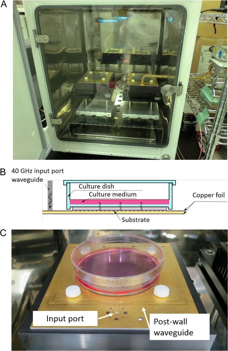
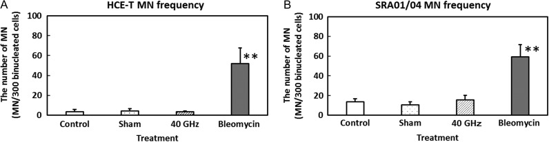
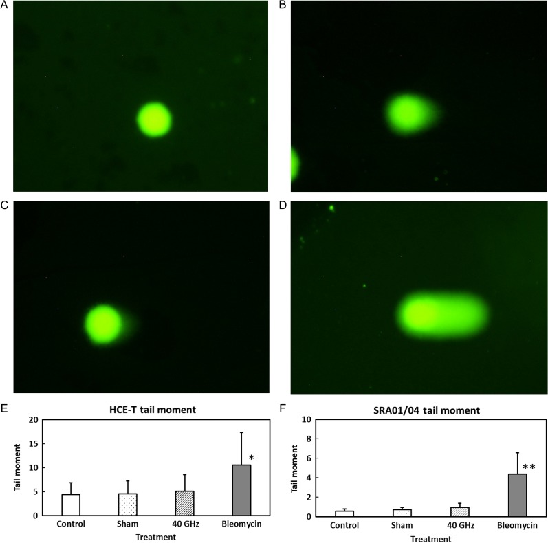
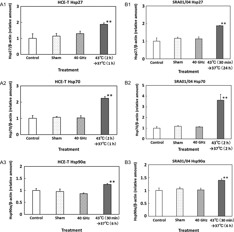
Millimeter waves are used in various fields, and the risks of this wavelength range for human health must be carefully evaluated. In this study, we investigated the effects of millimeter waves on genotoxicity and heat shock protein expression in human corneal epithelial (HCE-T) and human lens epithelial (SRA01/04) cells. We exposed the cells to 40-GHz millimeter waves at 1 mW/cm2 for 24 h. We observed no statistically significant increase in the micronucleus (MN) frequency or the level of DNA strand breaks in cells exposed to 40-GHz millimeter waves relative to sham-exposed and incubator controls. Heat shock protein (Hsp) expression also exhibited no statistically significant response to the 40-GHz exposure. These results indicate that exposure to 40 GHz millimeter waves under these conditions has little or no effect on MN formation, DNA strand breaks, or Hsp expression in HCE-T or SRA01/04 cells.
Overview
The claims and statistics of this paper haven’t been checked yet.
Run the check — free, no account needed. It reads the open-access text and checks the sources it cites.
Integrity signals
Registry
Cites
Statistics
Figures
Verification
Claims the paper makes
The claims and statistics of this paper haven’t been checked yet.
Run the check — free, no account needed. It reads the open-access text and checks the sources it cites.
Claims other papers make about this one
Each is checked against this paper’s own text: does it say what the citing paper says it does?
The source backs part of the claim, not all of it — or backs it for a different population, condition or comparison.
In the citing paper
“Despite the varied exposure models and methods used, no statistically significant evidence of DNA damage was identified in these studies.”
From 5G mobile networks and health—a state-of-the-science review of the research into low-level RF fields above 6 GHz (2021) · cites this paper as [17]
Experimental — accuracy not yet measured
Self-check
Percentages, sums and reported differences re-computed from the paper’s own numbers.
The self-check needs the paper’s full text.
Citations
Cites
No references could be resolved yet.
Research works printed but not linked: 32
- Koyama, 2016, Effects of long-term exposure to 60 GHz millimeter-waves on the genotoxicity and heat shock protein (Hsp) expression of cells derived from human eye, Int J Environ Res Public Health, 13, 802, 10.3390/ijerph13080802 · DOI · Paper page
- Wang, 2014, HSP27, 70 and 90, anti-apoptotic proteins, in clinical cancer therapy (Review), Int J Clin Oncol, 45, 18, 10.3892/ijo.2014.2399 · DOI
- Beneduci, 2005, Frequency and irradiation time-dependent antiprolifirative effect of low-power millimetre waves on RPMI 7932 human melanoma cell line, Anticancer Res, 25, 1023
- Korenstein-Ilan, 2008, Terahertz radiation increases genomic instability in human lymphocytes, Radiat Res, 170, 224, 10.1667/RR0944.1 · DOI · Paper page
- Alexandrov, 2011, Non-thermal effects of terahertz radiation on gene expression in mouse stem cells, Biomed Opt Express, 2, 2679, 10.1364/BOE.2.002679 · DOI
- Hintzsche, 2011, Terahertz radiation induces spindle disturbances in human–hamster hybrid cells, Radiat Res, 175, 569, 10.1667/RR2406.1 · DOI
- IARC (International Agency for Research on Cancer), 2013, IARC Monographs on the Evaluation of Carcinogenic Risks to Humans. Non-Ionizing Radiation, Part 2: Radiofrequency Electromagnetic Fields, 1
- Mahamoud, 2016, Additive effects of millimeter waves and 2-deoxyglucose co-exposure on the human keratinocyte transcriptome, PLoS One, 11, e0160810, 10.1371/journal.pone.0160810 · DOI
- Belpomme, 2018, Thermal and non-thermal effects of low intensity non-ionizing radiation: an international perspective, Environ Pollut, 242, 643, 10.1016/j.envpol.2018.07.019 · DOI
- Scarfì, 2003, THz exposure of whole blood for the study of biological effects on human lymphocytes, J Biol Phys, 29, 171, 10.1023/A:1024440708943 · DOI
- Bourne, 2008, The effects of terahertz radiation on human keratinocyte primary cultures and neural cell cultures, Altern Lab Anim, 36, 667, 10.1177/026119290803600610 · DOI
- Hintzsche, 2013, Terahertz radiation at 0.380 THz and 2.520 THz does not lead to DNA damage in skin cells in vitro, Radiat Res, 179, 38, 10.1667/RR3077.1 · DOI
- Gandhi, 1980, Millimeter wave absorption spectra of biological samples, Bioelectromagnetics, 1, 285, 10.1002/bem.2250010305 · DOI · Paper page
- Gandhi, 1983, Some basic properties of biological tissues for potential biomedical applications of millimeter waves, J Microw Power, 18, 295, 10.1080/16070658.1983.11689334 · DOI
- Shiina, 2015, High-efficiency applicator based on printed circuit board in millimeter-wave region, IEEE Trans Microw Theory Tech, 63, 3311, 10.1109/TMTT.2015.2472401 · DOI
- Shiina, 2016, Exposure characteristics of leaky wave applicator at millimeter-wave region, IEICE Trans Commun, J99-B, 106
- Araki-Sasaki, 1995, An SV40-immortalized human corneal epithelial cell line and its characterization, Invest Ophthalmol Vis Sci, 36, 614
- Koyama, 2004, Effects of 2.45-GHz electromagnetic fields with a wide range of SARs on micronucleus formation in CHO-K1 cells, ScientificWorldJournal, 4, 29, 10.1100/tsw.2004.176 · DOI
- Sakurai, 2009, Intermediate frequency magnetic fields generated by an induction heating (IH) cooktop do not affect genotoxicities and expression of heat shock proteins, Int J Radiat Biol, 85, 883, 10.1080/09553000903184358 · DOI
- Ramundo-Orlando, 2009, The response of giant phospholipid vesicles to millimeter waves radiation, Biochim Biophys Acta, 1788, 1497, 10.1016/j.bbamem.2009.04.006 · DOI
- Chidichimo, 2002, Selective inhibition of tumoral cells growth by low power millimeter waves, Anticancer Res, 22, 1681
- Beneduci, 2007, Antiproliferative effect of millimeter radiation on human erythromyeloid leukemia cell line K562 in culture: ultrastructural- and metabolic-induced changes, Bioelectrochemistry, 70, 214, 10.1016/j.bioelechem.2006.07.008 · DOI
- Haas, 2016, Impact of 60-GHz millimeter waves on stress and pain-related protein expression in differentiating neuron-like cells, Bioelectromagnetics, 37, 444, 10.1002/bem.21995 · DOI
- Haas, 2017, Effect of acute millimeter wave exposure on dopamine metabolism of NGF-treated PC12 cells, J Radiat Res, 58, 439, 10.1093/jrr/rrx004 · DOI
- Yaekashiwa, 2018, Investigation of the non-thermal effects of exposing cells to 70–300 GHz irradiation using a widely tunable source, J Radiat Res, 59, 116, 10.1093/jrr/rrx075 · DOI
- Vlasova, 2018, Extremely high-frequency electromagnetic radiation enhances neutrophil response to particulate agonists, Bioelectromagnetics, 39, 144, 10.1002/bem.22103 · DOI
- Markar, 2003, Effects of millimeter waves on cyclophosphamide induced suppression of T cell functions, Bioelectromagnetics, 24, 356, 10.1002/bem.10106 · DOI
- Markar, 2005, Effect of millimeter waves on natural killer cell activation, Bioelectromagnetics, 26, 10, 10.1002/bem.20046 · DOI
- Logani, 2006, Effect of millimeter wave irradiation on tumor metastatis, Bioelectromagnetics, 27, 258, 10.1002/bem.20208 · DOI
- Noviskova, 2008, Morphometric analysis of hypothalamic cells sowing c-Fos proteins after movement restriction and EHF-irradiation, Pathophysiology, 15, 19, 10.1016/j.pathophys.2007.11.001 · DOI
- Gapeyev, 2011, The role of fatty acids in anti-inflammatory effects of low-intensity extremely high-frequency electromagnetic radiation, Bioelectromagnetics, 32, 388, 10.1002/bem.20645 · DOI
- Gapeyev, 2015, Modifying effects of low-intensity extremely high-frequency electromagnetic radiation on content and composition of fatty acids in thymus of mice exposed to X-rays, Int J Radiat Biol, 91, 277, 10.3109/09553002.2014.980467 · DOI
Cited by
Sorted by citations
Perspective on Terahertz Applications in Bioscience and Biotechnology
Markelz, A. G., & Mittleman, D. M. (2022) · ACS Photonics · doi:10.1021/acsphotonics.2c00228 · Cited by 178
Karipidis, K., Mate, R., Urban, D., Tinker, R., & Wood, A. (2021) · Journal of Exposure Science & Environmental Epidemiology · doi:10.1038/s41370-021-00297-6 · Cited by 170
Metadata
- Authors
- Koyama, S., Narita, E., Suzuki, Y., Shiina, T., Taki, M., Shinohara, N., & Miyakoshi, J.
- Affiliations
- Kyoto University (JP)
- Tokyo Metropolitan University (JP)
- Corresponding author
- Miyakoshi, Junji
- Venue
- Journal of Radiation Research
- Publisher
- Oxford University Press (OUP)
- Published
- 2019-06-14
- Volume
- 60
- Issue
- 4
- Pages
- 417-423
- ISSN
- 0449-3060 (ISSN-L)
- 1349-9157
- PMC
- PMC6640906
- OpenAlex
- W2951389379
- Cited by
- 13
- References
- 0
- Citations by year
- 2024: 4 · 2023: 2 · 2022: 4 · 2021: 1 · 2020: 2
- FWCI
- 0.85
- Type
- journal article
- Language
- en
- Open access
- gold
- doi.org
- Licence
- Locations
- Journal of Radiation Research (publishedVersion, cc-by)
- PubMed Central (publishedVersion, cc-by)
- PubMed (publishedVersion)
- Institutional Repositories DataBase (IRDB) (submittedVersion, cc-by)
- Kyoto University Research Information Repository (Kyoto University) (submittedVersion, cc-by)
- Funders
- Ministry of Internal Affairs and Communications, Japan
- Topics
- Life Sciences › Biochemistry, Genetics and Molecular Biology › Biophysics › Electromagnetic Fields and Biological Effects
- Life Sciences › Biochemistry, Genetics and Molecular Biology › Cancer Research › Carcinogens and Genotoxicity Assessment
- Health Sciences › Medicine › Radiology, Nuclear Medicine and Imaging › Effects of Radiation Exposure
- Keywords
- millimeter wave, genotoxicity, heat shock proteins, human corneal epithelial cells, lens epithelial cells, micronucleus formation, DNA strand breaks, long-term exposure
- MeSH terms
- Cell Line, Cornea, DNA Damage, Electromagnetic Fields, Epithelial Cells, Gene Expression Regulation, Heat-Shock Proteins, Humans, Micronucleus Tests, Comet Assay
About these checks
Registry, retraction, Cites and statistics results are computed, not judged, with one exception: a printed reference that no index lists is read once by a model before it counts as a citation error. Claims are extracted by a model and kept only when their quote appears verbatim in the text; each cited source is then checked, with a model’s help, for whether it says what the claim relies on. Those verdicts can be wrong — read the claim’s quote and the cited source before relying on one.
Statistics and arithmetic are recomputed, not judged.
Confidence bands are shown only for fields calibrated on our annotated test set. None is calibrated yet, so no band is shown.
What counts as a claim ›How we measure confidence ›How we check this ›
Are you an author, or do you see a problem with this page? Request a review
How to cite
Cite CoChat for the checks on this page, and the paper for its own findings.
- This page
- CoChat. (2026). Citation check: Long-term exposure to a 40-GHz electromagnetic field does not affect genotoxicity or heat shock protein expression in HCE-T or SRA01/04 cells. https://app.cochat.ai/papers/10.1093/jrr/rrz017
- The paper
- Koyama, S., Narita, E., Suzuki, Y., Shiina, T., Taki, M., Shinohara, N., & Miyakoshi, J. (2019). Long-term exposure to a 40-GHz electromagnetic field does not affect genotoxicity or heat shock protein expression in HCE-T or SRA01/04 cells. Journal of Radiation Research. https://doi.org/10.1093/jrr/rrz017
