

The DOI matches its Crossref record; 2 of the 2 references it cites were found in a registry or an index.
3′-Terminal 2′-O-methylation of lung cancer miR-21-5p enhances its stability and association with Argonaute 2
Abstract
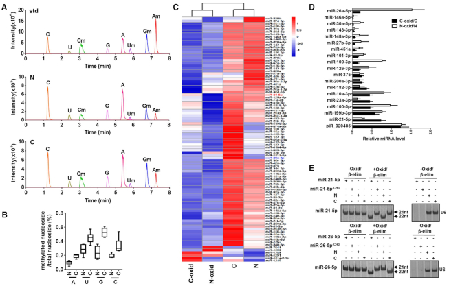
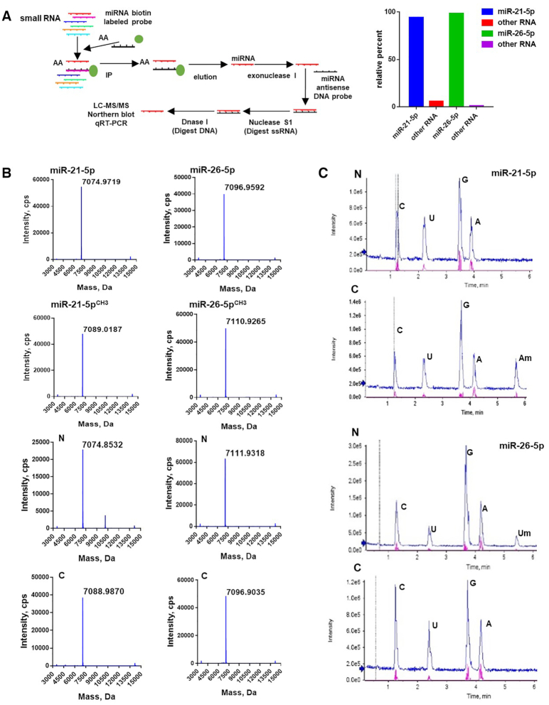
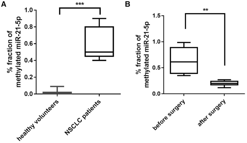
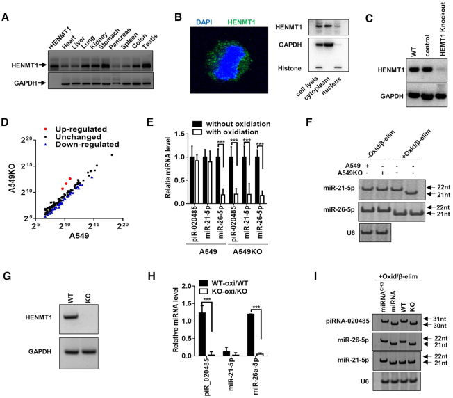
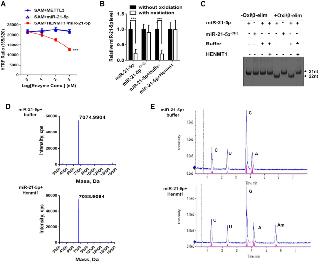
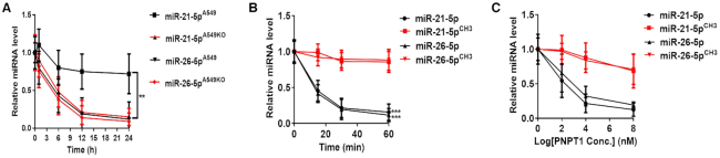
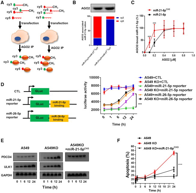
Methylation of miRNAs at the 2′-hydroxyl group on the ribose at 3′-end (2′-O-methylation, 2′Ome) is critical for miRNA function in plants and Drosophila. Whether this methylation phenomenon exists for mammalian miRNA remains unknown. Through LC–MS/MS analysis, we discover that majority of miR-21-5p isolated from human non-small cell lung cancer (NSCLC) tissue possesses 3′-terminal 2′Ome. Predominant 3′-terminal 2′Ome of miR-21-5p in cancer tissue is confirmed by qRT-PCR and northern blot after oxidation/β-elimination procedure. Cancerous and the paired non-cancerous lung tissue miRNAs display different pattern of 3′-terminal 2′Ome. We further identify HENMT1 as the methyltransferase responsible for 3′-terminal 2′Ome of mammalian miRNAs. Compared to non-methylated miR-21-5p, methylated miR-21-5p is more resistant to digestion by 3′→5′ exoribonuclease polyribonucleotide nucleotidyltransferase 1 (PNPT1) and has higher affinity to Argonaute-2, which may contribute to its higher stability and stronger inhibition on programmed cell death protein 4 (PDCD4) translation, respectively. Our findings reveal HENMT1-mediated 3′-terminal 2′Ome of mammalian miRNAs and highlight its role in enhancing miRNA’s stability and function.
Overview
The claims and statistics of this paper haven’t been checked yet.
Run the check — free, no account needed. It reads the open-access text and checks the sources it cites.
Integrity signals
Registry
Cites
Statistics
Figures
Verification
Claims the paper makes
The claims and statistics of this paper haven’t been checked yet.
Run the check — free, no account needed. It reads the open-access text and checks the sources it cites.
Claims other papers make about this one
Each is checked against this paper’s own text: does it say what the citing paper says it does?
The source backs part of the claim, not all of it — or backs it for a different population, condition or comparison.
In the citing paper
“Chemical modifications, such as 2′-sugar modifications like 2′-O-methyl and 2′-fluoro substitutions, enhance the stability200,201 of microRNAs, making it more resistant to degradation by nucleases and increasing stability without impeding RISC recognition.”
From MiR-128-3p – a gray eminence of the human central nervous system (2024) · cites this paper as [200]
Experimental — accuracy not yet measured
Self-check
Percentages, sums and reported differences re-computed from the paper’s own numbers.
The self-check needs the paper’s full text.
Citations
Cites
Frankel, L. B., Christoffersen, N. R., Jacobsen, A., Lindow, M., Krogh, A., & Lund, A. H. (2008) · Journal of Biological Chemistry · doi:10.1074/jbc.m707224200 · Cited by 1,141
METTL1 Promotes let-7 MicroRNA Processing via m7G Methylation
Pandolfini, L., Barbieri, I., Bannister, A. J., Hendrick, A., Andrews, B., Webster, N., Murat, P., Mach, P., Brandi, R., Robson, S. C., et al. (2019) · Molecular Cell · doi:10.1016/j.molcel.2019.03.040 · Cited by 491 · cited 1×
“There are different kinds of modification of miRNAs, including 2′-O-methylation (10) and m7G (11), which all can affect miRNA biogenesis and functions thereby playing a role in miRNA-based gene regulation network (10,11).”
Background
Research works printed but not linked: 34
- Chen, 2008, Characterization of microRNAs in serum: a novel class of biomarkers for diagnosis of cancer and other diseases, Cell Res., 18, 997, 10.1038/cr.2008.282 · DOI · Paper page
- Chen, 2012, Secreted microRNAs: a new form of intercellular communication, Trends Cell Biol., 22, 125, 10.1016/j.tcb.2011.12.001 · DOI · Paper page
- Zen, 2012, Circulating microRNAs: a novel class of biomarkers to diagnose and monitor human cancers, Med. Res. Rev., 32, 326, 10.1002/med.20215 · DOI
- Chen, 2012, Horizontal transfer of microRNAs: molecular mechanisms and clinical applications, Protein Cell, 3, 28, 10.1007/s13238-012-2003-z · DOI
- Cortez, 2011, MicroRNAs in body fluids–the mix of hormones and biomarkers, Nat. Rev. Clin. Oncol., 8, 467, 10.1038/nrclinonc.2011.76 · DOI
- Mori, 2019, Extracellular miRNAs: from biomarkers to mediators of physiology and disease, Cell Metab., 30, 656, 10.1016/j.cmet.2019.07.011 · DOI
- Turchinovich, 2016, Extracellular miRNA: a collision of two paradigms, Trends Biochem. Sci., 41, 883, 10.1016/j.tibs.2016.08.004 · DOI
- Quinn, 2015, Extracellular RNAs: development as biomarkers of human disease, J. Extracell. Vesicles, 4, 10.3402/jev.v4.27495 · DOI
- Li, 2012, Argonaute 2 complexes selectively protect the circulating microRNAs in cell-secreted microvesicles, PLoS One, 7, e46957, 10.1371/journal.pone.0046957 · DOI
- Yu, 2005, Methylation as a crucial step in plant microRNA biogenesis, Science, 307, 932, 10.1126/science.1107130 · DOI
- Abe, 2014, Impact of age-associated increase in 2′-O-methylation of miRNAs on aging and neurodegeneration in Drosophila, Genes Dev., 28, 44, 10.1101/gad.226654.113 · DOI · Paper page
- Hoerter, 2007, Chemical modification resolves the asymmetry of siRNA strand degradation in human blood serum, RNA, 13, 1887, 10.1261/rna.602307 · DOI
- Kirino, 2007, Mouse Piwi-interacting RNAs are 2′-O-methylated at their 3′ termini, Nat. Struct. Mol. Biol., 14, 347, 10.1038/nsmb1218 · DOI
- Ji, 2012, Regulation of small RNA stability: methylation and beyond, Cell Res., 22, 624, 10.1038/cr.2012.36 · DOI
- Lim, 2015, HENMT1 and piRNA stability are required for adult male germ cell transposon repression and to define the spermatogenic program in the mouse, PLos Genet., 11, e1005782, 10.1371/journal.pgen.1005782 · DOI
- Alberti, 2018, Cell-type specific sequencing of microRNAs from complex animal tissues, Nat. Methods, 15, 283, 10.1038/nmeth.4610 · DOI
- Ohara, 2007, The 3′ termini of mouse Piwi-interacting RNAs are 2′-O-methylated, Nat. Struct. Mol. Biol., 14, 349, 10.1038/nsmb1220 · DOI
- Wei, 2014, Importin 8 regulates the transport of mature MicroRNAs into the cell nucleus, J. Biol. Chem., 289, 10270, 10.1074/jbc.C113.541417 · DOI
- Wang, 2018, Direct quantification of 3′ terminal 2′-O-methylation of small RNAs by RT-qPCR, RNA, 24, 1520, 10.1261/rna.065144.117 · DOI
- Cheng, 2015, miR-19b downregulates intestinal SOCS3 to reduce intestinal inflammation in Crohn's disease, Sci. Rep., 5, 10397, 10.1038/srep10397 · DOI
- Chan, 2009, Structural and biochemical insights into 2′-O-methylation at the 3′-terminal nucleotide of RNA by Hen1, Proc. Natl Acad. Sci. U.S.A., 106, 17699, 10.1073/pnas.0907540106 · DOI
- Yan, 2013, A high-throughput quantitative approach reveals more small RNA modifications in mouse liver and their correlation with diabetes, Anal. Chem., 85, 12173, 10.1021/ac4036026 · DOI
- Kumarswamy, 2011, Regulation and function of miRNA-21 in health and disease, RNA Biol, 8, 706, 10.4161/rna.8.5.16154 · DOI
- Kirino, 2007, The mouse homolog of HEN1 is a potential methylase for Piwi-interacting RNAs, RNA, 13, 1397, 10.1261/rna.659307 · DOI
- Mickute, 2018, Animal Hen1 2′-O-methyltransferases as tools for 3′-terminal functionalization and labelling of single-stranded RNAs, Nucleic Acids Res., 46, e104, 10.1093/nar/gky514 · DOI
- Kimos, 2016, Development of an HTRF assay for the detection and characterization of inhibitors of catechol-O-methyltransferase, J. Biomol. Screen, 21, 490, 10.1177/1087057115616793 · DOI
- Ruegger, 2012, MicroRNA turnover: when, how, and why, Trends Biochem. Sci., 37, 436, 10.1016/j.tibs.2012.07.002 · DOI
- Kuhn, 2013, Eukaryotic Argonautes come into focus, Trends Biochem. Sci., 38, 263, 10.1016/j.tibs.2013.02.008 · DOI
- Elkayam, 2012, The structure of human argonaute-2 in complex with miR-20a, Cell, 150, 100, 10.1016/j.cell.2012.05.017 · DOI
- Jin, 2017, MiR-26 enhances chemosensitivity and promotes apoptosis of hepatocellular carcinoma cells through inhibiting autophagy, Cell Death. Dis., 8, e2540, 10.1038/cddis.2016.461 · DOI
- Zhang, 2012, MicroRNA degradation and turnover: regulating the regulators, Wiley Interdiscip. Rev. RNA, 3, 593, 10.1002/wrna.1114 · DOI
- Ameres, 2010, Target RNA-directed trimming and tailing of small silencing RNAs, Science, 328, 1534, 10.1126/science.1187058 · DOI · Paper page
- Ren, 2014, Methylation protects microRNAs from an AGO1-associated activity that uridylates 5′ RNA fragments generated by AGO1 cleavage, Proc. Natl Acad. Sci. U.S.A., 111, 6365, 10.1073/pnas.1405083111 · DOI
- Ren, 2014, Small RNAs meet their targets: when methylation defends miRNAs from uridylation, RNA Biol., 11, 1099, 10.4161/rna.36243 · DOI
Cited by
Sorted by citations
MiR-128-3p – a gray eminence of the human central nervous system
Kiel, K., Król, S. K., Bronisz, A., & Godlewski, J. (2024) · Molecular Therapy Nucleic Acids · doi:10.1016/j.omtn.2024.102141 · Cited by 15
Metadata
- Authors
- Liang, H., Jiao, Z., Rong, W., Qu, S., Liao, Z., Sun, X., Wei, Y., Zhao, Q., Wang, J., Liu, Y., Chen, X., Wang, T., Zhang, C.-Y., & Zen, K.
- Affiliations
- State Key Laboratory of Pharmaceutical Biotechnology, Department of Gastroenterology, Drum Tower Hospital, The Affiliated Hospital of Nanjing University Medical School, Nanjing, Jiangsu 210093, China
- Department of Cardiothoracic Surgery, Nanjing Drum Tower Hospital, Medical School of Nanjing University, Nanjing, China
- Department of Emergency Medicine, Nanjing Drum Tower Hospital, Medical School, Nanjing University, 210008 Nanjing, China
- Center for Inflammation, Immunity and Infectious Diseases, Georgia State University, Atlanta, GA 30032, USA
- ORCID
- Zen, Ke
- Venue
- Nucleic Acids Research
- Publisher
- Oxford University Press (OUP)
- Published
- 2020-06-15
- ISSN
- 0305-1048
- 1362-4962
- PMC
- PMC7367198
- Cited by
- 21
- References
- 2
- Language
- en
- Funders
- Ministry of Science and Technology (2018YFA0507100)
- National Natural Science Foundation of China (31801088)
- Natural Science Foundation of Jiangsu Province (BK20170076)
- China Postdoctoral Science Foundation (2018M642210)
- Jiangsu Province Postdoctoral Fund (2018K050A)
About these checks
Registry, retraction, Cites and statistics results are computed, not judged, with one exception: a printed reference that no index lists is read once by a model before it counts as a citation error. Claims are extracted by a model and kept only when their quote appears verbatim in the text; each cited source is then checked, with a model’s help, for whether it says what the claim relies on. Those verdicts can be wrong — read the claim’s quote and the cited source before relying on one.
Statistics and arithmetic are recomputed, not judged.
Confidence bands are shown only for fields calibrated on our annotated test set. None is calibrated yet, so no band is shown.
What counts as a claim ›How we measure confidence ›How we check this ›
Are you an author, or do you see a problem with this page? Request a review
