Cited by 128Prostate Cancer Treatment and ResearchCancer-related gene regulationEpigenetics and DNA Methylation

The DOI matches its Crossref record; 59 of the 59 references it cites were found in a registry or an index.

Regulation of the androgen receptor by SET9-mediated methylation.

Open access

Abstract

The androgen receptor (AR) is a member of the nuclear hormone receptor family of transcription factors that plays a critical role in regulating expression of genes involved in prostate development and transformation. Upon hormone binding, the AR associates with numerous co-regulator proteins that regulate the activation status of target genes via flux to the post-translational modification status of histones and the receptor. Here we show that the AR interacts with and is directly methylated by the histone methyltransferase enzyme SET9. Methylation of the AR on lysine 632 is necessary for enhancing transcriptional activity of the receptor by facilitating both inter-domain communication between the N- and C-termini and recruitment to androgen-target genes. We also show that SET9 is pro-proliferative and anti-apoptotic in prostate cancer cells and demonstrates up-regulated nuclear expression in prostate cancer tissue. In all, our date indicate a new mechanism of AR regulation that may be therapeutically exploitable for prostate cancer treatment.

Overview

The claims and statistics of this paper haven’t been checked yet.

Run the check — free, no account needed. It reads the open-access text and checks the sources it cites.

A few free checks a day · papers already verified open instantly · results are public

Integrity signals

Registry

Crossref · 10.1093/nar/gkq861

Cites

59 of 59 references verified0 citation errors · 0 not checkable

Statistics

Figures

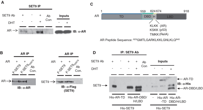
Figure 1. AR and SET9 interact in vitro and in vivo. (A) LNCaP cells grown in steroid-depleted media and treated with and without 10 nM DHT for 6 h were subject to IP using an anti-SET9 antibody followed by western analysis using an anti-AR antibody. (B) HEK293T cells were transiently transfected with pFlag-AR and either pFlag-SET9 or pFlag-SET9H297A for 48 h prior to IP using an anti-AR antibody followed by immunoblotting with anti-AR and -Flag antibodies. (C) Diagrammatic representation of the domains of the androgen receptor showing target lysine sequences of SET9 and sequence of AR peptide used in 3H methylation assays. (D) In vitro IP between His-tagged SET9 and N- and C-terminal fragments of the AR using anti-SET9 antibody followed by western analysis using an anti-His antibody.
Figure 1. AR and SET9 interact in vitro and in vivo. (A) LNCaP cells grown in steroid-depleted media and treated with and without 10 nM DHT for 6 h were subject to IP using an anti-SET9 antibody followed by western analysis using an anti-AR antibody. (B) HEK293T cells were transiently transfected with pFlag-AR and either pFlag-SET9 or pFlag-SET9H297A for 48 h prior to IP using an anti-AR antibody followed by immunoblotting with anti-AR and -Flag antibodies. (C) Diagrammatic representation of the domains of the androgen receptor showing target lysine sequences of SET9 and sequence of AR peptide used in 3H methylation assays. (D) In vitro IP between His-tagged SET9 and N- and C-terminal fragments of the AR using anti-SET9 antibody followed by western analysis using an anti-His antibody.CC BY-NC · PubMed Central
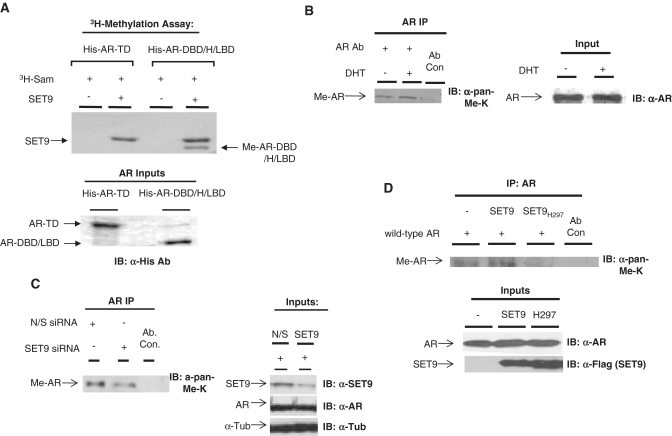
Figure 2. The AR is methylated by SET9 in vitro and in vivo. (A) An in vitro 3H-SAM methylation assay containing purified SET9 and N- and C-terminal fragments of the AR was incubated for 30 min and samples subject to PAGE, dried and exposed to X-ray film. (B) LNCaP cells grown in steroid-depleted media were treated for 6 h with 10 nM DHT prior to IP using an anti-AR antibody and western analysis using an anti-pan-methyl-lysine antibody (α-pan-methyl-K). (C) LNCaP cells grown in serum-containing media were transiently transfected with either non-silencing (N/S) or SET9 siRNAs for 72 h prior to IP and western analysis as in (B). (D) HEK293T cells were transiently transfected with AR and either SET9 or SET9H297A for 48 h prior to IP and western analysis as in (B).
Figure 2. The AR is methylated by SET9 in vitro and in vivo. (A) An in vitro 3H-SAM methylation assay containing purified SET9 and N- and C-terminal fragments of the AR was incubated for 30 min and samples subject to PAGE, dried and exposed to X-ray film. (B) LNCaP cells grown in steroid-depleted media were treated for 6 h with 10 nM DHT prior to IP using an anti-AR antibody and western analysis using an anti-pan-methyl-lysine antibody (α-pan-methyl-K). (C) LNCaP cells grown in serum-containing media were transiently transfected with either non-silencing (N/S) or SET9 siRNAs for 72 h prior to IP and western analysis as in (B). (D) HEK293T cells were transiently transfected with AR and either SET9 or SET9H297A for 48 h prior to IP and western analysis as in (B).CC BY-NC · PubMed Central
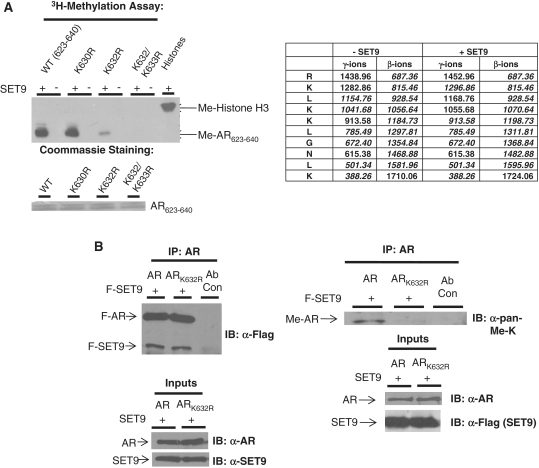
Figure 3. Lysine 632 of the AR is methylated by SET9. (A) Left panel: 3H-SAM methylation assay incorporating SET9 and wild-type or point mutants of AR623–640 peptides. Right panel: mass spectrometry analysis of AR peptide (623–640) with (+SET9) and without (−SET9) SET9-mediated methylation. Numbers in italics represent detected fragments in the spectra. A shift of 14 Da at lysine 632 represents methylation and is apparent in both γ and β ion analysis. (B) HEK293T cells transiently transfected with Flag-tagged SET9 and AR or ARK632R were subject to IP using an anti-AR antibody followed by western analysis using anti-Flag and anti-pan-methyl-lysine antibodies.
Figure 3. Lysine 632 of the AR is methylated by SET9. (A) Left panel: 3H-SAM methylation assay incorporating SET9 and wild-type or point mutants of AR623–640 peptides. Right panel: mass spectrometry analysis of AR peptide (623–640) with (+SET9) and without (−SET9) SET9-mediated methylation. Numbers in italics represent detected fragments in the spectra. A shift of 14 Da at lysine 632 represents methylation and is apparent in both γ and β ion analysis. (B) HEK293T cells transiently transfected with Flag-tagged SET9 and AR or ARK632R were subject to IP using an anti-AR antibody followed by western analysis using anti-Flag and anti-pan-methyl-lysine antibodies.CC BY-NC · PubMed Central
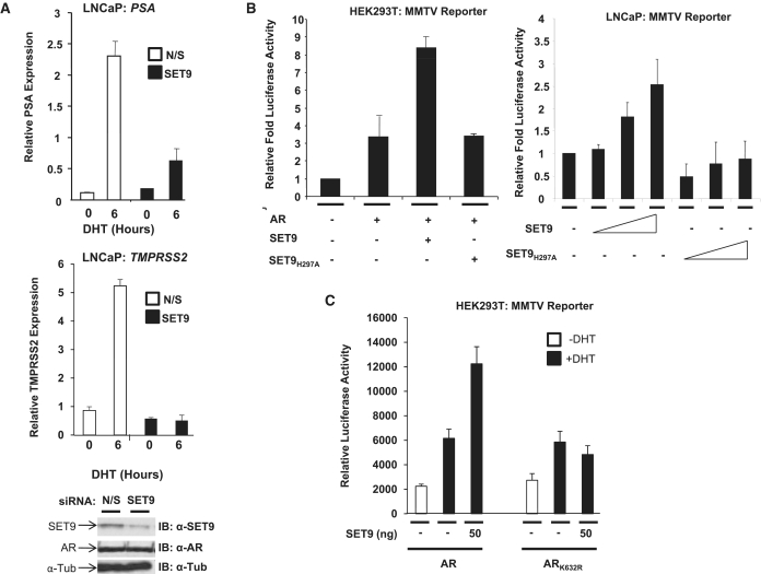
Figure 4. SET9 is an AR co-activator. (A) LNCaP cells grown in steroid-depleted media were transiently transfected with non-silencing (N/S) or SET9 siRNAs for 66 h prior to treatment with and without 10 nM DHT for 6 h followed by RNA extraction and real-time PCR analysis of PSA and TMPRSS2 mRNA expression. (B) HEK293T and LNCaP cells were transiently transfected for 48 h with AR (HEK293T cells only) and either SET9 or SET9H297A together with both MMTV-luciferase and β-galactosidase reporters prior to luciferase and β-galactosidase expression analyses. (C) HEK293T cells were transiently transfected and analysed as in (B) with the inclusion of ARK632R. Data represent average of three independent repeats ± standard error (A–C).
Figure 4. SET9 is an AR co-activator. (A) LNCaP cells grown in steroid-depleted media were transiently transfected with non-silencing (N/S) or SET9 siRNAs for 66 h prior to treatment with and without 10 nM DHT for 6 h followed by RNA extraction and real-time PCR analysis of PSA and TMPRSS2 mRNA expression. (B) HEK293T and LNCaP cells were transiently transfected for 48 h with AR (HEK293T cells only) and either SET9 or SET9H297A together with both MMTV-luciferase and β-galactosidase reporters prior to luciferase and β-galactosidase expression analyses. (C) HEK293T cells were transiently transfected and analysed as in (B) with the inclusion of ARK632R. Data represent average of three independent repeats ± standard error (A–C).CC BY-NC · PubMed Central
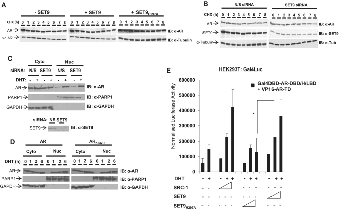
Figure 5. SET9 enhances AR N- and C-terminal interaction without affecting receptor stability or nuclear shuttling. (A) HEK293T cells transiently transfected with empty vector, SET9 or SET9H297A were treated with 1 µM cycloheximide (CHX) for up to 8 h and then subject to western analysis using anti-AR and anti-α-tubulin antibodies. (B) LNCaP cells transiently transfected with non-silencing (N/S) or SET9 siRNAs were treated with 1 µM CHX and subject to western analysis as in (A). (C) LNCaP cells transiently transfected with N/S or SET9 siRNAs in steroid-depleted media were treated with and without 10 nM DHT for 6 h prior to nuclear-cytoplasmic extraction and western analysis using anti-AR, -PARP1 and -GAPDH antibodies. (D) HEK293T cells transiently transfected with AR or ARK632R were subject to the same experimental procedure as in (C) with the inclusion of an additional 10 nM DHT time-point at 2 h. (E) Mammalian two-hybrid analysis of AR-TD and AR-DBD/H/LBD fragment interaction in HEK293T cells transiently transfected with either SET9, SET9H297A or SRC-1 and Gal4-luciferase and β-galactosidase reporters. Cells were treated with and without 10 nM DHT for 24-h prior to harvesting, and luciferase and β-galactosidase analyses. Data represents the average of three independent experiments performed in quadruplicate ± standard error (asterisk represents statistical significance <0.05).
Figure 5. SET9 enhances AR N- and C-terminal interaction without affecting receptor stability or nuclear shuttling. (A) HEK293T cells transiently transfected with empty vector, SET9 or SET9H297A were treated with 1 µM cycloheximide (CHX) for up to 8 h and then subject to western analysis using anti-AR and anti-α-tubulin antibodies. (B) LNCaP cells transiently transfected with non-silencing (N/S) or SET9 siRNAs were treated with 1 µM CHX and subject to western analysis as in (A). (C) LNCaP cells transiently transfected with N/S or SET9 siRNAs in steroid-depleted media were treated with and without 10 nM DHT for 6 h prior to nuclear-cytoplasmic extraction and western analysis using anti-AR, -PARP1 and -GAPDH antibodies. (D) HEK293T cells transiently transfected with AR or ARK632R were subject to the same experimental procedure as in (C) with the inclusion of an additional 10 nM DHT time-point at 2 h. (E) Mammalian two-hybrid analysis of AR-TD and AR-DBD/H/LBD fragment interaction in HEK293T cells transiently transfected with either SET9, SET9H297A or SRC-1 and Gal4-luciferase and β-galactosidase reporters. Cells were treated with and without 10 nM DHT for 24-h prior to harvesting, and luciferase and β-galactosidase analyses. Data represents the average of three independent experiments performed in quadruplicate ± standard error (asterisk represents statistical significance <0.05).CC BY-NC · PubMed Central
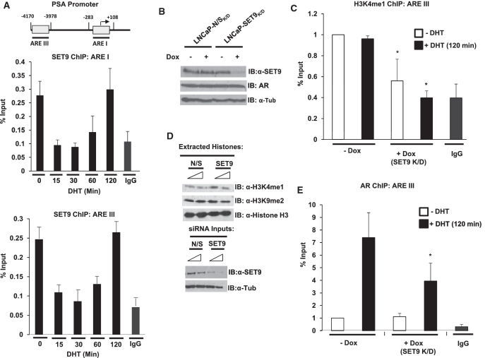
Figure 6. SET9 is recruited to the AR-regulated PSA promoter to methylate histone H3K4 and facilitate AR recruitment. (A) LNCaP cells grown in steroid-depleted media for 72-h were treated with 10 nM DHT over a time-course of 0–120 min and then subject to ChIP analysis using an anti-SET9 antibody, or non-specific isotype control, followed by quantitative PCR analysis using primers specific to the proximal (ARE I) and distal (ARE III) regions of the PSA promoter. (B) Representative SET9 knockdown and AR levels from the stable doxycycline-inducible SET9 knockdown LNCaP cell line (LNCaP-SET9K/D) compared to the equivalent non-silencing (N/S) cell line (LNCaP-N/SK/D). (C) ChIP analysis of histone H3K4 mono-methylation (H3K4me1) at ARE III in LNCaP-SET9K/D cells treated with and without doxycycline for 48 h and 10 nM DHT for 0 or 120 min. (D) Representative western analysis of global histone H3K4me1 and H3K9me2 modifications in LNCaP cells transiently transfected with 25 and 50 nM N/S or SET9 siRNAs. (E) An anti-AR antibody was used in ChIP analysis as in (B). Data represents three independent repeats ± standard error (A and E).
Figure 6. SET9 is recruited to the AR-regulated PSA promoter to methylate histone H3K4 and facilitate AR recruitment. (A) LNCaP cells grown in steroid-depleted media for 72-h were treated with 10 nM DHT over a time-course of 0–120 min and then subject to ChIP analysis using an anti-SET9 antibody, or non-specific isotype control, followed by quantitative PCR analysis using primers specific to the proximal (ARE I) and distal (ARE III) regions of the PSA promoter. (B) Representative SET9 knockdown and AR levels from the stable doxycycline-inducible SET9 knockdown LNCaP cell line (LNCaP-SET9K/D) compared to the equivalent non-silencing (N/S) cell line (LNCaP-N/SK/D). (C) ChIP analysis of histone H3K4 mono-methylation (H3K4me1) at ARE III in LNCaP-SET9K/D cells treated with and without doxycycline for 48 h and 10 nM DHT for 0 or 120 min. (D) Representative western analysis of global histone H3K4me1 and H3K9me2 modifications in LNCaP cells transiently transfected with 25 and 50 nM N/S or SET9 siRNAs. (E) An anti-AR antibody was used in ChIP analysis as in (B). Data represents three independent repeats ± standard error (A and E).CC BY-NC · PubMed Central
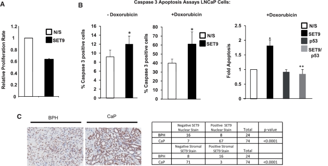
Figure 7. SET9 regulates LNCaP cell proliferation, apoptosis and is aberrantly expressed in prostate cancer. (A) WST-1 proliferation assays were conducted in SET9-depleted and non-depleted LNCaP cells grown in steroid-depleted media supplemented with 10 nM DHT for 72 h. Data represents the mean of three independent experiments. (B) LNCaP cells grown in serum-containing media and transiently transfected with non-silencing (N/S), SET9 and/or p53 siRNAs were treated with and without 0.5 µM doxorubicin for 24-h prior to caspase 3 analysis. Data represents the mean of three independent repeats ± standard error (asterisk represents statistical significance <0.05). (C) Representative SET9 staining of prostate tissue using an anti-SET9 antibody in immunohistochemistry (left panel) and table summarizing nuclear and stromal staining pattern of SET9 in cancer and normal tissue (right panel).
Figure 7. SET9 regulates LNCaP cell proliferation, apoptosis and is aberrantly expressed in prostate cancer. (A) WST-1 proliferation assays were conducted in SET9-depleted and non-depleted LNCaP cells grown in steroid-depleted media supplemented with 10 nM DHT for 72 h. Data represents the mean of three independent experiments. (B) LNCaP cells grown in serum-containing media and transiently transfected with non-silencing (N/S), SET9 and/or p53 siRNAs were treated with and without 0.5 µM doxorubicin for 24-h prior to caspase 3 analysis. Data represents the mean of three independent repeats ± standard error (asterisk represents statistical significance <0.05). (C) Representative SET9 staining of prostate tissue using an anti-SET9 antibody in immunohistochemistry (left panel) and table summarizing nuclear and stromal staining pattern of SET9 in cancer and normal tissue (right panel).CC BY-NC · PubMed Central
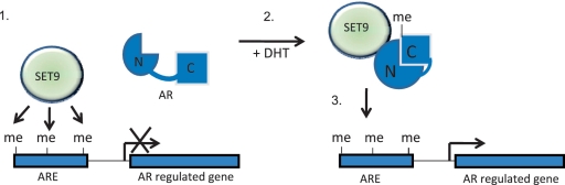
Figure 8. Postulated model of SET9-mediated regulation of the AR. Our working model is that SET9 enhances AR activity by methylating the receptor and histone H3-K4. Step 1: SET9 is present at androgen response elements (AREs) in the absence of active AR to mono-methylate H3-K4. Step 2: Activation of the AR via dihydrotestosterone (DHT) binding enables interaction of SET9 and the AR followed by methylation of the receptor on lysine 632 that facilitates inter-domain interaction of the N- and C-termini of the AR. Step 3: Methylated AR associates with target genes and enhances transcription.
Figure 8. Postulated model of SET9-mediated regulation of the AR. Our working model is that SET9 enhances AR activity by methylating the receptor and histone H3-K4. Step 1: SET9 is present at androgen response elements (AREs) in the absence of active AR to mono-methylate H3-K4. Step 2: Activation of the AR via dihydrotestosterone (DHT) binding enables interaction of SET9 and the AR followed by methylation of the receptor on lysine 632 that facilitates inter-domain interaction of the N- and C-termini of the AR. Step 3: Methylated AR associates with target genes and enhances transcription.CC BY-NC · PubMed Central

Verification

Claims the paper makes

The claims and statistics of this paper haven’t been checked yet.

Run the check — free, no account needed. It reads the open-access text and checks the sources it cites.

A few free checks a day · papers already verified open instantly · results are public

Claims other papers make about this one

Each is checked against this paper’s own text: does it say what the citing paper says it does?

Self-check

Percentages, sums and reported differences re-computed from the paper’s own numbers.

The self-check needs the paper’s full text.

Citations

Cites

Show all 59 references

Cited by

Sorted by citations

See all 128

Metadata

Authors
Gaughan, L., Stockley, J., Wang, N., McCracken, S. R. C., Treumann, A., Armstrong, K., Shaheen, F., Watt, K., McEwan, I. J., Wang, C., Pestell, R. G., & Robson, C. N.
Affiliations
Thomas Jefferson University (US)
Corresponding author
Gaughan, Luke
Venue
Nucleic Acids Research
Published
2010-10-19
Volume
39
Issue
4
Pages
1266-1279
ISSN
  • 0305-1048 (ISSN-L)
  • 1362-4962
OpenAlex
W2129651872
Cited by
127
References
59
Citations by year
2026: 3 · 2025: 3 · 2024: 3 · 2023: 11 · 2022: 12 · 2021: 6 · 2020: 4 · 2019: 5 · 2018: 10 · 2017: 12 · 2016: 8 · 2015: 7 · 2014: 8 · 2013: 15 · 2012: 10
FWCI
9.34
Type
journal article
Language
en
Open access
Locations
  • Nucleic Acids Research (publishedVersion, cc-by-nc)
  • PubMed Central (publishedVersion, cc-by-nc)
  • PubMed (publishedVersion)
  • The Medicine Forum (submittedVersion)
  • Newcastle University ePrints (Newcastle Univesity) (cc-by-nc)
Funders
  • Cancer Research UK
  • Academy of Medical Sciences (AMS-SGCL4-McCracken)
Topics
  • Life Sciences › Biochemistry, Genetics and Molecular Biology › Cancer Research › Prostate Cancer Treatment and Research
  • Life Sciences › Biochemistry, Genetics and Molecular Biology › Molecular Biology › Cancer-related gene regulation
  • Life Sciences › Biochemistry, Genetics and Molecular Biology › Molecular Biology › Epigenetics and DNA Methylation
Keywords
androgen receptor, histone methyltransferase, lysine methylation, receptor regulation, transcriptional activation, prostate cancer, apoptosis, cell proliferation, nuclear receptors
MeSH terms
Cell Nucleus, Histones, Humans, Lysine, Male, Methylation, Prostatic Neoplasms, Histone-Lysine N-Methyltransferase, Receptors, Androgen, Transcriptional Activation, Prostate-Specific Antigen, Active Transport, Cell Nucleus, Cell Line, Tumor, Cell Proliferation

About these checks

Registry, retraction, Cites and statistics results are computed, not judged, with one exception: a printed reference that no index lists is read once by a model before it counts as a citation error. Claims are extracted by a model and kept only when their quote appears verbatim in the text; each cited source is then checked, with a model’s help, for whether it says what the claim relies on. Those verdicts can be wrong — read the claim’s quote and the cited source before relying on one.

Statistics and arithmetic are recomputed, not judged.

Confidence bands are shown only for fields calibrated on our annotated test set. None is calibrated yet, so no band is shown.

What counts as a claim ›How we measure confidence ›How we check this ›

Are you an author, or do you see a problem with this page? Request a review