

The DOI matches its Crossref record; 39 of the 39 references it cites were found in a registry or an index.
The E3 ubiquitin ligase UBE3A is an integral component of the molecular circadian clock through regulating the BMAL1 transcription factor
Open accessAbstract
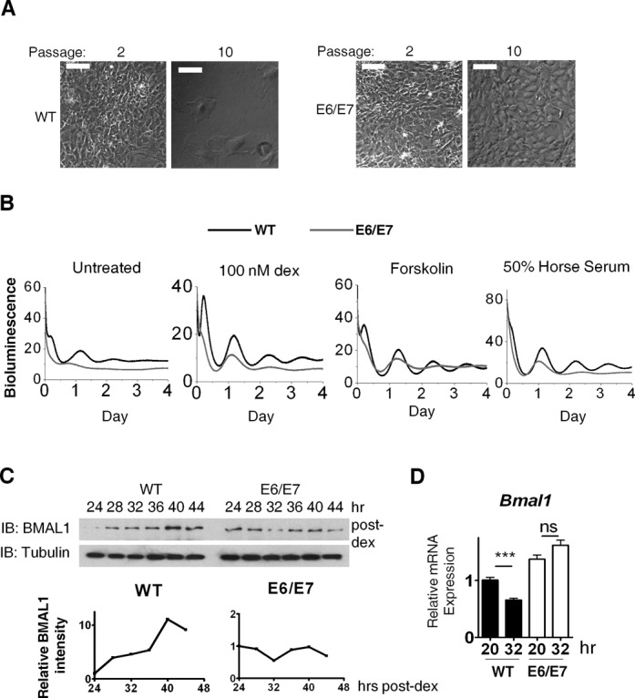
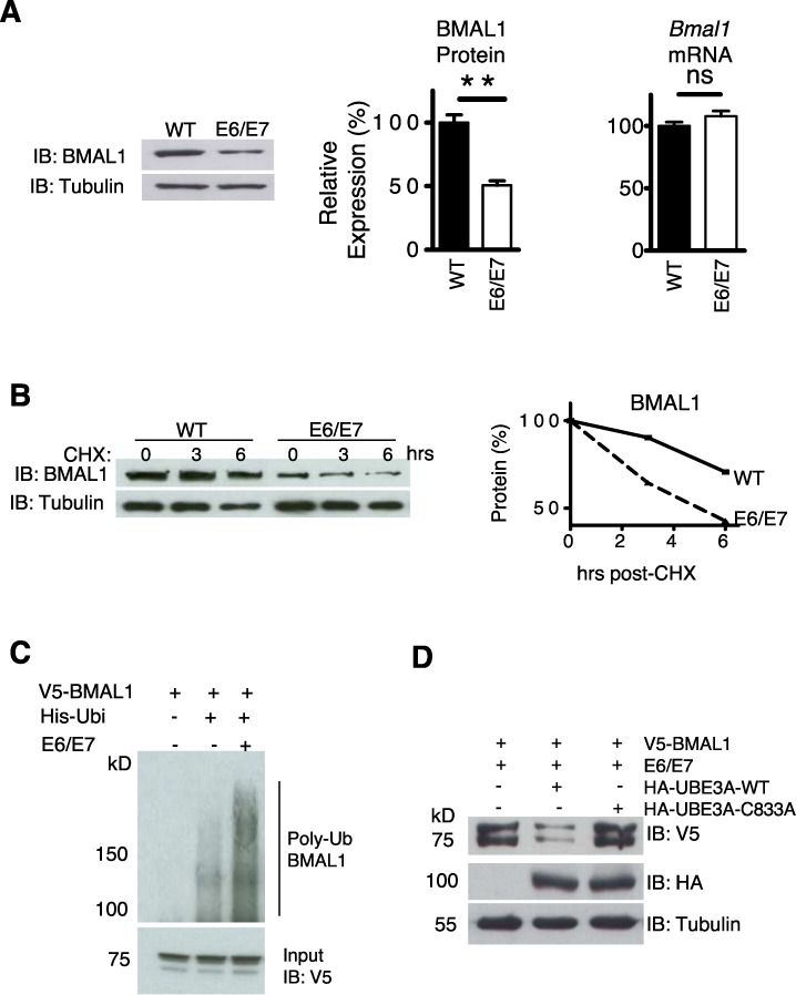
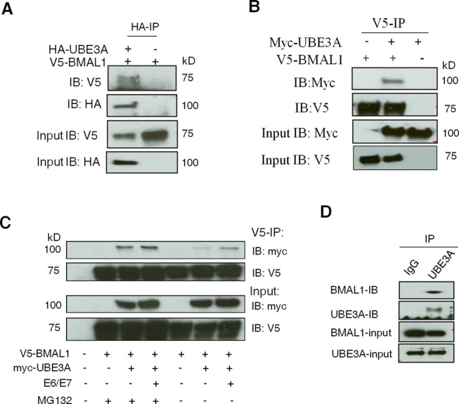
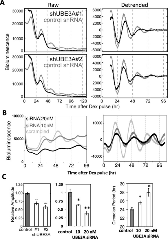
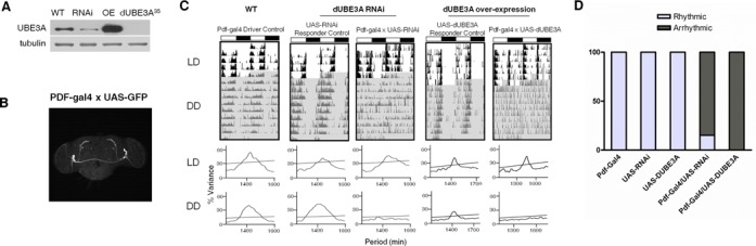
Post-translational modifications (such as ubiquitination) of clock proteins are critical in maintaining the precision and robustness of the evolutionarily conserved circadian clock. Ubiquitination of the core clock transcription factor BMAL1 (brain and muscle Arnt-like 1) has recently been reported. However, it remains unknown whether BMAL1 ubiquitination affects circadian pacemaking and what ubiquitin ligase(s) is involved. Here, we show that activating UBE3A (by expressing viral oncogenes E6/E7) disrupts circadian oscillations in mouse embryonic fibroblasts, measured using PER2::Luc dynamics, and rhythms in endogenous messenger ribonucleic acid and protein levels of BMAL1. Over-expression of E6/E7 reduced the level of BMAL1, increasing its ubiquitination and proteasomal degradation. UBE3A could bind to and degrade BMAL1 in a ubiquitin ligase-dependent manner. This occurred both in the presence and absence of E6/E7. We provide in vitro (knockdown/over-expression in mammalian cells) and in vivo (genetic manipulation in Drosophila) evidence for an endogenous role of UBE3A in regulating circadian dynamics and rhythmic locomotor behaviour. Together, our data reveal an essential and conserved role of UBE3A in the regulation of the circadian system in mammals and flies and identify a novel mechanistic link between oncogene E6/E7-mediated cell transformation and circadian (BMAL1) disruption.
Overview
The claims and statistics of this paper haven’t been checked yet.
Run the check — free, no account needed. It reads the open-access text and checks the sources it cites.
Integrity signals
Registry
Cites
Statistics
Figures
Verification
Claims the paper makes
The claims and statistics of this paper haven’t been checked yet.
Run the check — free, no account needed. It reads the open-access text and checks the sources it cites.
Claims other papers make about this one
Each is checked against this paper’s own text: does it say what the citing paper says it does?
In the citing paper
“UBE3A targets the circadian transcription factor BMAL1 for proteasomal degradation and thus is crucial for maintaining the pace of molecular clock timing117.”
From Rhythms of life: circadian disruption and brain disorders across the lifespan (2019) · cites this paper as [117]
Self-check
Percentages, sums and reported differences re-computed from the paper’s own numbers.
The self-check needs the paper’s full text.
Citations
Cites
Coordination of circadian timing in mammals
Reppert, S. M., & Weaver, D. R. (2002) · Nature · doi:10.1038/nature00965 · Cited by 4,379
The HPV-16 E6 and E6-AP complex functions as a ubiquitin-protein ligase in the ubiquitination of p53
Scheffner, M., Huibregtse, J. M., Vierstra, R. D., & Howley, P. M. (1993) · Cell · doi:10.1016/0092-8674(93)90384-3 · Cited by 2,416
Yoo, S.-H., Yamazaki, S., Lowrey, P. L., Shimomura, K., Ko, C. H., Buhr, E. D., Siepka, S. M., Hong, H.-K., Oh, W. J., Yoo, O. J., et al. (2004) · Proceedings of the National Academy of Sciences · doi:10.1073/pnas.0308709101 · Cited by 2,329
Resetting central and peripheral circadian oscillators in transgenic rats
Yamazaki, S., Numano, R., Abe, M., Hida, A., Takahashi, R.-I., Ueda, M., Block, G. D., Sakaki, Y., Menaker, M., & Tei, H. (2000) · Science · doi:10.1126/science.288.5466.682 · Cited by 1,907
Mop3 is an essential component of the master circadian pacemaker in mammals.
Bunger, M. K., Wilsbacher, L. D., Moran, S. M., Clendenin, C., Radcliffe, L. A., Hogenesch, J. B., Simon, M. C., Takahashi, J. S., & Bradfield, C. A. (2000) · Cell · doi:10.1016/s0092-8674(00)00205-1 · Cited by 1,621
The genetics of mammalian circadian order and disorder: implications for physiology and disease
Takahashi, J. S., Hong, H.-K., Ko, C. H., & McDearmon, E. L. (2008) · Nature Reviews Genetics · doi:10.1038/nrg2430 · Cited by 1,611
Münger, K., Phelps, W. C., Bubb, V. J., Howley, P. M., & Schlegel, R. (1989) · Journal of Virology · doi:10.1128/jvi.63.10.4417-4421.1989 · Cited by 1,335
UBE3A/E6-AP mutations cause Angelman syndrome
Kishino, T., Lalande, M., & Wagstaff, J. E. (1997) · Nature Genetics · doi:10.1038/ng0197-70 · Cited by 1,304
Fu, L., Pelicano, H., Liu, J., Huang, P., & Lee, C. C. (2002) · Cell · doi:10.1016/s0092-8674(02)00961-3 · Cited by 1,268
Control Mechanism of the Circadian Clock for Timing of Cell Division in Vivo
Matsuo, T., Yamaguchi, S., Mitsui, S., Emi, A., Shimoda, F., & Okamura, H. (2003) · Science · doi:10.1126/science.1086271 · Cited by 1,132
Cited by
Sorted by citations
Molecular mechanisms and physiological importance of circadian rhythms
Patke, A., Young, M. W., & Axelrod, S. (2020) · Nature Reviews Molecular Cell Biology · doi:10.1038/s41580-019-0179-2 · Cited by 1,388
Rhythms of life: circadian disruption and brain disorders across the lifespan
Logan, R. W., & McClung, C. A. (2019) · Nature Reviews Neuroscience · doi:10.1038/s41583-018-0088-y · Cited by 713
Cellular degradation systems in ferroptosis
Chen, X., Yu, C., Kang, R., Kroemer, G., & Tang, D. (2021) · Cell Death and Differentiation · doi:10.1038/s41418-020-00728-1 · Cited by 671
Cancer and the Circadian Clock
Shafi, A. A., & Knudsen, K. E. (2019) · Cancer Research · doi:10.1158/0008-5472.can-19-0566 · Cited by 264
A splice variant of human Bmal1 acts as a negative regulator of the molecular circadian clock
Lee, J., Park, E., Kim, G. H., Kwon, I., & Kim, K. (2018) · Experimental & Molecular Medicine · doi:10.1038/s12276-018-0187-x · Cited by 200
Metadata
- Authors
- Gossan, N. C., Zhang, F., Guo, B., Jin, D., Yoshitane, H., Yao, A., Glossop, N., Zhang, Y. Q., Fukada, Y., & Meng, Q.-J.
- Affiliations
- University of Manchester (GB)
- The University of Tokyo (JP)
- Chinese Academy of Sciences (CN)
- Corresponding author
- Meng, Qing-Jun
- Venue
- Nucleic Acids Research
- Publisher
- Oxford University Press (OUP)
- Published
- 2014-04-11
- Volume
- 42
- Issue
- 9
- Pages
- 5765-5775
- ISSN
- 0305-1048 (ISSN-L)
- 1362-4962
- PMC
- PMC4027211
- OpenAlex
- W2156456978
- Cited by
- 106
- References
- 39
- Citations by year
- 2026: 3 · 2025: 6 · 2024: 4 · 2023: 7 · 2022: 8 · 2021: 8 · 2020: 11 · 2019: 12 · 2018: 15 · 2017: 9 · 2016: 6 · 2015: 12 · 2014: 3
- FWCI
- 4.87
- Type
- journal article
- Language
- en
- Open access
- gold
- doi.org
- Locations
- Nucleic Acids Research (publishedVersion, cc-by)
- Europe PMC (PubMed Central) (publishedVersion, cc-by)
- PubMed Central (publishedVersion, cc-by)
- PubMed (publishedVersion)
- Research Explorer (The University of Manchester) (publishedVersion, other-oa)
- Funders
- Wellcome Trust
- University of Manchester
- National Natural Science Foundation of China
- Medical Research Council (G0900414)
- Biotechnology and Biological Sciences Research Council (BB/H531600/1)
- Japan Society for the Promotion of Science (24227001)
- Topics
- Life Sciences › Neuroscience › Endocrine and Autonomic Systems › Circadian rhythm and melatonin
- Life Sciences › Biochemistry, Genetics and Molecular Biology › Molecular Biology › Ubiquitin and proteasome pathways
- Life Sciences › Agricultural and Biological Sciences › Plant Science › Light effects on plants
- Keywords
- UBE3A, Bmal1, circadian clock, ubiquitination, E6 and E7 oncogenes, proteasomal degradation, clock proteins, locomotor rhythm, cell transformation, mouse embryonic fibroblasts
- MeSH terms
- Animals, Drosophila melanogaster, Gene Expression Regulation, Gene Expression, Drosophila Proteins, NIH 3T3 Cells, Ubiquitin-Protein Ligases, Proteasome Endopeptidase Complex, Mice, Ubiquitination, ARNTL Transcription Factors, Circadian Clocks, Proteolysis
About these checks
Registry, retraction, Cites and statistics results are computed, not judged, with one exception: a printed reference that no index lists is read once by a model before it counts as a citation error. Claims are extracted by a model and kept only when their quote appears verbatim in the text; each cited source is then checked, with a model’s help, for whether it says what the claim relies on. Those verdicts can be wrong — read the claim’s quote and the cited source before relying on one.
Statistics and arithmetic are recomputed, not judged.
Confidence bands are shown only for fields calibrated on our annotated test set. None is calibrated yet, so no band is shown.
What counts as a claim ›How we measure confidence ›How we check this ›
Are you an author, or do you see a problem with this page? Request a review
