Cited by 338CRISPR and Genetic EngineeringDNA Repair MechanismsRNA regulation and disease

The DOI matches its Crossref record; 56 of the 56 references it cites were found in a registry or an index.

Knock-in of large reporter genes in human cells via CRISPR/Cas9-induced homology-dependent and independent DNA repair

Open access

Abstract

CRISPR/Cas9-induced site-specific DNA double-strand breaks (DSBs) can be repaired by homology-directed repair (HDR) or non-homologous end joining (NHEJ) pathways. Extensive efforts have been made to knock-in exogenous DNA to a selected genomic locus in human cells; which, however, has focused on HDR-based strategies and was proven inefficient. Here, we report that NHEJ pathway mediates efficient rejoining of genome and plasmids following CRISPR/Cas9-induced DNA DSBs, and promotes high-efficiency DNA integration in various human cell types. With this homology-independent knock-in strategy, integration of a 4.6 kb promoterless ires-eGFP fragment into the GAPDH locus yielded up to 20% GFP+ cells in somatic LO2 cells, and 1.70% GFP+ cells in human embryonic stem cells (ESCs). Quantitative comparison further demonstrated that the NHEJ-based knock-in is more efficient than HDR-mediated gene targeting in all human cell types examined. These data support that CRISPR/Cas9-induced NHEJ provides a valuable new path for efficient genome editing in human ESCs and somatic cells.

Overview

The claims and statistics of this paper haven’t been checked yet.

Run the check — free, no account needed. It reads the open-access text and checks the sources it cites.

A few free checks a day · papers already verified open instantly · results are public

Integrity signals

Registry

Crossref · 10.1093/nar/gkw064

Cites

56 of 56 references verified0 citation errors · 0 not checkable

Statistics

Figures

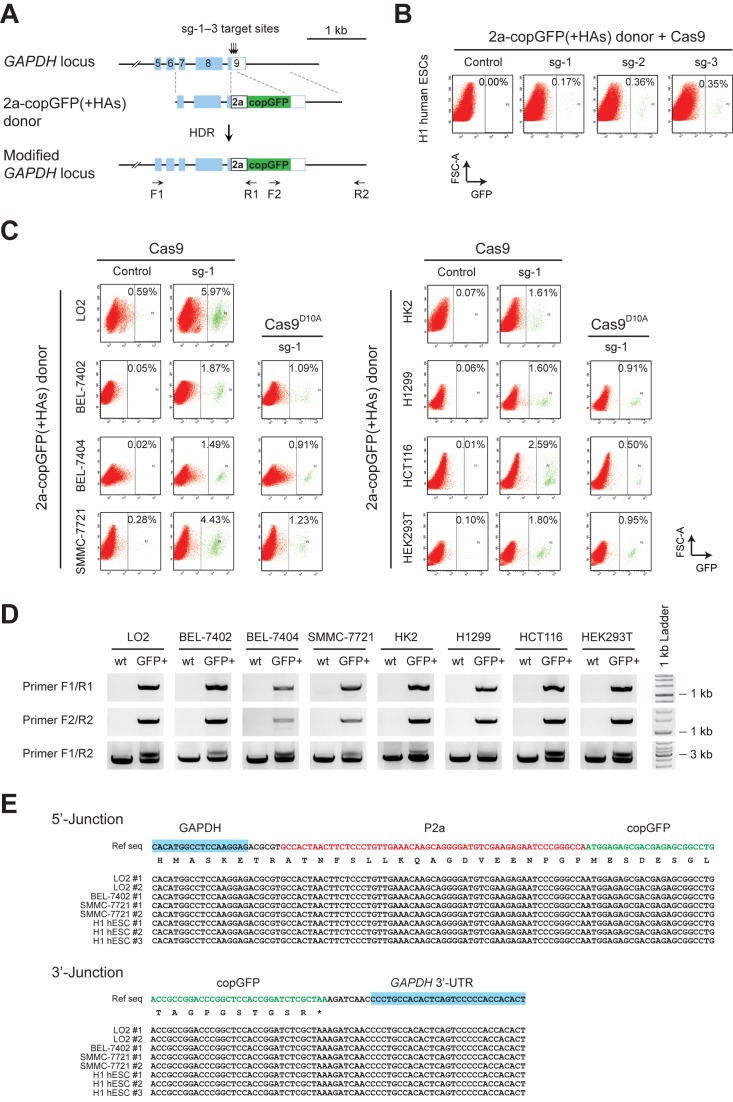
Figure 1. Varied frequency of HDR-mediated gene targeting in different human cell lines. (A) Schematics of the donor plasmid and targeting strategy for HDR-mediated knock-in of the 2a-copGFP reporter at GAPDH 3′-UTR. Dashed lines indicate sections of homology between the genomic locus and plasmid DNA. Positions of PCR primers used for detection of reporter knock-in are shown. (B) FACS analysis of H1 human ESCs, showing HDR-mediated integration of 2a-copGFP in the presence of Cas9 and sg-1, 2 or 3. Human ESCs were co-transfected with donor/Cas9/sgRNAs by nucleofection, and analyzed four days after transfection. X and Y-axis denote levels of GFP signal and forward scatter area, respectively. (C) FACS results showing varied frequencies of HDR-mediated 2a-copGFP knock-in in different human cell lines. 2a-copGFP(+HAs) donor and Cas9 were co-transfected, with or without sg-1, into eight human cell lines. GFP+ cells from the Cas9/sg-1 targeted cells (green signals gated to the right of the dashed line in each panel) were sorted for further analysis. To compare with the fully functional Cas9, nickase Cas9D10A were co-transfected with 2a-copGFP(+HAs) donor and sg-1 in selected cell lines, and the FACS results were shown in the column 3 and 6. (D) Genome PCR analysis of GFP+ cells produced with Cas9/sg-1 in C. Primer binding sites are indicated in A. Primer pair F1/R1 and F2/R2 amplify the 5′-junction (1350 bp) and the 3′-junction (1473 bp) of the 2a-copGFP integration, respectively. Primers F1/R2 amplified two DNA fragments that represent the wild type (2480 bp) and modified alleles (3241 bp). All amplified DNA fragments exhibited the expected sizes, indicating correct integration of 2a-copGFP via HDR into the genome. (E) Sequencing results of the PCR fragments amplified from the junctions. Expected modifications were confirmed at both 5′- and 3′-junctions, indicating precise integration of 2a-copGFP through HDR-mediated repair.
Figure 1. Varied frequency of HDR-mediated gene targeting in different human cell lines. (A) Schematics of the donor plasmid and targeting strategy for HDR-mediated knock-in of the 2a-copGFP reporter at GAPDH 3′-UTR. Dashed lines indicate sections of homology between the genomic locus and plasmid DNA. Positions of PCR primers used for detection of reporter knock-in are shown. (B) FACS analysis of H1 human ESCs, showing HDR-mediated integration of 2a-copGFP in the presence of Cas9 and sg-1, 2 or 3. Human ESCs were co-transfected with donor/Cas9/sgRNAs by nucleofection, and analyzed four days after transfection. X and Y-axis denote levels of GFP signal and forward scatter area, respectively. (C) FACS results showing varied frequencies of HDR-mediated 2a-copGFP knock-in in different human cell lines. 2a-copGFP(+HAs) donor and Cas9 were co-transfected, with or without sg-1, into eight human cell lines. GFP+ cells from the Cas9/sg-1 targeted cells (green signals gated to the right of the dashed line in each panel) were sorted for further analysis. To compare with the fully functional Cas9, nickase Cas9D10A were co-transfected with 2a-copGFP(+HAs) donor and sg-1 in selected cell lines, and the FACS results were shown in the column 3 and 6. (D) Genome PCR analysis of GFP+ cells produced with Cas9/sg-1 in C. Primer binding sites are indicated in A. Primer pair F1/R1 and F2/R2 amplify the 5′-junction (1350 bp) and the 3′-junction (1473 bp) of the 2a-copGFP integration, respectively. Primers F1/R2 amplified two DNA fragments that represent the wild type (2480 bp) and modified alleles (3241 bp). All amplified DNA fragments exhibited the expected sizes, indicating correct integration of 2a-copGFP via HDR into the genome. (E) Sequencing results of the PCR fragments amplified from the junctions. Expected modifications were confirmed at both 5′- and 3′-junctions, indicating precise integration of 2a-copGFP through HDR-mediated repair.CC BY-NC · PubMed Central
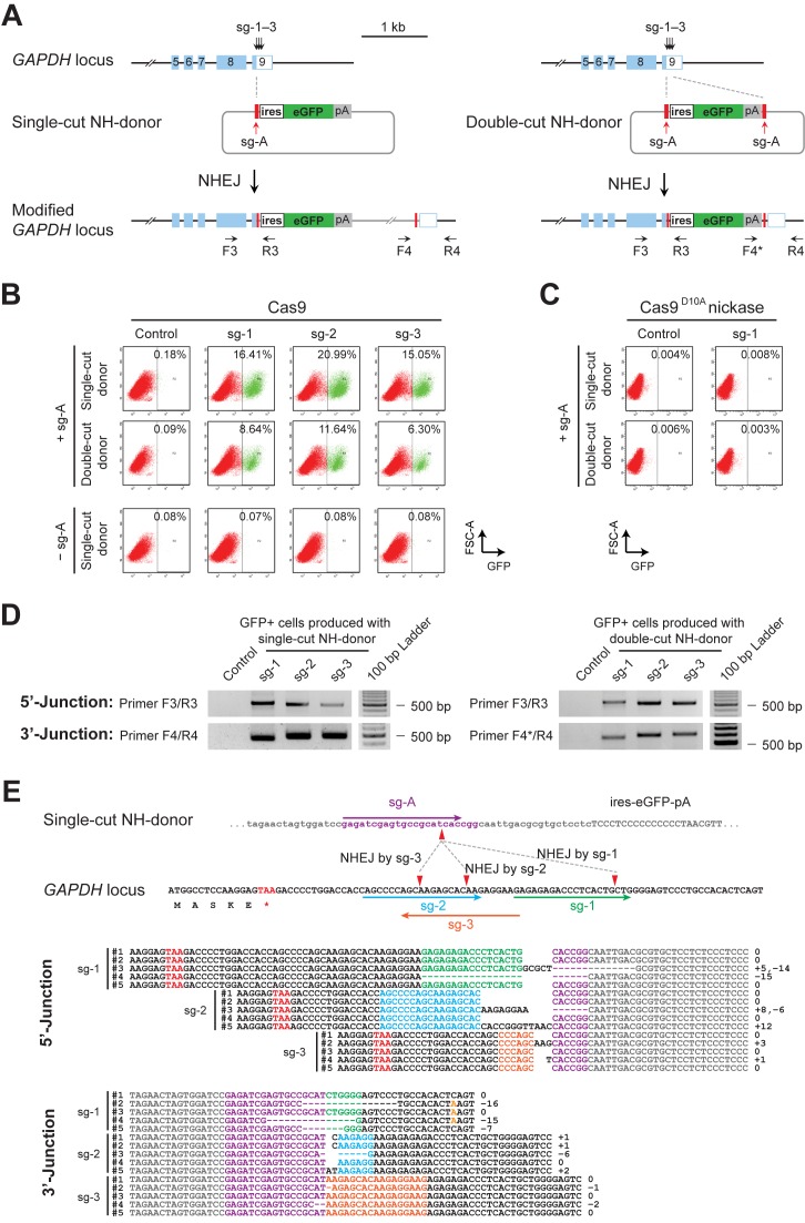
Figure 2. Homology-independent knock-in of reporter genes into CRISPR/Cas9-induced DSBs in genome. (A) Schematics of the donor plasmids and targeting strategy for CRISPR/Cas9-induced homology-independent insertion of the ires-eGFP reporter at the GAPDH 3′-UTR. Two NH-donor plasmids were generated; one plasmid (left panel) carries a single sg-A target site at the 5′ of ires-eGFP reporter (single-cut donor), and the other (right panel) carries two sg-A target sites at both the 5′ and 3′ sides of ires-eGFP (double-cut donor). Dashed lines indicate positions of DSBs introduced and the cleaved genome and plasmids rejoined. (B) FACS analysis of LO2 cells showing homology-independent knock-in of the ires-eGFP reporter, induced by different combinations of donor plasmids and sgRNAs in the presence of fully functional Cas9. The top row shows the targeting results produced with single-cut NH-donor/Cas9/sg-A/sg-1, 2 or 3, while the middle row shows the results obtained when the double-cut donor plasmid was used. Controls without sg-A are shown in the bottom row. GFP+ cells are gated to the right of the dashed line in each panel. (C) FACS analysis showing no detectable reporter integration in LO2 cells when nickase Cas9D10A was used instead of fully functional Cas9, either with the single- or the double-cut NH-donor plasmid. (D) Genomic PCR of GFP+ cells sorted from samples in B. Positions of primers used for junction detection are indicated in A. PCR amplifications showed DNA fragments at expected sizes, indicating correct integration of the ires-eGFP donors at the GAPDH 3′-UTR. (E) The upper panel shows the schematics of sg-1, 2 and 3 target sites in the GAPDH genomic locus, sg-A target site in the single-cut NH-donor, and the positions of cleavage and rejoining between the genome and the donor DNAs. The lower panel shows sequences of the integration junctions amplified from GFP+ cells produced with the single-cut donor (in D, left panel). 5′- and 3′-junctions of sg-1, 2 or 3-induced integrations were analyzed separately. For each junction, multiple sequences are shown. Nucleotides of different sgRNA target sites and PAMs are color-coded. Sequences from donor templates are shown in gray and the genomic DNA sequences flanking the integration junction are shown in black.
Figure 2. Homology-independent knock-in of reporter genes into CRISPR/Cas9-induced DSBs in genome. (A) Schematics of the donor plasmids and targeting strategy for CRISPR/Cas9-induced homology-independent insertion of the ires-eGFP reporter at the GAPDH 3′-UTR. Two NH-donor plasmids were generated; one plasmid (left panel) carries a single sg-A target site at the 5′ of ires-eGFP reporter (single-cut donor), and the other (right panel) carries two sg-A target sites at both the 5′ and 3′ sides of ires-eGFP (double-cut donor). Dashed lines indicate positions of DSBs introduced and the cleaved genome and plasmids rejoined. (B) FACS analysis of LO2 cells showing homology-independent knock-in of the ires-eGFP reporter, induced by different combinations of donor plasmids and sgRNAs in the presence of fully functional Cas9. The top row shows the targeting results produced with single-cut NH-donor/Cas9/sg-A/sg-1, 2 or 3, while the middle row shows the results obtained when the double-cut donor plasmid was used. Controls without sg-A are shown in the bottom row. GFP+ cells are gated to the right of the dashed line in each panel. (C) FACS analysis showing no detectable reporter integration in LO2 cells when nickase Cas9D10A was used instead of fully functional Cas9, either with the single- or the double-cut NH-donor plasmid. (D) Genomic PCR of GFP+ cells sorted from samples in B. Positions of primers used for junction detection are indicated in A. PCR amplifications showed DNA fragments at expected sizes, indicating correct integration of the ires-eGFP donors at the GAPDH 3′-UTR. (E) The upper panel shows the schematics of sg-1, 2 and 3 target sites in the GAPDH genomic locus, sg-A target site in the single-cut NH-donor, and the positions of cleavage and rejoining between the genome and the donor DNAs. The lower panel shows sequences of the integration junctions amplified from GFP+ cells produced with the single-cut donor (in D, left panel). 5′- and 3′-junctions of sg-1, 2 or 3-induced integrations were analyzed separately. For each junction, multiple sequences are shown. Nucleotides of different sgRNA target sites and PAMs are color-coded. Sequences from donor templates are shown in gray and the genomic DNA sequences flanking the integration junction are shown in black.CC BY-NC · PubMed Central
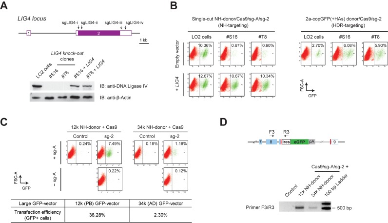
Figure 3. Conventional NHEJ repair mediates efficient knock-in of large reporter genes. (A) Upper panel shows the schematics of the sgLIG4-i–iv target positions at the LIG4 locus. These sgRNAs were combined and co-transfected with Cas9 into LO2 cells to generate LIG4 knock-out clones. Lower panel is the western blot, showing the loss of DNA ligase IV in the obtained LIG4 null clones, and LIG4 expression introduced by transfection of LIG4 cDNA construct into these cells. (B) FACS analysis of the LIG4 knock-out LO2 cells. Homology-independent knock-in was induced by single-cut NH-donor/Cas9/sg-A/sg-2, and HDR-based knock-in was introduced using 2a-copGFP(+HAs) donor/Cas9/sg-2, in both wild type as well as LIG4 knock-out LO2 cells. Drastic decrease of NH-targeting and rescue by LIG4 overexpression were observed in both LIG4 null clone #S16 and #T8 (left panel). Significant increase of HDR knock-in was also observed in LIG4 null cells (right panel). (C) FACS results showing NHEJ-mediated knock-in with large size donors. 12k and 34k NH-donors were co-transfected with Cas9/sg-A/sg-2 into wild type LO2 cells. Controls were transfected without sg-2 or sg-A. GFP+ cells are gated to the right of the dashed line in each panel. At the same time, constant GFP-expressing 12k (PB) and 34k (AD) GFP-vectors were transfected in parallel; and the transfection efficiencies examined at day 2 by FACS are shown at the lower panel. (D) PCR detection of the reporter integration in the transfected cells (unsorted) in C. Primer pair F3/R3 detected the 5′-junctions of the12k and 34k NH-donors integrated at GAPDH 3′-UTR. PCR amplifications showed DNA fragments at expected sizes.
Figure 3. Conventional NHEJ repair mediates efficient knock-in of large reporter genes. (A) Upper panel shows the schematics of the sgLIG4-i–iv target positions at the LIG4 locus. These sgRNAs were combined and co-transfected with Cas9 into LO2 cells to generate LIG4 knock-out clones. Lower panel is the western blot, showing the loss of DNA ligase IV in the obtained LIG4 null clones, and LIG4 expression introduced by transfection of LIG4 cDNA construct into these cells. (B) FACS analysis of the LIG4 knock-out LO2 cells. Homology-independent knock-in was induced by single-cut NH-donor/Cas9/sg-A/sg-2, and HDR-based knock-in was introduced using 2a-copGFP(+HAs) donor/Cas9/sg-2, in both wild type as well as LIG4 knock-out LO2 cells. Drastic decrease of NH-targeting and rescue by LIG4 overexpression were observed in both LIG4 null clone #S16 and #T8 (left panel). Significant increase of HDR knock-in was also observed in LIG4 null cells (right panel). (C) FACS results showing NHEJ-mediated knock-in with large size donors. 12k and 34k NH-donors were co-transfected with Cas9/sg-A/sg-2 into wild type LO2 cells. Controls were transfected without sg-2 or sg-A. GFP+ cells are gated to the right of the dashed line in each panel. At the same time, constant GFP-expressing 12k (PB) and 34k (AD) GFP-vectors were transfected in parallel; and the transfection efficiencies examined at day 2 by FACS are shown at the lower panel. (D) PCR detection of the reporter integration in the transfected cells (unsorted) in C. Primer pair F3/R3 detected the 5′-junctions of the12k and 34k NH-donors integrated at GAPDH 3′-UTR. PCR amplifications showed DNA fragments at expected sizes.CC BY-NC · PubMed Central
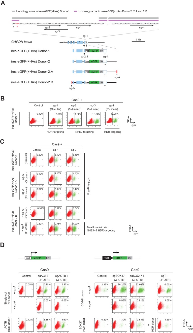
Figure 4. Comparison between HDR- and NHEJ-mediated reporter knock-in. (A) Schematics showing a zoomed-in view of the sg-1–4 target sites and their positions on the genomic GAPDH locus, as well as the design of ires-eGFP(+HAs) Donor-1, 2, 2.A and 2.B plasmids. Homology arm regions used in the ires-eGFP(+HAs) donor-1 are highlighted in gray, and the HAs used in donor-2, 2.A and 2.B are highlighted in purple. Donor-2.A carries a single sg-A target site at the 3′, and Donor-2.B carries a sg-A target site at the 5′ of the ires-eGFP(+HAs) cassette. (B) FACS analysis of LO2 cells transfected with the ires-eGFP(+HAs) donor-1/Cas9 and sg-1, 2, 3 or 4. Due to the different target positions on the genome and the donor, sg-1 induced HDR-mediated knock-in; sg-2 and sg-3 induced NHEJ-based knock-in; and sg-4 mainly produced GFP+ cells via the HDR-based knock-in through the intact 5′ homology arm. (C) FACS analysis showing HDR-mediated knock-in with circular and linear donor templates. The ires-eGFP(+HAs) Donor-2, 2.A or 2.B were transfected together with Cas9/sg-1 or Cas9/sg-2. The Donor-2.A and 2.B were both examined in the presence of sg-A (linear) as well as in the absence of sg-A (circular). Cas9/sg-A cleaves the Donor-2.A at 3′ of the ires-eGFP(+HAs) cassette and the linearized Donor 2.A produced GFP+ cells via HDR-mediated knock-in. Distinctly, Cas9/sg-A cleaves the Donor-2.B at 5′ of the ires-eGFP(+HAs) cassette, and the linearized Donor 2.B produced high proportion of GFP+ cells via both NHEJ- and HDR-mediated knock-in. (D) FACS results showing NHEJ- and HDR-mediated reporter knock-in at ACTB, SOX17 and T gene loci. Upper panel shows the schematics of ires-eGFP and PGK-eGFP reporters used for knock-in at ACTB and SOX17 or T gene loci, respectively. Single-cut NH-donor was co-transfected with Cas9/sg-A/sgACTB-i or sgACTB-ii to target the ACTB locus (lower left panel, top two rows); while the CE NH-donor was co-transfected with Cas9/sg-A/sgSOX17-i, sgSOX17-ii or sgT-i to target the SOX17 or T gene loci (lower right panel, top two rows). ACTB HDR-donor carrying ires-eGFP, and SOX17 and T HDR-donors containing PGK-eGFP, were co-transfected with Cas9 and corresponding sgRNAs to examine the HDR-based knock-in (lower panel, bottom row). Control samples were transfected without gene-specific sgRNA or sg-A. FACS analysis for the tests at ACTB locus was performed at day 5 after transfection. Cells transfected with PGK-eGFP containing donors for the tests at SOX17 and T loci, were maintained for five passages before FACS analysis. GFP+ cells are gated to the right of the dashed line in each panel.
Figure 4. Comparison between HDR- and NHEJ-mediated reporter knock-in. (A) Schematics showing a zoomed-in view of the sg-1–4 target sites and their positions on the genomic GAPDH locus, as well as the design of ires-eGFP(+HAs) Donor-1, 2, 2.A and 2.B plasmids. Homology arm regions used in the ires-eGFP(+HAs) donor-1 are highlighted in gray, and the HAs used in donor-2, 2.A and 2.B are highlighted in purple. Donor-2.A carries a single sg-A target site at the 3′, and Donor-2.B carries a sg-A target site at the 5′ of the ires-eGFP(+HAs) cassette. (B) FACS analysis of LO2 cells transfected with the ires-eGFP(+HAs) donor-1/Cas9 and sg-1, 2, 3 or 4. Due to the different target positions on the genome and the donor, sg-1 induced HDR-mediated knock-in; sg-2 and sg-3 induced NHEJ-based knock-in; and sg-4 mainly produced GFP+ cells via the HDR-based knock-in through the intact 5′ homology arm. (C) FACS analysis showing HDR-mediated knock-in with circular and linear donor templates. The ires-eGFP(+HAs) Donor-2, 2.A or 2.B were transfected together with Cas9/sg-1 or Cas9/sg-2. The Donor-2.A and 2.B were both examined in the presence of sg-A (linear) as well as in the absence of sg-A (circular). Cas9/sg-A cleaves the Donor-2.A at 3′ of the ires-eGFP(+HAs) cassette and the linearized Donor 2.A produced GFP+ cells via HDR-mediated knock-in. Distinctly, Cas9/sg-A cleaves the Donor-2.B at 5′ of the ires-eGFP(+HAs) cassette, and the linearized Donor 2.B produced high proportion of GFP+ cells via both NHEJ- and HDR-mediated knock-in. (D) FACS results showing NHEJ- and HDR-mediated reporter knock-in at ACTB, SOX17 and T gene loci. Upper panel shows the schematics of ires-eGFP and PGK-eGFP reporters used for knock-in at ACTB and SOX17 or T gene loci, respectively. Single-cut NH-donor was co-transfected with Cas9/sg-A/sgACTB-i or sgACTB-ii to target the ACTB locus (lower left panel, top two rows); while the CE NH-donor was co-transfected with Cas9/sg-A/sgSOX17-i, sgSOX17-ii or sgT-i to target the SOX17 or T gene loci (lower right panel, top two rows). ACTB HDR-donor carrying ires-eGFP, and SOX17 and T HDR-donors containing PGK-eGFP, were co-transfected with Cas9 and corresponding sgRNAs to examine the HDR-based knock-in (lower panel, bottom row). Control samples were transfected without gene-specific sgRNA or sg-A. FACS analysis for the tests at ACTB locus was performed at day 5 after transfection. Cells transfected with PGK-eGFP containing donors for the tests at SOX17 and T loci, were maintained for five passages before FACS analysis. GFP+ cells are gated to the right of the dashed line in each panel.CC BY-NC · PubMed Central
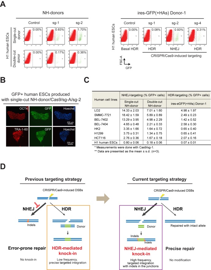
Figure 5. CRISPR/Cas9-coupled NHEJ mediates more efficient reporter knock-in than HDR in human ESCs and somatic cell lines. (A) Knock-in of ires-eGFP reporter via CRISPR/Cas9-coupled NHEJ repair in human ESCs. Left panel shows the NHEJ-mediated knock-in of ires-eGFP reporter. Both single-cut and double-cut NH- donors were examined in combination with sg-1 or sg-2. Right panel shows the reporter integration introduced with the ires-eGFP(+HAs) donor-1 and Cas9/sg-1, 2 or 4. Sg-1 and sg-4 mainly produced GFP+ cells via the HDR-based knock-in; while sg-2 induced NHEJ-based knock-in, due to the presence of sg-2 target site at the 5′-HA in the donor. FACS analyses were performed four days after nucleofection. GFP+ cells are gated to the right of the dashed line in each panel. (B) Immunofluorescence detection of pluripotency markers OCT4 (upper panel) and TRA-1–60 (lower panel) in the GFP+ cells sorted from H1 human ESCs transfected with single-cut donor/Cas9/sg-A/sg-2. Nuclei were counterstained with Hoechst. (C) Summary of Cas9/sg-1-induced NHEJ- and HDR-targeting in human ESCs and somatic cell lines examined. Single-cut and double-cut NH-donor were used for NHEJ-based targeting. The ires-eGFP (+HAs) donor-1 plasmid was used for HDR-based targeting. Percentages of GFP+ cells, presented as the mean ± s.d., were derived from three independent experiments. (D) Schematic diagram illustrating the balance between NHEJ- and HDR-mediated DSB repair in human cells, as well as the strategies (boxes) used for gene targeting.
Figure 5. CRISPR/Cas9-coupled NHEJ mediates more efficient reporter knock-in than HDR in human ESCs and somatic cell lines. (A) Knock-in of ires-eGFP reporter via CRISPR/Cas9-coupled NHEJ repair in human ESCs. Left panel shows the NHEJ-mediated knock-in of ires-eGFP reporter. Both single-cut and double-cut NH- donors were examined in combination with sg-1 or sg-2. Right panel shows the reporter integration introduced with the ires-eGFP(+HAs) donor-1 and Cas9/sg-1, 2 or 4. Sg-1 and sg-4 mainly produced GFP+ cells via the HDR-based knock-in; while sg-2 induced NHEJ-based knock-in, due to the presence of sg-2 target site at the 5′-HA in the donor. FACS analyses were performed four days after nucleofection. GFP+ cells are gated to the right of the dashed line in each panel. (B) Immunofluorescence detection of pluripotency markers OCT4 (upper panel) and TRA-1–60 (lower panel) in the GFP+ cells sorted from H1 human ESCs transfected with single-cut donor/Cas9/sg-A/sg-2. Nuclei were counterstained with Hoechst. (C) Summary of Cas9/sg-1-induced NHEJ- and HDR-targeting in human ESCs and somatic cell lines examined. Single-cut and double-cut NH-donor were used for NHEJ-based targeting. The ires-eGFP (+HAs) donor-1 plasmid was used for HDR-based targeting. Percentages of GFP+ cells, presented as the mean ± s.d., were derived from three independent experiments. (D) Schematic diagram illustrating the balance between NHEJ- and HDR-mediated DSB repair in human cells, as well as the strategies (boxes) used for gene targeting.CC BY-NC · PubMed Central

Verification

Claims the paper makes

The claims and statistics of this paper haven’t been checked yet.

Run the check — free, no account needed. It reads the open-access text and checks the sources it cites.

A few free checks a day · papers already verified open instantly · results are public

Claims other papers make about this one

Each is checked against this paper’s own text: does it say what the citing paper says it does?

Self-check

Percentages, sums and reported differences re-computed from the paper’s own numbers.

The self-check needs the paper’s full text.

Citations

Cites

Show all 56 references

Cited by

Sorted by citations

See all 338

Metadata

Authors
He, X., Tan, C., Wang, F., Wang, Y., Zhou, R., Cui, D., You, W., Zhao, H., Ren, J., & Feng, B.
Affiliations
Chinese University of Hong Kong (HK)
Corresponding author
Feng, Bo
Venue
Nucleic Acids Research
Published
2016-02-04
Volume
44
Issue
9
Pages
e85-e85
ISSN
  • 0305-1048 (ISSN-L)
  • 1362-4962
OpenAlex
W2267596434
Cited by
338
References
56
Citations by year
2026: 4 · 2025: 18 · 2024: 29 · 2023: 25 · 2022: 36 · 2021: 42 · 2020: 56 · 2019: 35 · 2018: 43 · 2017: 35 · 2016: 15
FWCI
22.17
Type
journal article
Language
en
Open access
Locations
  • Nucleic Acids Research (publishedVersion, cc-by-nc)
  • Europe PMC (PubMed Central) (publishedVersion, cc-by-nc)
  • PubMed Central (publishedVersion, cc-by-nc)
  • PubMed (publishedVersion)
  • HighWire Press Open Archive (submittedVersion)
Funders
  • National Natural Science Foundation of China (T13-607/12R, 2015CB964700, NSFC 31171433, JCYJ20140425184428469)
  • Chinese University of Hong Kong
  • Hong Kong University of Science and Technology
  • National Key Research and Development Program of China (2015CB964700)
Topics
  • Life Sciences › Biochemistry, Genetics and Molecular Biology › Molecular Biology › CRISPR and Genetic Engineering
  • Life Sciences › Biochemistry, Genetics and Molecular Biology › Molecular Biology › DNA Repair Mechanisms
  • Life Sciences › Biochemistry, Genetics and Molecular Biology › Molecular Biology › RNA regulation and disease
Keywords
CRISPR/Cas9, non-homologous end joining, homology-directed repair, knock-in, DNA double-strand breaks, human embryonic stem cells, somatic cells, genome editing efficiency
MeSH terms
Human Embryonic Stem Cells, Gene Editing, RNA, Guide, CRISPR-Cas Systems, DNA, Humans, Genes, Reporter, Glyceraldehyde-3-Phosphate Dehydrogenase (Phosphorylating), HCT116 Cells, Cell Line, Tumor, Green Fluorescent Proteins, DNA Breaks, Double-Stranded, Gene Knock-In Techniques, HEK293 Cells, DNA End-Joining Repair, Recombinational DNA Repair, Clustered Regularly Interspaced Short Palindromic Repeats, CRISPR-Cas Systems

About these checks

Registry, retraction, Cites and statistics results are computed, not judged, with one exception: a printed reference that no index lists is read once by a model before it counts as a citation error. Claims are extracted by a model and kept only when their quote appears verbatim in the text; each cited source is then checked, with a model’s help, for whether it says what the claim relies on. Those verdicts can be wrong — read the claim’s quote and the cited source before relying on one.

Statistics and arithmetic are recomputed, not judged.

Confidence bands are shown only for fields calibrated on our annotated test set. None is calibrated yet, so no band is shown.

What counts as a claim ›How we measure confidence ›How we check this ›

Are you an author, or do you see a problem with this page? Request a review