

The DOI matches its Crossref record; 1 of the 1 references it cites were found in a registry or an index.
Mechanistic Insights into the Function of 14-3-3 Proteins as Negative Regulators of Brassinosteroid Signaling in Arabidopsis
Open accessPublished version of a preprint
Abstract
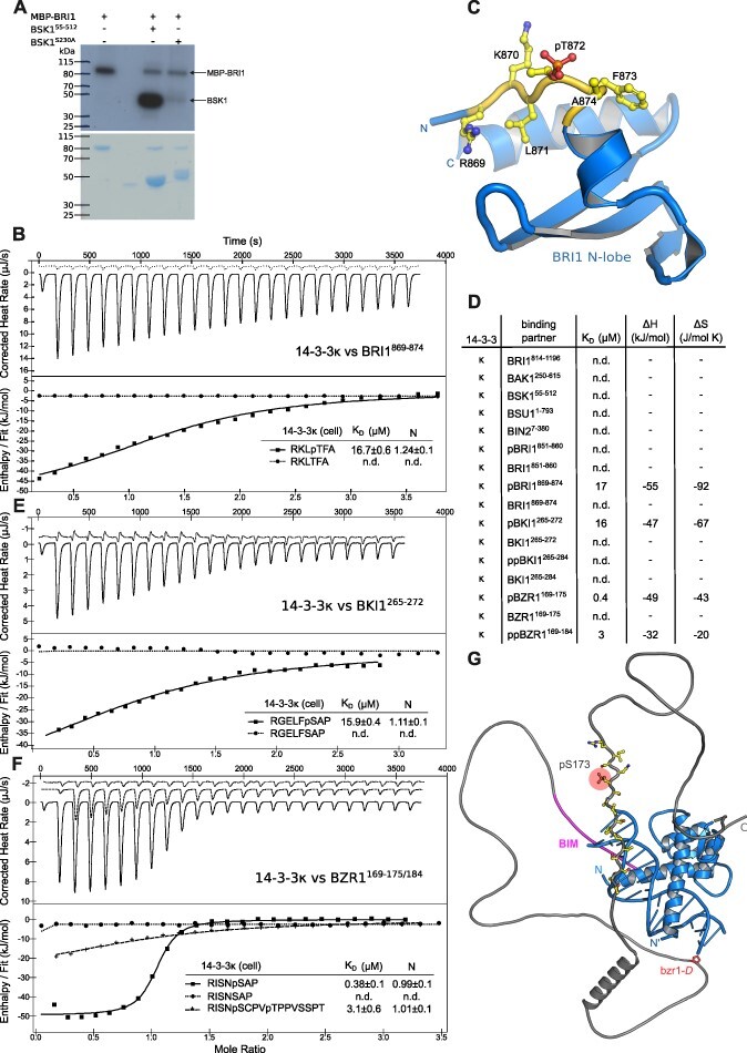
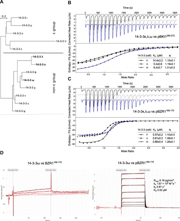
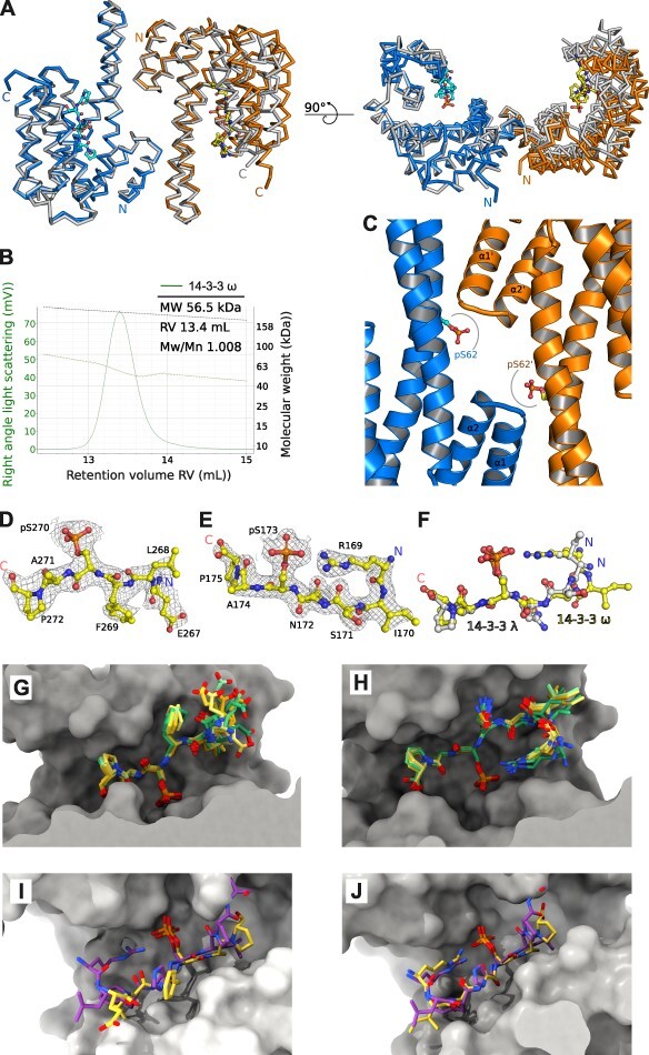
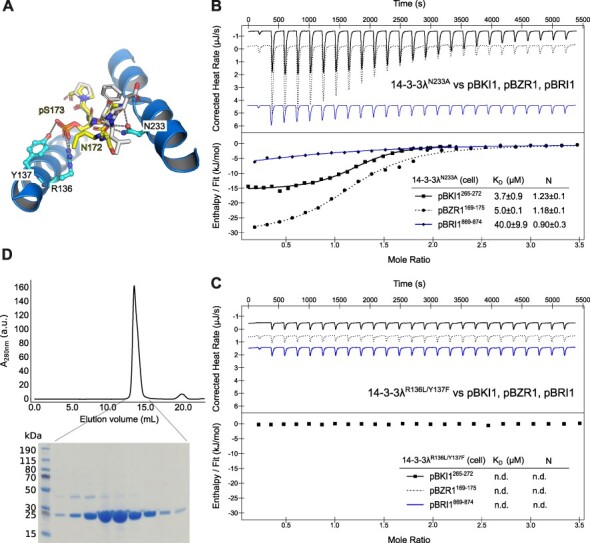
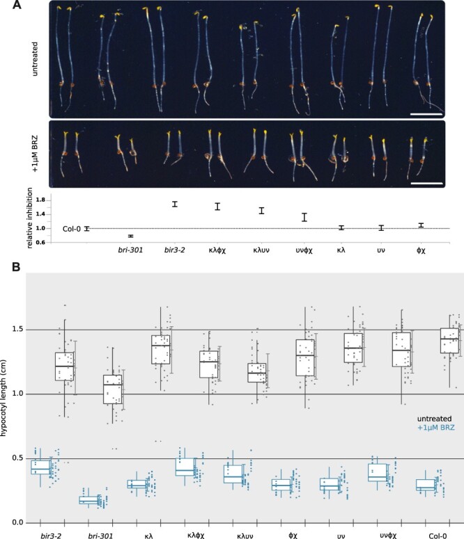
Brassinosteroids (BRs) are vital plant steroid hormones sensed at the cell surface by a membrane signaling complex comprising the receptor kinase BRI1 and a SERK family co-receptor kinase. Activation of this complex lead to dissociation of the inhibitor protein BKI1 from the receptor and to differential phosphorylation of BZR1/BES1 transcription factors by the glycogen synthase kinase 3 protein BIN2. Many phosphoproteins of the BR signaling pathway, including BRI1, SERKs, BKI1 and BZR1/BES1 can associate with 14-3-3 proteins. In this study, we use quantitative ligand binding assays to define the minimal 14-3-3 binding sites in the N-terminal lobe of the BRI1 kinase domain, in BKI1, and in BZR1 from Arabidopsis thaliana. All three motifs require to be phosphorylated to specifically bind 14-3-3s with mid- to low-micromolar affinity. BR signaling components display minimal isoform preference within the 14-3-3 non-ε subgroup. 14-3-3λ and 14-3-3 ω isoform complex crystal structures reveal that BKI1 and BZR1 bind as canonical type II 14-3-3 linear motifs. Disruption of key amino acids in the phosphopeptide binding site through mutation impairs the interaction of 14-3-3λ with all three linear motifs. Notably, quadruple loss-of-function mutants from the non-ε group exhibit gain-of-function BR signaling phenotypes, suggesting a role for 14-3-3 proteins as overall negative regulators of the BR pathway. Collectively, our work provides further mechanistic and genetic evidence for the regulatory role of 14-3-3 proteins at various stages of the BR signaling cascade.
Overview
The claims and statistics of this paper haven’t been checked yet.
Run the check — free, no account needed. It reads the open-access text and checks the sources it cites.
Integrity signals
Registry
Cites
Statistics
Figures
Verification
Claims the paper makes
The claims and statistics of this paper haven’t been checked yet.
Run the check — free, no account needed. It reads the open-access text and checks the sources it cites.
Self-check
Percentages, sums and reported differences re-computed from the paper’s own numbers.
The self-check needs the paper’s full text.
Citations
Cites
Highly accurate protein structure prediction with AlphaFold
Jumper, J., Evans, R., Pritzel, A., Green, T., Figurnov, M., Ronneberger, O., Tunyasuvunakool, K., Bates, R., Žídek, A., Potapenko, A., et al. (2021) · Nature · doi:10.1038/s41586-021-03819-2 · Cited by 43,764
Research works printed but not linked: 109
- Adams, 2010, PHENIX: a comprehensive Python-based system for macromolecular structure solution, Acta Crystallogr. D Biol. Crystallogr., 66, 213, 10.1107/S0907444909052925 · DOI · Paper page
- Afonine, 2012, Towards automated crystallographic structure refinement with phenix.refine, Acta Cryst. D, 68, 352, 10.1107/S0907444912001308 · DOI · Paper page
- Amorim-Silva, 2019, TTL proteins scaffold brassinosteroid signaling components at the plasma membrane to optimize signal transduction in Arabidopsis, Plant Cell, 31, 1807, 10.1105/tpc.19.00150 · DOI
- Asami, 2000, Characterization of brassinazole, a triazole-type brassinosteroid biosynthesis inhibitor, Plant Physiol., 123, 93, 10.1104/pp.123.1.93 · DOI
- Bai, 2007, Functions of OsBZR1 and 14-3-3 proteins in brassinosteroid signaling in rice, Proc. Natl. Acad. Sci. U.S.A., 104, 13839, 10.1073/pnas.0706386104 · DOI
- Ballone, 2018, 14-3-3: A case study in PPI modulation, Molecules, 23, 10.3390/molecules23061386 · DOI
- Benzinger, 2005, The crystal structure of the non-liganded 14-3-3σ protein: insights into determinants of isoform specific ligand binding and dimerization, Cell Res., 15, 219, 10.1038/sj.cr.7290290 · DOI
- Bieniossek, 2012, MultiBac: expanding the research toolbox for multiprotein complexes, Trends Biochem. Sci., 37, 49, 10.1016/j.tibs.2011.10.005 · DOI
- Bojar, 2014, Crystal structures of the phosphorylated BRI1 kinase domain and implications for brassinosteroid signal initiation, Plant J., 78, 31, 10.1111/tpj.12445 · DOI
- Bücherl, 2013, Visualization of BRI1 and BAK1(SERK3) membrane receptor heterooligomers during brassinosteroid signaling, Plant Physiol., 162, 1911, 10.1104/pp.113.220152 · DOI
- Chaiwanon, 2016, Immunophilin-like FKBP42/TWISTED DWARF1 interacts with the receptor kinase BRI1 to regulate brassinosteroid signaling in Arabidopsis, Mol. Plant, 9, 593, 10.1016/j.molp.2016.01.008 · DOI
- Chang, 2009, Proteomic profiling of tandem affinity purified 14-3-3 protein complexes in Arabidopsis thaliana, Proteomics, 9, 2967, 10.1002/pmic.200800445 · DOI
- Clouse, 1996, A brassinosteroid-insensitive mutant in arabidopsis thaliana exhibits multiple defects in growth and development, Plant Physiol., 111, 671, 10.1104/pp.111.3.671 · DOI
- Davis, 2007, MolProbity: all-atom contacts and structure validation for proteins and nucleic acids, Nucleic Acids Res., 35, W375, 10.1093/nar/gkm216 · DOI
- Denison, 2014, Phosphorylation-related modification at the dimer interface of 14-3-3ω dramatically alters monomer interaction dynamics, Arch. Biochem. Biophys., 541, 1, 10.1016/j.abb.2013.10.025 · DOI
- Denison, 2011, 14-3-3 proteins in plant physiology, Semin. Cell Dev. Biol., 22, 720, 10.1016/j.semcdb.2011.08.006 · DOI
- De Rybel, 2009, Chemical inhibition of a subset of Arabidopsis thaliana GSK3-like kinases activates brassinosteroid signaling, Chem. Biol., 16, 594, 10.1016/j.chembiol.2009.04.008 · DOI
- Edgar, 2004, MUSCLE: a multiple sequence alignment method with reduced time and space complexity, BMC Bioinf., 5, 10.1186/1471-2105-5-113 · DOI
- Ehsan, 2005, Interaction of Arabidopsis BRASSINOSTEROID-INSENSITIVE 1 receptor kinase with a homolog of mammalian TGF-beta receptor interacting protein, Plant J., 43, 251, 10.1111/j.1365-313X.2005.02448.x · DOI
- Emsley, 2004, Coot: model-building tools for molecular graphics, Acta Crystallogr. D Biol. Crystallogr., 60, 2126, 10.1107/S0907444904019158 · DOI · Paper page
- Friedrichsen, 2000, Brassinosteroid-insensitive-1 is a ubiquitously expressed leucine-rich repeat receptor serine/threonine kinase, Plant Physiol., 123, 1247, 10.1104/pp.123.4.1247 · DOI
- Fuglsang, 2003, The binding site for regulatory 14-3-3 protein in plant plasma membrane H+-ATPase: Involvement Of A Region Promoting Phosphorylation-Independent Interaction In Addition To The Phosphorylation-Dependent C-Terminal End *, J. Biol. Chem., 278, 42266, 10.1074/jbc.M306707200 · DOI
- Fu, 2000, Proteins: structure, function, and regulation, Annu. Rev. Pharmacol. Toxicol., 40, 617, 10.1146/annurev.pharmtox.40.1.617 · DOI
- Gampala, 2007, An essential role for 14-3-3 proteins in brassinosteroid signal transduction in Arabidopsis, Dev. Cell, 13, 177, 10.1016/j.devcel.2007.06.009 · DOI
- Gao, 2021, Ion homeostasis and metabolome analysis of Arabidopsis 14-3-3 quadruple mutants to salt stress, Front. Plant Sci., 12, 10.3389/fpls.2021.697324 · DOI
- Gao, 2021, Physiological and interactomic analysis reveals versatile functions of Arabidopsis 14-3-3 quadruple mutants in response to Fe deficiency, Sci. Rep., 11
- Gao, 2014, Light modulated activity of root alkaline/neutral invertase involves the interaction with 14-3-3 proteins, Plant J., 80, 785, 10.1111/tpj.12677 · DOI
- Goddard, 2018, UCSF ChimeraX: meeting modern challenges in visualization and analysis, Protein Sci., 27, 14, 10.1002/pro.3235 · DOI · Paper page
- Gökirmak, 2015, Phosphomimetic mutation of a conserved serine residue in Arabidopsis thaliana 14-3-3ω suggests a regulatory role of phosphorylation in dimerization and target interactions, Plant Physiol. Biochem., 97, 296, 10.1016/j.plaphy.2015.10.022 · DOI
- Gou, 2012, Genetic evidence for an indispensable role of somatic embryogenesis receptor kinases in brassinosteroid signaling, PLoS Genet., 8, 10.1371/journal.pgen.1002452 · DOI
- Grove, 1979, Brassinolide, a plant growth-promoting steroid isolated from Brassica napus pollen, Nature, 281, 216, 10.1038/281216a0 · DOI
- Guindon, 2010, New algorithms and methods to estimate maximum-likelihood phylogenies: assessing the performance of PhyML 3.0, Syst. Biol., 59, 307, 10.1093/sysbio/syq010 · DOI · Paper page
- Hashimoto, 2010, Ao38, a new cell line from eggs of the black witch moth, ascalapha odorata (lepidoptera: noctuidae), is permissive for AcMNPV infection and produces high levels of recombinant proteins, BMC Biotechnol., 10, 10.1186/1472-6750-10-50 · DOI
- He, 2002, The GSK3-like kinase BIN2 phosphorylates and destabilizes BZR1, a positive regulator of the brassinosteroid signaling pathway in Arabidopsis, Proc. Natl. Acad. Sci. U.S.A., 99, 10185, 10.1073/pnas.152342599 · DOI
- He, 2000, Perception of brassinosteroids by the extracellular domain of the receptor kinase BRI1, Science, 288, 2360, 10.1126/science.288.5475.2360 · DOI
- Hohmann, 2018, The SERK3 elongated allele defines a role for BIR ectodomains in brassinosteroid signalling, Nat. Plants, 4, 345, 10.1038/s41477-018-0150-9 · DOI
- Hohmann, 2020, Constitutive activation of leucine-rich repeat receptor kinase signaling pathways by BAK1-INTERACTING RECEPTOR-LIKE KINASE3 chimera, Plant Cell, 32, 3311, 10.1105/tpc.20.00138 · DOI
- Hohmann, 2018, Mechanistic basis for the activation of plant membrane receptor kinases by SERK-family coreceptors, Pnas, 115, 3488, 10.1073/pnas.1714972115 · DOI
- Hothorn, 2011, Structural basis of steroid hormone perception by the receptor kinase BRI1, Nature, 474, 467, 10.1038/nature10153 · DOI
- Hothorn, 2008, Simultaneous inference in general parametric models, Biom J., 50, 346, 10.1002/bimj.200810425 · DOI
- Huang, 2018, Shade-induced nuclear localization of PIF7 is regulated by phosphorylation and 14-3-3 proteins in Arabidopsis, eLife, 7, 10.7554/eLife.31636 · DOI
- Imkampe, 2017, The arabidopsis leucine-rich repeat receptor kinase BIR3 negatively regulates BAK1 receptor complex formation and stabilizes BAK1, Plant Cell, 29, 2285, 10.1105/tpc.17.00376 · DOI
- Jaillais, 2011, Tyrosine phosphorylation controls brassinosteroid receptor activation by triggering membrane release of its kinase inhibitor, Genes Dev., 25, 232, 10.1101/gad.2001911 · DOI
- Jiang, 2024, Molecular evolution and interaction of 14-3-3 proteins with H+-ATPases in plant abiotic stresses, J. Exp. Bot., 75, 689, 10.1093/jxb/erad414 · DOI
- Jiang, 2015, The intrinsically disordered protein BKI1 is essential for inhibiting BRI1 signaling in plants, Mol. Plant, 8, 1675, 10.1016/j.molp.2015.07.012 · DOI
- Kabsch, 1993, Automatic processing of rotation diffraction data from crystals of initially unknown symmetry and cell constants, J. Appl. Crystallogr., 26, 795, 10.1107/S0021889893005588 · DOI
- Karlova, 2006, The Arabidopsis SOMATIC EMBRYOGENESIS RECEPTOR-LIKE KINASE1 protein complex includes BRASSINOSTEROID-INSENSITIVE1, Plant Cell, 18, 626, 10.1105/tpc.105.039412 · DOI
- Kim, 2009, Brassinosteroid signal transduction from cell-surface receptor kinases to nuclear transcription factors, Nat. Cell Biol., 11, 1254, 10.1038/ncb1970 · DOI · Paper page
- Kim, 2023, Mapping the signaling network of BIN2 kinase using TurboID-mediated biotin labeling and phosphoproteomics, Plant Cell, 35, 975, 10.1093/plcell/koad013 · DOI
- Kitsche, 2014, Testing for qualitative interaction using ratios of treatment differences, Stat. Med., 33, 1477, 10.1002/sim.6048 · DOI
- Konagaya, 2004, Members of 14-3-3 protein isoforms interacting with the resistance gene product N and the elicitor of Tobacco mosaic virus, J. Gen. Plant Pathol., 70, 221, 10.1007/s10327-003-0113-4 · DOI
- Latz, 2007, TPK1, a Ca2+-regulated Arabidopsis vacuole two-pore K+ channel is activated by 14-3-3 proteins, Plant J., 52, 449, 10.1111/j.1365-313X.2007.03255.x · DOI
- Lee, 2020, proteins contribute to leaf and root development via brassinosteroid insensitive 1 in Arabidopsis thaliana, Genes Genom., 42, 347, 10.1007/s13258-019-00909-4 · DOI
- Li, 1997, A putative leucine-rich repeat receptor kinase involved in brassinosteroid signal transduction, Cell, 90, 929, 10.1016/S0092-8674(00)80357-8 · DOI
- Li, 2023, Scaffold protein RACK1 regulates BR signaling by modulating the nuclear localization of BZR1, New Phytol., 239, 1804, 10.1111/nph.19049 · DOI
- Li, 2002, Regulation of brassinosteroid signaling by a GSK3/SHAGGY-like kinase, Science, 295, 1299, 10.1126/science.1065769 · DOI
- Li, 2001, BIN2, a new brassinosteroid-insensitive locus in Arabidopsis, Plant Physiol., 127, 14, 10.1104/pp.127.1.14 · DOI
- Li, 2002, BAK1, an Arabidopsis LRR receptor-like protein kinase, interacts with BRI1 and modulates brassinosteroid signaling, Cell, 110, 213, 10.1016/S0092-8674(02)00812-7 · DOI
- Lozano-Durán, 2014, The bacterial effector HopM1 suppresses PAMP-triggered oxidative burst and stomatal immunity, New Phytol., 202, 259, 10.1111/nph.12651 · DOI
- McCoy, 2007, Phaser crystallographic software, J. Appl. Crystallogr., 40, 658, 10.1107/S0021889807021206 · DOI · Paper page
- Modi, 2020, Two negatively charged invariant residues influence ligand binding and conformational dynamics of 14-3-3ζ, FEBS Lett., 594, 878, 10.1002/1873-3468.13662 · DOI
- Mora-García, 2004, Nuclear protein phosphatases with Kelch-repeat domains modulate the response to brassinosteroids in Arabidopsis, Genes Dev., 18, 448, 10.1101/gad.1174204 · DOI
- Muslin, 1996, Interaction of 14-3-3 with signaling proteins is mediated by the recognition of phosphoserine, Cell, 84, 889, 10.1016/S0092-8674(00)81067-3 · DOI
- Nam, 2002, BRI1/BAK1, a receptor kinase pair mediating brassinosteroid signaling, Cell, 110, 203, 10.1016/S0092-8674(02)00814-0 · DOI · Paper page
- Nosaki, 2018, Structural basis for brassinosteroid response by BIL1/BZR1, Nat. Plants, 4, 771, 10.1038/s41477-018-0255-1 · DOI
- Novikova, 2022, Meet your MAKR: the membrane-associated kinase regulator protein family in the regulation of plant development, FEBS J., 289, 6172, 10.1111/febs.16132 · DOI
- Oh, 2000, Recombinant brassinosteroid insensitive 1 receptor-like kinase autophosphorylates on serine and threonine residues and phosphorylates a conserved peptide motif in vitro, Plant Physiol., 124, 751, 10.1104/pp.124.2.751 · DOI
- Oh, 2009, Tyrosine phosphorylation of the BRI1 receptor kinase emerges as a component of brassinosteroid signaling in Arabidopsis, Proc. Natl. Acad. Sci. U.S.A., 106, 658, 10.1073/pnas.0810249106 · DOI
- Ottmann, 2007, Structure of a 14-3-3 coordinated hexamer of the plant plasma membrane H+ -ATPase by combining X-ray crystallography and electron cryomicroscopy, Mol. Cell, 25, 427, 10.1016/j.molcel.2006.12.017 · DOI · Paper page
- Peng, 2010, A direct docking mechanism for a plant GSK3-like kinase to phosphorylate its substrates, J. Biol. Chem., 285, 24646, 10.1074/jbc.M110.142547 · DOI
- Perrière, 1996, On-line tools for sequence retrieval and multivariate statistics in molecular biology, Comput. Appl. Biosci., 12, 63
- Prado, 2019, Oscillating aquaporin phosphorylation and 14-3-3 proteins mediate the circadian regulation of leaf hydraulics, Plant Cell, 31, 417, 10.1105/tpc.18.00804 · DOI
- Puntervoll, 2003, ELM server: a new resource for investigating short functional sites in modular eukaryotic proteins, Nucleic Acids Res., 31, 3625, 10.1093/nar/gkg545 · DOI
- Reuter, 2021, Light-triggered and phosphorylation-dependent 14-3-3 association with NON-PHOTOTROPIC HYPOCOTYL 3 is required for hypocotyl phototropism, Nat. Commun., 12, 10.1038/s41467-021-26332-6 · DOI · Paper page
- Rienties, 2005, The arabidopsis SERK1 protein interacts with the AAA-ATPase AtCDC48, the 14-3-3 protein GF14lambda and the PP2C phosphatase KAPP, Planta, 221, 394, 10.1007/s00425-004-1447-7 · DOI
- Rittinger, 1999, Structural analysis of 14-3-3 phosphopeptide complexes identifies a dual role for the nuclear export signal of 14-3-3 in ligand binding, Mol. Cell, 4, 153, 10.1016/S1097-2765(00)80363-9 · DOI
- Ryu, 2010, Phosphorylation dependent nucleocytoplasmic shuttling of BES1 is a key regulatory event in brassinosteroid signaling, Mol. Cells, 29, 283, 10.1007/s10059-010-0035-x · DOI
- Ryu, 2007, Nucleocytoplasmic shuttling of BZR1 mediated by phosphorylation is essential in Arabidopsis brassinosteroid signaling, Plant Cell, 19, 2749, 10.1105/tpc.107.053728 · DOI
- Santiago, 2013, Molecular mechanism for plant steroid receptor activation by somatic embryogenesis co-receptor kinases, Science, 341, 889, 10.1126/science.1242468 · DOI · Paper page
- Schindelin, 2012, Fiji: an open-source platform for biological-image analysis, Nat. Methods, 9, 676, 10.1038/nmeth.2019 · DOI · Paper page
- Schmidt, 1997, A leucine-rich repeat containing receptor-like kinase marks somatic plant cells competent to form embryos, Development, 124, 2049, 10.1242/dev.124.10.2049 · DOI
- She, 2011, Structural insight into brassinosteroid perception by BRI1, Nature, 474, 472, 10.1038/nature10178 · DOI
- Sirichandra, 2010, The Arabidopsis ABA-activated kinase OST1 phosphorylates the bZIP transcription factor ABF3 and creates a 14-3-3 binding site involved in its turnover, PLoS One, 5, 10.1371/journal.pone.0013935 · DOI
- Stanislas, 2009, Quantitative proteomics reveals a dynamic association of proteins to detergent-resistant membranes upon elicitor signaling in Tobacco *, Mol. Cell. Proteomics, 8, 2186, 10.1074/mcp.M900090-MCP200 · DOI
- Sullivan, 2009, Interaction specificity of Arabidopsis 14-3-3 proteins with phototropin receptor kinases, Febs. Lett., 583, 2187, 10.1016/j.febslet.2009.06.011 · DOI
- Sun, 2017, Scanning for new BRI1 mutations via TILLING analysis, Plant Physiol., 174, 1881, 10.1104/pp.17.00118 · DOI
- Tang, 2011, PP2A activates brassinosteroid-responsive gene expression and plant growth by dephosphorylating BZR1, Nat. Cell Biol., 13, 124, 10.1038/ncb2151 · DOI
- Taoka, 2011, 14-3-3 proteins act as intracellular receptors for rice Hd3a florigen, Nature, 476, 332, 10.1038/nature10272 · DOI
- Tseng, 2012, The role of a 14-3-3 protein in stomatal opening mediated by PHOT2 in Arabidopsis, Plant Cell, 24, 1114, 10.1105/tpc.111.092130 · DOI
- van Kleeff, 2014, Higher order Arabidopsis 14-3-3 mutants show 14-3-3 involvement in primary root growth both under control and abiotic stress conditions, J. Exp. Bot., 65, 5877, 10.1093/jxb/eru338 · DOI
- Vert, 2006, Downstream nuclear events in brassinosteroid signalling, Nature, 441, 96, 10.1038/nature04681 · DOI
- Waksman, 2023, Phototropin phosphorylation of ROOT PHOTOTROPISM 2 and its role in mediating phototropism, leaf positioning, and chloroplast accumulation movement in Arabidopsis, Plant J., 114, 390, 10.1111/tpj.16144 · DOI
- Wang, 2006, Brassinosteroids regulate dissociation of BKI1, a negative regulator of BRI1 signaling, from the plasma membrane, Science, 313, 1118, 10.1126/science.1127593 · DOI
- Wang, 2005, Identification and functional analysis of in vivo phosphorylation sites of the Arabidopsis BRASSINOSTEROID-INSENSITIVE1 receptor kinase, Plant Cell, 17, 1685, 10.1105/tpc.105.031393 · DOI
- Wang, 2014, Structural insights into the negative regulation of BRI1 signaling by BRI1-interacting protein BKI1, Cell Res., 24, 1328, 10.1038/cr.2014.132 · DOI
- Wang, 2008, Sequential transphosphorylation of the BRI1/BAK1 receptor kinase complex impacts early events in brassinosteroid signaling, Dev. Cell, 15, 220, 10.1016/j.devcel.2008.06.011 · DOI
- Wang, 2002, Nuclear-localized BZR1 mediates brassinosteroid-induced growth and feedback suppression of brassinosteroid biosynthesis, Dev. Cell, 2, 505, 10.1016/S1534-5807(02)00153-3 · DOI
- Wang, 2001, BRI1 is a critical component of a plasma-membrane receptor for plant steroids, Nature, 410, 380, 10.1038/35066597 · DOI · Paper page
- Wang, 2013, Identification of BZR1-interacting proteins as potential components of the brassinosteroid signaling pathway in arabidopsis through tandem affinity purification, Mol. Cell. Proteomics, 12, 3653, 10.1074/mcp.M113.029256 · DOI
- Wang, 2011, Dual role of BKI1 and 14-3-3 s in brassinosteroid signaling to link receptor with transcription factors, Dev. Cell, 21, 825, 10.1016/j.devcel.2011.08.018 · DOI
- Xu, 2008, Is kinase activity essential for biological functions of BRI1?, Cell Res., 18, 472, 10.1038/cr.2008.36 · DOI
- Xu, 2012, TFT6 and TFT7, two different members of tomato 14-3-3 gene family, play distinct roles in plant adaption to low phosphorus stress, Plant Cell Environ., 35, 1393, 10.1111/j.1365-3040.2012.02497.x · DOI
- Yaffe, 1997, The structural basis for 14-3-3: phosphopeptide binding specificity, Cell, 91, 961, 10.1016/S0092-8674(00)80487-0 · DOI
- Yang, 2006, Structural basis for protein–protein interactions in the 14-3-3 protein family, Proc. Natl. Acad. Sci., 103, 17237, 10.1073/pnas.0605779103 · DOI
- Yang, 2019, Calcium-activated 14-3-3 proteins as a molecular switch in salt stress tolerance, Nat. Commun., 10
- Yin, 2002, BES1 accumulates in the nucleus in response to brassinosteroids to regulate gene expression and promote stem elongation, Cell, 109, 181, 10.1016/S0092-8674(02)00721-3 · DOI
- Yu, 2023, Auxin promotes hypocotyl elongation by enhancing BZR1 nuclear accumulation in Arabidopsis, Sci. Adv., 9, 10.1126/sciadv.ade2493 · DOI
- Zhang, 2018, A temperature-sensitive misfolded bri1-301 receptor requires its kinase activity to promote growth1 [OPEN], Plant Physiol., 178, 1704, 10.1104/pp.18.00452 · DOI
- Zhao, 2002, Two putative BIN2 substrates are nuclear components of brassinosteroid signaling, Plant Physiol., 130, 1221, 10.1104/pp.102.010918 · DOI
Metadata
- Authors
- Obergfell, E., Hohmann, U., Moretti, A., Chen, H., & Hothorn, M.
- Affiliations
- University of Geneva (CH)
- Corresponding author
- Hothorn, Michael
- Venue
- Plant And Cell Physiology
- Publisher
- Oxford University Press (OUP)
- Published
- 2024-05-23
- Volume
- 65
- Issue
- 10
- Pages
- 1674-1688
- ISSN
- 0032-0781 (ISSN-L)
- 1471-9053
- PMC
- PMC11558545
- OpenAlex
- W4398782129
- Cited by
- 10
- References
- 1
- Citations by year
- 2026: 2 · 2025: 6 · 2024: 2
- FWCI
- 1.36
- Type
- journal article
- Language
- en
- Open access
- hybrid
- doi.org
- Licence
- Locations
- Plant and Cell Physiology (publishedVersion, cc-by)
- bioRxiv (Cold Spring Harbor Laboratory) (acceptedVersion, cc-by)
- PubMed Central (publishedVersion, cc-by)
- PubMed (publishedVersion)
- Archive ouverte UNIGE (University of Geneva) (submittedVersion, cc-by)
- DORA PSI (Paul Scherrer Institute) (submittedVersion, cc-by)
- Funders
- Howard Hughes Medical Institute (55008733)
- Schweizerischer Nationalfonds zur Förderung der Wissenschaftlichen Forschung (310030_205201)
- Topics
- Life Sciences › Biochemistry, Genetics and Molecular Biology › Molecular Biology › 14-3-3 protein interactions
- Life Sciences › Agricultural and Biological Sciences › Plant Science › Plant Molecular Biology Research
- Life Sciences › Agricultural and Biological Sciences › Plant Science › Light effects on plants
- Keywords
- 14-3-3 proteins, brassinosteroid signaling, BRI1, BZR1, BES1, BIN2, 14-3-3 binding motif, phosphopeptide binding, negative regulation, ligand binding assay, crystal structure, Arabidopsis thaliana, receptor kinase signaling
- MeSH terms
- Binding Sites, DNA-Binding Proteins, Phosphorylation, Protein Binding, Protein Kinases, Signal Transduction, Arabidopsis, Arabidopsis Proteins, 14-3-3 Proteins, Brassinosteroids
About these checks
Registry, retraction, Cites and statistics results are computed, not judged, with one exception: a printed reference that no index lists is read once by a model before it counts as a citation error. Claims are extracted by a model and kept only when their quote appears verbatim in the text; each cited source is then checked, with a model’s help, for whether it says what the claim relies on. Those verdicts can be wrong — read the claim’s quote and the cited source before relying on one.
Statistics and arithmetic are recomputed, not judged.
Confidence bands are shown only for fields calibrated on our annotated test set. None is calibrated yet, so no band is shown.
What counts as a claim ›How we measure confidence ›How we check this ›
Are you an author, or do you see a problem with this page? Request a review
