CoChat

RNA polymerase errors cause splicing defects and can be regulated by differential expression of RNA polymerase subunits

Open access
Carey, Lucas B. · 2015 · eLife

Preprint — published as 10.7554/elife.09945 · Paper page

The DOI matches its Crossref record; 1 of the 1 references it cites were found in a registry or an index.

Abstract

Errors during transcription may play an important role in determining cellular phenotypes: the RNA polymerase error rate is >4 orders of magnitude higher than that of DNA polymerase and errors are amplified >1000-fold due to translation. However, current methods to measure RNA polymerase fidelity are low-throughout, technically challenging, and organism specific. Here I show that changes in RNA polymerase fidelity can be measured using standard RNA sequencing protocols. I find that RNA polymerase is error-prone, and these errors can result in splicing defects. Furthermore, I find that differential expression of RNA polymerase subunits causes changes in RNA polymerase fidelity, and that coding sequences may have evolved to minimize the effect of these errors. These results suggest that errors caused by RNA polymerase may be a major source of stochastic variability at the level of single cells.

Overview

The claims and statistics of this paper haven’t been checked yet.

Free: it reads the open-access text and checks the first 5 claims that cite a source we can check. Create a free account to check every claim.

A few free checks a day · papers already verified open instantly · results are public

Integrity signals

Registry

Crossref · 10.1101/026799

Cites

1 of 1 references verified0 citation errors · 0 not checkable

Statistics

Figures

Figure 1. A computational framework to measure relative changes in RNA polymerase fidelity. (a) Pipeline to identify potential RNA polymerase errors in RNA-seq data. High quality full-length RNA-seq reads are mapped to the reference genome or transcriptome using bwa, and only reads that map completely with two or fewer mismatches are kept. (b) Then 10 bp from the front and 10 bp from the end of the read are discarded as these regions have high error rates and are prone to poor quality local alignments. (c) Errors that occur multiple times (purple boxes) are discarded, as these are likely due to subclonal DNA mutations or motifs that sequence poorly on the HiSeq. Unique errors in the middle of reads (cyan box) are kept and counted. DOI: http://dx.doi.org/10.7554/eLife.09945.003
Figure 1. A computational framework to measure relative changes in RNA polymerase fidelity. (a) Pipeline to identify potential RNA polymerase errors in RNA-seq data. High quality full-length RNA-seq reads are mapped to the reference genome or transcriptome using bwa, and only reads that map completely with two or fewer mismatches are kept. (b) Then 10 bp from the front and 10 bp from the end of the read are discarded as these regions have high error rates and are prone to poor quality local alignments. (c) Errors that occur multiple times (purple boxes) are discarded, as these are likely due to subclonal DNA mutations or motifs that sequence poorly on the HiSeq. Unique errors in the middle of reads (cyan box) are kept and counted. DOI: http://dx.doi.org/10.7554/eLife.09945.003CC BY · PubMed Central
Figure 1—figure supplement 1. Cycle-specific error rates and better differentiation of genetically determined error rates using base quality value cutoffs. Six yeast RNA-cDNA libraries were sequenced on the same lane in a HiSeq. (a) The average mismatch rate (across the six cDNA libraries) to the reference genome at each position was determined using different minimum base-quality thresholds using GATK ErrorRatePerCycle. Independent of the quality threshold, cycles at the ends, as well as some cycles in the middle, have high error rates. (b) The measured error rate for each sample using a minimum base quality of 10. (c) The measured error rate for each sample using a minimum base quality of 39. DOI: http://dx.doi.org/10.7554/eLife.09945.004
Figure 1—figure supplement 1. Cycle-specific error rates and better differentiation of genetically determined error rates using base quality value cutoffs. Six yeast RNA-cDNA libraries were sequenced on the same lane in a HiSeq. (a) The average mismatch rate (across the six cDNA libraries) to the reference genome at each position was determined using different minimum base-quality thresholds using GATK ErrorRatePerCycle. Independent of the quality threshold, cycles at the ends, as well as some cycles in the middle, have high error rates. (b) The measured error rate for each sample using a minimum base quality of 10. (c) The measured error rate for each sample using a minimum base quality of 39. DOI: http://dx.doi.org/10.7554/eLife.09945.004CC BY · PubMed Central
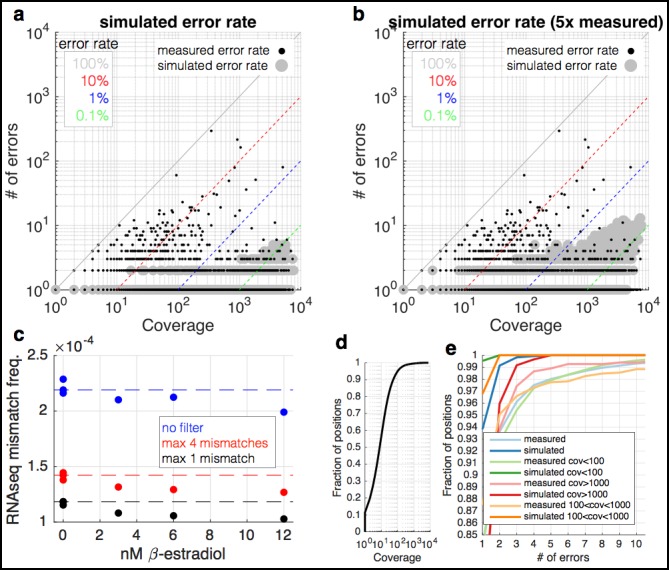
Figure 1—figure supplement 2. RNA-seq data are enriched for mismatches to the reference genome that occur far more often than expected. (a) At each coverage (x-axis), a point is shown if there is any position in the genome with the observed number of errors (y-axis) (small black dots). The diagonal lines show mismatch frequencies of 100%, 10%, 1%, and 0.1% – any point falling on these lines has the given mismatch frequency. With large grey circles are shown simulated data in which the same coverage as the yeast RNA-seq data are used, but with a mismatch frequency identical to the measured overall mismatch frequency of the yeast data. Locations in the graph in which a black point occurs but there is no grey point are locations in which there are more mismatches than expected by change. Note that at a coverage of <100, we expect to see no mismatches more than twice, and 0.5% of positions with two observances of identical mismatches. (b) Identical to (a) but with the simulated mismatch frequency five times the observed. (c) Shown are measured mismatch frequencies for the yeast RPB9 and DST1 induction data at different β-estradiol concentrations, at different filters for the maximal allowed number of observed identical mismatches. The dashed lines show the average mismatch frequency for the 0 nM condition. For all filters, low β-estradiol conditions have higher RNA-seq mismatch frequencies. (d) The coverage of the yeast RNA-seq data; ~95% of the genome is covered by <100 reads. (e) Shown are the fraction of positions in the genome (y-axis) with X errors (x-axis) for the yeast RNA-seq data (cyan) and simulated data (blue). Also shown are the same data for positions of the genome with different coverage. For positions covered by <1000 reads (95% of the genome) the expectation is 0 or 1 sequence mismatch (blue and orange lines). Positions with far greater numbers of mismatches are likely due to subclonal mutations and technical bias. DOI: http://dx.doi.org/10.7554/eLife.09945.005
Figure 1—figure supplement 2. RNA-seq data are enriched for mismatches to the reference genome that occur far more often than expected. (a) At each coverage (x-axis), a point is shown if there is any position in the genome with the observed number of errors (y-axis) (small black dots). The diagonal lines show mismatch frequencies of 100%, 10%, 1%, and 0.1% – any point falling on these lines has the given mismatch frequency. With large grey circles are shown simulated data in which the same coverage as the yeast RNA-seq data are used, but with a mismatch frequency identical to the measured overall mismatch frequency of the yeast data. Locations in the graph in which a black point occurs but there is no grey point are locations in which there are more mismatches than expected by change. Note that at a coverage of <100, we expect to see no mismatches more than twice, and 0.5% of positions with two observances of identical mismatches. (b) Identical to (a) but with the simulated mismatch frequency five times the observed. (c) Shown are measured mismatch frequencies for the yeast RPB9 and DST1 induction data at different β-estradiol concentrations, at different filters for the maximal allowed number of observed identical mismatches. The dashed lines show the average mismatch frequency for the 0 nM condition. For all filters, low β-estradiol conditions have higher RNA-seq mismatch frequencies. (d) The coverage of the yeast RNA-seq data; ~95% of the genome is covered by <100 reads. (e) Shown are the fraction of positions in the genome (y-axis) with X errors (x-axis) for the yeast RNA-seq data (cyan) and simulated data (blue). Also shown are the same data for positions of the genome with different coverage. For positions covered by <1000 reads (95% of the genome) the expectation is 0 or 1 sequence mismatch (blue and orange lines). Positions with far greater numbers of mismatches are likely due to subclonal mutations and technical bias. DOI: http://dx.doi.org/10.7554/eLife.09945.005CC BY · PubMed Central
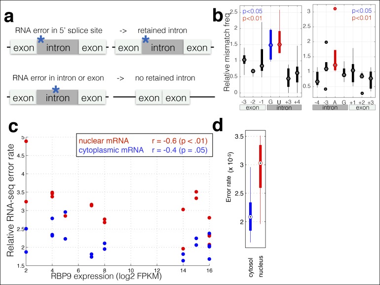
Figure 2. RNA polymerase errors cause intron retention and error rates are correlated with RPB9 expression. (a) RNA polymerase errors at the splice junction should result in intron retention, as DNA mutations at the 5’ donor site are known to cause intron retention. (b) Shown are the RNA-seq mismatch rates at each position relative to the 5’ donor splice site, for sequencing reads that span an exon–intron junction. Mismatch rates from chromatin-associated and nuclear RNAs are higher at the 5’ and 3’ splice sites, suggesting that RNA polymerase errors at this site result in intron retention. (c) For all ENCODE cell lines, RPB9 expression was determined from whole-cell RNA-seq data, and the RNA-seq error rate was measured separately for the cytoplasmic and nuclear fractions. (d) The RNA-seq error rate is higher (paired t-test, p=0.0019) in the nuclear than the cytoplasmic fraction, suggesting that quality-control mechanism may block nuclear export of low quality mRNAs. DOI: http://dx.doi.org/10.7554/eLife.09945.006
Figure 2. RNA polymerase errors cause intron retention and error rates are correlated with RPB9 expression. (a) RNA polymerase errors at the splice junction should result in intron retention, as DNA mutations at the 5’ donor site are known to cause intron retention. (b) Shown are the RNA-seq mismatch rates at each position relative to the 5’ donor splice site, for sequencing reads that span an exon–intron junction. Mismatch rates from chromatin-associated and nuclear RNAs are higher at the 5’ and 3’ splice sites, suggesting that RNA polymerase errors at this site result in intron retention. (c) For all ENCODE cell lines, RPB9 expression was determined from whole-cell RNA-seq data, and the RNA-seq error rate was measured separately for the cytoplasmic and nuclear fractions. (d) The RNA-seq error rate is higher (paired t-test, p=0.0019) in the nuclear than the cytoplasmic fraction, suggesting that quality-control mechanism may block nuclear export of low quality mRNAs. DOI: http://dx.doi.org/10.7554/eLife.09945.006CC BY · PubMed Central
Figure 2—figure supplement 1. RNA-seq mismatch rates for all trinucleotides in chromatin-associated and nuclear RNAs. (a,b) The 5’ and 3’ splicing motifs in the human genome. (c) The RNA-seq mismatch frequencies for all single nucleotides. (d) The RNA-seq mismatch rate to the reference genome for each trinucleotide, normalized to the average mismatch rate across all trinucleotides. For each trinucleotide, red shows the mismatch frequency at the first base, blue at the second, and green at the third. Error bars are standard deviation across all samples. DOI: http://dx.doi.org/10.7554/eLife.09945.007
Figure 2—figure supplement 1. RNA-seq mismatch rates for all trinucleotides in chromatin-associated and nuclear RNAs. (a,b) The 5’ and 3’ splicing motifs in the human genome. (c) The RNA-seq mismatch frequencies for all single nucleotides. (d) The RNA-seq mismatch rate to the reference genome for each trinucleotide, normalized to the average mismatch rate across all trinucleotides. For each trinucleotide, red shows the mismatch frequency at the first base, blue at the second, and green at the third. Error bars are standard deviation across all samples. DOI: http://dx.doi.org/10.7554/eLife.09945.007CC BY · PubMed Central
Figure 2—figure supplement 2. RBP9 expression negatively correlates with RNA-seq mismatch rates. The mismatch frequency is shown across all cells lines. (a) RPB9 mRNA expression is normalized by the median expression level of all subunits. (b) RPB9 mRNA expression is normalized by RBP3 (POLR2C) expression. DOI: http://dx.doi.org/10.7554/eLife.09945.008
Figure 2—figure supplement 2. RBP9 expression negatively correlates with RNA-seq mismatch rates. The mismatch frequency is shown across all cells lines. (a) RPB9 mRNA expression is normalized by the median expression level of all subunits. (b) RPB9 mRNA expression is normalized by RBP3 (POLR2C) expression. DOI: http://dx.doi.org/10.7554/eLife.09945.008CC BY · PubMed Central
Figure 3. RNA polymerase error rate is determined by the expression level of RPB9 and DST1. (a) RNA-seq error rates I re-measured for two strains (Z3EVpr-RPB9, black points, Z3EVpr-DST1, blue points) grown at different concentrations of β-estradiol. The points show the relationship between RPB9 expression levels (determined by RNA-seq) and RNA-seq error rates. The blue points show RPB9 expression levels for the Z3EVpr-DST1 strain, in which DST1 expression ranges from 16 fragments per kilobase per million (FPKM) at 0 nM β-estradiol to 120 FPKM native expression to 756 FPKM at 25 nM β-estradiol. Low induction of both DST1 or RPB9 results in high RNA-seq error rates (red box), while wild-type and higher induction levels result in low RNA-seq error rates (black box). (b) Across all genes, the intron retention rate is higher in conditions with low RNA polymerase fidelity (t-test between high and low error rate samples, p=0.029), consistent with the hypothesis that RNA polymerase errors result in splicing defects. (c) The error rate for each of the 12 single base changes are shown for induction experiments that gave high (red) or low (black) RNA-seq error rates. Transitions (G<–>A, C<–>U) are marked with green boxes and transversions (A<–>C, G<–>U) with purple. DOI: http://dx.doi.org/10.7554/eLife.09945.009
Figure 3. RNA polymerase error rate is determined by the expression level of RPB9 and DST1. (a) RNA-seq error rates I re-measured for two strains (Z3EVpr-RPB9, black points, Z3EVpr-DST1, blue points) grown at different concentrations of β-estradiol. The points show the relationship between RPB9 expression levels (determined by RNA-seq) and RNA-seq error rates. The blue points show RPB9 expression levels for the Z3EVpr-DST1 strain, in which DST1 expression ranges from 16 fragments per kilobase per million (FPKM) at 0 nM β-estradiol to 120 FPKM native expression to 756 FPKM at 25 nM β-estradiol. Low induction of both DST1 or RPB9 results in high RNA-seq error rates (red box), while wild-type and higher induction levels result in low RNA-seq error rates (black box). (b) Across all genes, the intron retention rate is higher in conditions with low RNA polymerase fidelity (t-test between high and low error rate samples, p=0.029), consistent with the hypothesis that RNA polymerase errors result in splicing defects. (c) The error rate for each of the 12 single base changes are shown for induction experiments that gave high (red) or low (black) RNA-seq error rates. Transitions (G<–>A, C<–>U) are marked with green boxes and transversions (A<–>C, G<–>U) with purple. DOI: http://dx.doi.org/10.7554/eLife.09945.009CC BY · PubMed Central
Figure 3—figure supplement 1. Mutations that affect transcription elongation do not affect measured RNA-seq mismatch frequencies. Two separate experiments were performed with wild-type controls and mutants involved in transcription elongation. Individual bars show the RNA-seq mismatch frequency of biological replicates. DOI: http://dx.doi.org/10.7554/eLife.09945.010
Figure 3—figure supplement 1. Mutations that affect transcription elongation do not affect measured RNA-seq mismatch frequencies. Two separate experiments were performed with wild-type controls and mutants involved in transcription elongation. Individual bars show the RNA-seq mismatch frequency of biological replicates. DOI: http://dx.doi.org/10.7554/eLife.09945.010CC BY · PubMed Central
Figure 3—figure supplement 2. Decreases in RPB9 and DST1 expression in yeast results in more single base insertions in RNA-seq data. For each RNA-seq dataset, the number of inserts (+N) or deletions (–N) in the mpileup output (N is the number of bases in the indel) were counted, and this number divided by the total number of mapped reads in each sample. On the right are the same data but zoomed in on each metric to better show the comparison between the two sets of samples. DOI: http://dx.doi.org/10.7554/eLife.09945.011
Figure 3—figure supplement 2. Decreases in RPB9 and DST1 expression in yeast results in more single base insertions in RNA-seq data. For each RNA-seq dataset, the number of inserts (+N) or deletions (–N) in the mpileup output (N is the number of bases in the indel) were counted, and this number divided by the total number of mapped reads in each sample. On the right are the same data but zoomed in on each metric to better show the comparison between the two sets of samples. DOI: http://dx.doi.org/10.7554/eLife.09945.011CC BY · PubMed Central
Figure 4. In-frame stop codons are less likely to be created by polymerase errors. For all genes in yeast, I calculated the number of codons which are one polymerase error from a stop codon. (a) Fewer in-frame codons can be turned into a stop codon by a single-nucleotide change, compared to out-of-frame codons. (b) Codons that are one error away from generating an in-frame stop codon are more likely to be found at the ends of the open reading frames (ORFs), compared to the beginning of the ORF. DOI: http://dx.doi.org/10.7554/eLife.09945.012
Figure 4. In-frame stop codons are less likely to be created by polymerase errors. For all genes in yeast, I calculated the number of codons which are one polymerase error from a stop codon. (a) Fewer in-frame codons can be turned into a stop codon by a single-nucleotide change, compared to out-of-frame codons. (b) Codons that are one error away from generating an in-frame stop codon are more likely to be found at the ends of the open reading frames (ORFs), compared to the beginning of the ORF. DOI: http://dx.doi.org/10.7554/eLife.09945.012CC BY · PubMed Central

Verification

Claims the paper makes

The claims and statistics of this paper haven’t been checked yet.

Free: it reads the open-access text and checks the first 5 claims that cite a source we can check. Create a free account to check every claim.

A few free checks a day · papers already verified open instantly · results are public

Self-check

Percentages, sums and reported differences re-computed from the paper’s own numbers.

The self-check needs the paper’s full text.

Citations

Cites

Show all 1 references

Research works printed but not linked: 37

  1. 10.1093/gbe/evr066 · DOI · Paper page
  2. 10.1073/pnas.1323011111 · DOI · Paper page
  3. 10.1534/genetics.107.071506 · DOI · Paper page
  4. 10.1073/pnas.1309843110 · DOI
  5. 10.1016/j.tig.2010.05.003 · DOI · Paper page
  6. 10.1074/jbc.M202059200 · DOI · Paper page
  7. 10.1111/j.1432-1033.1992.tb16900.x · DOI
  8. 10.1128/MCB.19.11.7357 · DOI · Paper page
  9. 10.1371/journal.pone.0015442 · DOI · Paper page
  10. 10.1016/0092-8674(77)90032-0 · DOI · Paper page
  11. 10.1101/gr.110882.110 · DOI · Paper page
  12. 10.1371/journal.pgen.1003595 · DOI
  13. 10.1016/j.mib.2015.01.010 · DOI
  14. 10.1371/journal.pgen.1004532 · DOI
  15. 10.1093/nar/gkt698 · DOI
  16. Kleinman, C. L. & Majewski, J. Comment on “Widespread RNA and DNA sequence differences in the human transcriptome”. Science 335, 1302-author reply 1302 (2012). · DOI · Paper page
  17. Pickrell, J. K. , Gilad, Y. & Pritchard, J. K. Comment on “Widespread RNA and DNA sequence differences in the human transcriptome”. Science 335, 1302-author reply 1302 (2012). · DOI · Paper page
  18. 10.1126/science.1207018 · DOI · Paper page
  19. 10.1186/1471-2105-12-451 · doi:10.1186/1471-2105-12-451 · Paper page
  20. 10.1101/gr.134445.111 · DOI · Paper page
  21. 10.1073/pnas.0813128106 · DOI
  22. 10.1021/bi4009566 · DOI
  23. 10.1016/S0960-9822(01)00036-7 · DOI
  24. 10.1073/pnas.0511330103 · DOI · Paper page
  25. 10.1074/jbc.M109.006908 · DOI · Paper page
  26. 10.1016/j.molcel.2008.04.017 · DOI · Paper page
  27. 10.1093/nar/gkt1402 · DOI
  28. McIsaac, R. S. , Oakes, B. L. , Botstein, D. & Noyes, M. B. Rapid synthesis and screening of chemically activated transcription factors with GFP-based reporters. Journal of visualized experiments: JoVE e51153–e51153 (2013). · DOI
  29. 10.1093/bioinformatics/btp352 · DOI · Paper page
  30. 10.1093/nar/gkh103 · DOI · Paper page
  31. 10.1093/bioinformatics/btq033 · DOI · Paper page
  32. 10.1093/nar/gks1313 · DOI · Paper page
  33. 10.1093/bioinformatics/btp324 · DOI · Paper page
  34. 10.1038/nature11247 · DOI · Paper page
  35. Jung, H. et al. Intron retention is a widespread mechanism of tumorsuppressor inactivation. Nat. Genet. (2015). · DOI · Paper page
  36. 2015, Microprocessor mediates transcriptional termination of long noncoding RNA transcripts hosting microRNAs, Nat. Struct. Mol. Biol, 22, 319, 10.1038/nsmb.2982 · DOI · Paper page
  37. 10.1074/jbc.M109.006908 · DOI · Paper page

Metadata

Authors
Carey, L. B.
Affiliations
Max Planck Society (DE)
Corresponding author
Carey, Lucas B.
Venue
eLife
Publisher
openRxiv
Published
2015-10-26
Volume
4
ISSN
2050-084X (ISSN-L)
OpenAlex
W2193989147
Cited by
51
References
1
Citations by year
2026: 4 · 2025: 1 · 2024: 5 · 2023: 4 · 2022: 2 · 2021: 6 · 2020: 6 · 2019: 6 · 2018: 6 · 2017: 7 · 2016: 4
FWCI
2.46
Type
journal article
Language
en
Open access
Locations
  • eLife (publishedVersion, cc-by)
  • bioRxiv (Cold Spring Harbor Laboratory) (acceptedVersion)
  • PubMed Central (publishedVersion, cc-by)
  • PubMed (publishedVersion)
  • Repositori digital de la UPF (Universitat Pompeu Fabra) (publishedVersion, cc-by)
  • DOAJ (DOAJ: Directory of Open Access Journals) (submittedVersion, cc-by-sa)
  • RECERCAT (Consorci de Serveis Universitaris de Catalunya) (submittedVersion, cc-by)
Funders
Agència de Gestió d'Ajuts Universitaris i de Recerca (2014SGR0974)
Topics
  • Life Sciences › Biochemistry, Genetics and Molecular Biology › Molecular Biology › RNA Research and Splicing
  • Life Sciences › Biochemistry, Genetics and Molecular Biology › Molecular Biology › RNA and protein synthesis mechanisms
  • Life Sciences › Biochemistry, Genetics and Molecular Biology › Molecular Biology › Genomics and Chromatin Dynamics
Keywords
transcription errors, aberrant splicing, RNA polymerase subunits, differential gene expression, stochastic variability, cell-to-cell variability
MeSH terms
Mutation, DNA-Directed RNA Polymerases, RNA Splicing, Saccharomyces cerevisiae, Transcription, Genetic, Sequence Analysis, DNA, Mutation Rate

About these checks

Registry, retraction, Cites and statistics results are computed, not judged, with one exception: a printed reference that no index lists is read once by a model before it counts as a citation error. Claims are extracted by a model and kept only when their quote appears verbatim in the text; each cited source is then checked, with a model’s help, for whether it says what the claim relies on. Those verdicts can be wrong — read the claim’s quote and the cited source before relying on one.

Statistics and arithmetic are recomputed, not judged.

Confidence bands are shown only for fields calibrated on our annotated test set. None is calibrated yet, so no band is shown.

What counts as a claim ›How we measure confidence ›How we check this ›

Are you an author, or do you see a problem with this page? Request a review

How to cite

Cite CoChat for the checks on this page, and the paper for its own findings.

This page
CoChat. (2026). Citation check: RNA polymerase errors cause splicing defects and can be regulated by differential expression of RNA polymerase subunits. https://app.cochat.ai/papers/10.1101/026799
The paper
Carey, L. B. (2015). RNA polymerase errors cause splicing defects and can be regulated by differential expression of RNA polymerase subunits. eLife. https://doi.org/10.1101/026799

Data sources

Bibliographic data: Semantic Scholar, OpenAlex, Crossref.

Contains information from the Semantic Scholar Academic Graph Datasets, made available under the ODC Attribution License (ODC-BY 1.0).

Semantic Scholar’s data is described in: Kinney, R., et al. (2023). The Semantic Scholar Open Data Platform. arXiv:2301.10140.