CoChat

DNA segment capture by Smc5/6 holo-complexes

Open access
Taschner, Michael, Gruber, Stephan · 2022 · Nature Structural & Molecular Biology

Preprint — published as 10.1038/s41594-023-00956-2 · Paper page

The DOI matches its Crossref record; 1 of the 1 references it cites were found in a registry or an index.

Abstract

Three distinct SMC complexes facilitate chromosome folding and segregation in eukaryotes, presumably by DNA translocation and loop extrusion. How SMCs interact with DNA is however not well understood. Among the SMC complexes, Smc5/6 has dedicated roles in DNA repair and in preventing a lethal buildup of aberrant DNA junctions. Here, we describe the reconstitution of ATP-dependent topological DNA loading by Smc5/6 rings. By inserting cysteine residues at selected protein interfaces, we obtained covalently closed compartments upon chemical cross-linking. We show that two SMC subcompartments and the kleisin compartment topologically entrap a plasmid molecule, but not the full SMC compartment. This is explained by a looped DNA segment inserting into the SMC compartment with the kleisin neck gate locking the loop in place when passing between the two DNA flanks and closing. This DNA segment capture strictly requires the Nse5/6 loader, which opens the neck gate prior to DNA passage. Similar segment capture events without gate opening may provide the power stroke for DNA translocation/loop extrusion in subsequent ATP hydrolysis cycles. Our biochemical experiments thus offer a unifying principle for SMC ATPase function in loading and translocation/extrusion, which is likely relevant to other members of the family of SMC proteins too.

Overview

The claims and statistics of this paper haven’t been checked yet.

Free: it reads the open-access text and checks the first 5 claims that cite a source we can check. Create a free account to check every claim.

A few free checks a day · papers already verified open instantly · results are public

Integrity signals

Registry

Crossref · 10.1101/2022.10.09.511515

Cites

1 of 1 references verified0 citation errors · 0 not checkable

Statistics

Figures

Fig. 1 DNA entrapment in Smc5/6. a, Schematic view of a supercoiled circular DNA substrate (left) bound to SK rings (with topological DNA entrapment, top; nontopological DNA entrapment, middle; no DNA entrapment, bottom) and of the Smc5/6 ring subunits (middle). The positions of the cysteines that were engineered for crosslinking of the hinge, Smc6–Nse4 and Smc5–Nse4 interfaces are indicated in green, purple and orange, respectively. Close-up views of the positions of cysteines are shown at the Smc6–Nse4 (top right) and Smc5–Nse4 (bottom right) interfaces. b, Two schematic representations of the SK compartment depending on the positioning of the Nse4 kleisin subunit. Three cysteine pairs—necessary to covalently close it—are indicated with colored handlebars. A cartoon showing the crosslinked complex with the colored compartment after denaturation is shown in the dashed box. Positions of hinge as well as N- and C-terminal ATPase domains are still shown as half-ovals and circles, respectively. c, Coisolation of crosslinked Smc5/6 proteins with plasmid DNA by agarose gel electrophoresis. The results were obtained with an octameric Smc5/6 holocomplex harboring cysteine pairs for crosslinking of the SK ring, comparing linear and circular DNA substrates. Traces of contaminating linear DNA from E. coli are marked with an asterisk. BsaI, BsaI restriction endonuclease. d, As in c but with titration of the protein complex and comparison of entrapment efficiencies between the wild-type (WT) and ATP-hydrolysis-deficient (EQ/EQ) complexes. Controls without either ATP or the Nse5/6 loader are also included. The plasmid substrate is largely supercoiled (sc), but a relaxed (Rel) form is also visible. Source data
Fig. 1 DNA entrapment in Smc5/6. a, Schematic view of a supercoiled circular DNA substrate (left) bound to SK rings (with topological DNA entrapment, top; nontopological DNA entrapment, middle; no DNA entrapment, bottom) and of the Smc5/6 ring subunits (middle). The positions of the cysteines that were engineered for crosslinking of the hinge, Smc6–Nse4 and Smc5–Nse4 interfaces are indicated in green, purple and orange, respectively. Close-up views of the positions of cysteines are shown at the Smc6–Nse4 (top right) and Smc5–Nse4 (bottom right) interfaces. b, Two schematic representations of the SK compartment depending on the positioning of the Nse4 kleisin subunit. Three cysteine pairs—necessary to covalently close it—are indicated with colored handlebars. A cartoon showing the crosslinked complex with the colored compartment after denaturation is shown in the dashed box. Positions of hinge as well as N- and C-terminal ATPase domains are still shown as half-ovals and circles, respectively. c, Coisolation of crosslinked Smc5/6 proteins with plasmid DNA by agarose gel electrophoresis. The results were obtained with an octameric Smc5/6 holocomplex harboring cysteine pairs for crosslinking of the SK ring, comparing linear and circular DNA substrates. Traces of contaminating linear DNA from E. coli are marked with an asterisk. BsaI, BsaI restriction endonuclease. d, As in c but with titration of the protein complex and comparison of entrapment efficiencies between the wild-type (WT) and ATP-hydrolysis-deficient (EQ/EQ) complexes. Controls without either ATP or the Nse5/6 loader are also included. The plasmid substrate is largely supercoiled (sc), but a relaxed (Rel) form is also visible. Source dataCC BY · PubMed Central
Fig. 2 DNA entrapment in Smc5/6 subcompartments. a, Schematic representation of subcompartments formed during ATP-dependent head engagement. The scheme on the left shows the complete Smc5/6 hexamer with the SK compartment highlighted as in Fig. 1. Combinations of cysteine pairs (colored handlebars as indicated) lead to covalent closure of K and S compartments, with the latter being split into upper (Sup) and lower (Slo) compartments by Nse3 crosslinking as indicated. Note that the schemes on the right show only the crosslinked subunits that remain attached after denaturation. Schemes in dashed boxes indicate compartments after denaturation as in Fig. 1b. XL, Crosslink; E, Engaged. b, Coisolation of crosslinked Smc5/6 proteins with plasmid DNA by agarose gel electrophoresis. Results obtained with protein preparations harboring cysteine pairs for crosslinking of the K (top), S (second top), Sup (third top) or Slo (bottom) compartments, as in Fig. 1d. Hex, Hexamer. c, Schematic representation of a DNA segment-capture state explaining the findings in b. Source data
Fig. 2 DNA entrapment in Smc5/6 subcompartments. a, Schematic representation of subcompartments formed during ATP-dependent head engagement. The scheme on the left shows the complete Smc5/6 hexamer with the SK compartment highlighted as in Fig. 1. Combinations of cysteine pairs (colored handlebars as indicated) lead to covalent closure of K and S compartments, with the latter being split into upper (Sup) and lower (Slo) compartments by Nse3 crosslinking as indicated. Note that the schemes on the right show only the crosslinked subunits that remain attached after denaturation. Schemes in dashed boxes indicate compartments after denaturation as in Fig. 1b. XL, Crosslink; E, Engaged. b, Coisolation of crosslinked Smc5/6 proteins with plasmid DNA by agarose gel electrophoresis. Results obtained with protein preparations harboring cysteine pairs for crosslinking of the K (top), S (second top), Sup (third top) or Slo (bottom) compartments, as in Fig. 1d. Hex, Hexamer. c, Schematic representation of a DNA segment-capture state explaining the findings in b. Source dataCC BY · PubMed Central
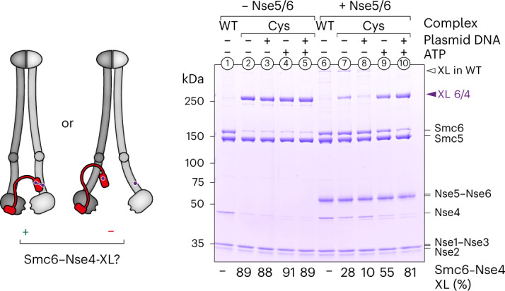
Fig. 3 Opening of the neck gate by Nse5/6. Crosslinking of purified Smc5/6 hexamers with cysteines at the Smc6–Nse4 interface is shown in the presence and absence of ligands. Detection of crosslinked species was by SDS–PAGE and Coomassie staining. Loss of crosslinking suggests that the gate was open. XL, Crosslink. Equivalent experiments for this interface containing ATPase head mutations as well as for other cysteine pairs are shown in Extended Data Fig. 6. Source data
Fig. 3 Opening of the neck gate by Nse5/6. Crosslinking of purified Smc5/6 hexamers with cysteines at the Smc6–Nse4 interface is shown in the presence and absence of ligands. Detection of crosslinked species was by SDS–PAGE and Coomassie staining. Loss of crosslinking suggests that the gate was open. XL, Crosslink. Equivalent experiments for this interface containing ATPase head mutations as well as for other cysteine pairs are shown in Extended Data Fig. 6. Source dataCC BY · PubMed Central
Fig. 4 DNA clamping is essential for DNA entrapment. Identification of putative DNA-binding residues in Smc5 and Smc6. a, The positions of the selected residues schematically displayed for the DNA-clamping state (Extended Data Fig. 7a). b, Positively charged residues on the Smc5 head mutated to alanine (A) or glutamate (E) in isolation or in combination as indicated. The mutant alleles were tested for function by plasmid shuffling. Counterselection against a pCEN(URA3) plasmid carrying a wild-type (WT) SMC5 allele by addition of 5-FOA revealed smc5 mutants resulting in a growth defect. c, As in b for residues in Smc6. d, Salt-stable DNA binding with wild-type and mutant Smc5/6 as determined by protein immobilization using a TwinStrep tag on Smc6 (bottom) and detection of coisolated plasmid DNA (‘recovered plasmid’) by agarose gel electrophoresis (top). e, DNA entrapment in the SK ring of wild-type, DNA-binding and ATP-hydrolysis-defective Smc5/6 mutants. Standard DNA entrapment salt conditions (150 mM NaCl) were used as in Fig. 1d (top) or buffer with reduced ionic strength (150 mM KOAc) as for the salt-stable DNA-binding assay in b. Source data
Fig. 4 DNA clamping is essential for DNA entrapment. Identification of putative DNA-binding residues in Smc5 and Smc6. a, The positions of the selected residues schematically displayed for the DNA-clamping state (Extended Data Fig. 7a). b, Positively charged residues on the Smc5 head mutated to alanine (A) or glutamate (E) in isolation or in combination as indicated. The mutant alleles were tested for function by plasmid shuffling. Counterselection against a pCEN(URA3) plasmid carrying a wild-type (WT) SMC5 allele by addition of 5-FOA revealed smc5 mutants resulting in a growth defect. c, As in b for residues in Smc6. d, Salt-stable DNA binding with wild-type and mutant Smc5/6 as determined by protein immobilization using a TwinStrep tag on Smc6 (bottom) and detection of coisolated plasmid DNA (‘recovered plasmid’) by agarose gel electrophoresis (top). e, DNA entrapment in the SK ring of wild-type, DNA-binding and ATP-hydrolysis-defective Smc5/6 mutants. Standard DNA entrapment salt conditions (150 mM NaCl) were used as in Fig. 1d (top) or buffer with reduced ionic strength (150 mM KOAc) as for the salt-stable DNA-binding assay in b. Source dataCC BY · PubMed Central
Fig. 5 DNA passes through the Smc6–Nse4 gate. a, Coisolation of crosslinked Smc5/6 proteins with plasmid DNA by agarose gel electrophoresis. Results were obtained with protein preparations containing combinations of cysteine pairs and an Smc6–Nse4 fusion protein. Schematic drawings of crosslinked and denatured protein species are shown below the gel, with the closed compartments indicated. Different observed step sizes of DNA ladders are caused by differences in the size of the crosslinked protein species. XL, Crosslink. b, Schematic representation of DNA clamping in the presence of an Smc6–Nse4 fusion protein. The length of the linker allows it to wrap around the DNA strand clamped on ATP-engaged heads. c, As in a with Smc5/6 holocomplexes at 400 nM, and with or without post-entrapment opening of the linker using the human rhinovirus 3C protease. d, Coisolation of crosslinked Smc5/6 proteins with plasmid DNA by agarose gel electrophoresis, comparing a protein preparation harboring cysteine pairs for crosslinking of the SK ring with one in which the hinge cysteine pair is replaced by a SpyTag–SpyCatcher fusion. Topological DNA entrapment in Smc5/6 is not prevented by the hinge fusion, unlike what is shown for cohesin. Source data
Fig. 5 DNA passes through the Smc6–Nse4 gate. a, Coisolation of crosslinked Smc5/6 proteins with plasmid DNA by agarose gel electrophoresis. Results were obtained with protein preparations containing combinations of cysteine pairs and an Smc6–Nse4 fusion protein. Schematic drawings of crosslinked and denatured protein species are shown below the gel, with the closed compartments indicated. Different observed step sizes of DNA ladders are caused by differences in the size of the crosslinked protein species. XL, Crosslink. b, Schematic representation of DNA clamping in the presence of an Smc6–Nse4 fusion protein. The length of the linker allows it to wrap around the DNA strand clamped on ATP-engaged heads. c, As in a with Smc5/6 holocomplexes at 400 nM, and with or without post-entrapment opening of the linker using the human rhinovirus 3C protease. d, Coisolation of crosslinked Smc5/6 proteins with plasmid DNA by agarose gel electrophoresis, comparing a protein preparation harboring cysteine pairs for crosslinking of the SK ring with one in which the hinge cysteine pair is replaced by a SpyTag–SpyCatcher fusion. Topological DNA entrapment in Smc5/6 is not prevented by the hinge fusion, unlike what is shown for cohesin. Source dataCC BY · PubMed Central
Fig. 6 Smc5/6 entraps DNA in vivo. The scheme on the left shows an outline of the procedure. Results from southern blotting on the right show that crosslinkable versions (6C) of both cohesin and Smc5/6 entrap the mini-chromosome (catenated monomers (CM)), but only cohesin does so in a cohesive manner (catenated dimers (CD)). Control strains lacking one of the ring cysteines (5C) abolish entrapment in both cases. Source data
Fig. 6 Smc5/6 entraps DNA in vivo. The scheme on the left shows an outline of the procedure. Results from southern blotting on the right show that crosslinkable versions (6C) of both cohesin and Smc5/6 entrap the mini-chromosome (catenated monomers (CM)), but only cohesin does so in a cohesive manner (catenated dimers (CD)). Control strains lacking one of the ring cysteines (5C) abolish entrapment in both cases. Source dataCC BY · PubMed Central
Fig. 7 Model for chromosome loading and DNA translocation by alternating between a holding and a segment-capture state. Schematic representation of the main findings presented in the present study. The segment-capture state (middle) is an essential intermediate in both DNA loading (left) and DNA translocation (right). Note that two possibilities for this state are indicated, differing in their degree of arm opening toward the hinge.
Fig. 7 Model for chromosome loading and DNA translocation by alternating between a holding and a segment-capture state. Schematic representation of the main findings presented in the present study. The segment-capture state (middle) is an essential intermediate in both DNA loading (left) and DNA translocation (right). Note that two possibilities for this state are indicated, differing in their degree of arm opening toward the hinge.CC BY · PubMed Central
Extended Data Fig. 1 Schematics of Smc5/6 complex architecture. (a) Overall architecture of the hexameric Smc5/6 core complex and the Nse5/6 dimer (adapted from20). For simplicity the Nse5/6 complex is shown separately with multiple arrows denoting various contact points with the core complex. (b) Simplified schematics focusing only on changes in Smc5/6 dimer architecture during the ATPase cycle and upon binding to the Nse5/6 loader and the DNA substrate.
Extended Data Fig. 1 Schematics of Smc5/6 complex architecture. (a) Overall architecture of the hexameric Smc5/6 core complex and the Nse5/6 dimer (adapted from20). For simplicity the Nse5/6 complex is shown separately with multiple arrows denoting various contact points with the core complex. (b) Simplified schematics focusing only on changes in Smc5/6 dimer architecture during the ATPase cycle and upon binding to the Nse5/6 loader and the DNA substrate.CC BY · PubMed Central
Extended Data Fig. 2 Analysis of Smc5/6 complexes harboring multiple engineered cysteines. (a) Crosslinking of Smc5/6 hexamers harboring multiple engineered cysteines as indicated. Identification of covalently closed ring species and intermediary crosslinking products by protein gel analysis on 2 types of gels (left: 4–12 % (w/v) gradient gel, right: 3–8 % (w/v) gradient gel) for proper separation of small and large proteins and detection by Coomassie staining. Note the presence of small amounts of ring species in two of the three control samples lacking one of the six cysteines (lanes 5 and 7), indicating minor off-target crosslinking. (b) ATPase activity assays with selected hexameric Smc5/6 complexes relevant for this study. Combinations of certain modifications lead to a reduction of ATPase activity. Error bars show standard deviations from the mean for three technical replicates. (c) Control experiments for entrapment experiments shown in Fig. 1 with preparations lacking a selected cysteine (5C). Note that low levels of co-entrapment detected with two of the three 5C samples are likely explained by weak off-target crosslinking. (d) Under conditions promoting DNA entrapment the ring species is visible in a protein gel in the presence of a linear (lane 1) but not a circular (lane 2) DNA substrate, presumably due to co-retention of the ring species with circular DNA in the loading well (see scheme on the right). The ring species re-appears after digestion of the circular substrate (lane 3). (e) Time-dependence of DNA entrapment in the SK ring (top panel) and the K compartment (bottom panel). Crosslinker was added to a sample aliquot at the indicated time points. Source data
Extended Data Fig. 2 Analysis of Smc5/6 complexes harboring multiple engineered cysteines. (a) Crosslinking of Smc5/6 hexamers harboring multiple engineered cysteines as indicated. Identification of covalently closed ring species and intermediary crosslinking products by protein gel analysis on 2 types of gels (left: 4–12 % (w/v) gradient gel, right: 3–8 % (w/v) gradient gel) for proper separation of small and large proteins and detection by Coomassie staining. Note the presence of small amounts of ring species in two of the three control samples lacking one of the six cysteines (lanes 5 and 7), indicating minor off-target crosslinking. (b) ATPase activity assays with selected hexameric Smc5/6 complexes relevant for this study. Combinations of certain modifications lead to a reduction of ATPase activity. Error bars show standard deviations from the mean for three technical replicates. (c) Control experiments for entrapment experiments shown in Fig. 1 with preparations lacking a selected cysteine (5C). Note that low levels of co-entrapment detected with two of the three 5C samples are likely explained by weak off-target crosslinking. (d) Under conditions promoting DNA entrapment the ring species is visible in a protein gel in the presence of a linear (lane 1) but not a circular (lane 2) DNA substrate, presumably due to co-retention of the ring species with circular DNA in the loading well (see scheme on the right). The ring species re-appears after digestion of the circular substrate (lane 3). (e) Time-dependence of DNA entrapment in the SK ring (top panel) and the K compartment (bottom panel). Crosslinker was added to a sample aliquot at the indicated time points. Source dataCC BY · PubMed Central
Extended Data Fig. 3 Schematic overview of assays used to examine the nature of Smc5/6 association with its DNA substrate. (a) In the topological loading assay, a crosslinkable version of the Smc5/6 hexamer (‘6C’) is incubated with a small supercoiled (‘sc’) plasmid substrate in the absence or presence of ATP and Nse5/6. Upon crosslinking with BMOE and protein denaturation (‘SDS’) only fully crosslinked rings that were topologically associated with the substrate are retained, leading to a characteristic laddering pattern in agarose gels. (b) The salt-stable binding assay does not involve crosslinking. Complexes are first incubated with the substrate under low-salt conditions before the salt concentration is increased to 1 M NaCl by buffer changes. Smc5/6 complexes are immobilized on beads using a TwinStrep tag on Smc6, and co-purified plasmid substrate is examined on an agarose gel.
Extended Data Fig. 3 Schematic overview of assays used to examine the nature of Smc5/6 association with its DNA substrate. (a) In the topological loading assay, a crosslinkable version of the Smc5/6 hexamer (‘6C’) is incubated with a small supercoiled (‘sc’) plasmid substrate in the absence or presence of ATP and Nse5/6. Upon crosslinking with BMOE and protein denaturation (‘SDS’) only fully crosslinked rings that were topologically associated with the substrate are retained, leading to a characteristic laddering pattern in agarose gels. (b) The salt-stable binding assay does not involve crosslinking. Complexes are first incubated with the substrate under low-salt conditions before the salt concentration is increased to 1 M NaCl by buffer changes. Smc5/6 complexes are immobilized on beads using a TwinStrep tag on Smc6, and co-purified plasmid substrate is examined on an agarose gel.CC BY · PubMed Central
Extended Data Fig. 4 Analysis of Nse3 crosslinking to SMC arms. Crosslinking of purified Smc5/6 hexamers with cysteines at the Smc6/Nse3 interface (a) or at the Smc5/Nse3 interface (b) in the presence and absence of ligands. Detection of crosslinked species by SDS-Page and Coomassie staining. Numbers below the gel indicate the efficiency of the respective crosslink. Source data
Extended Data Fig. 4 Analysis of Nse3 crosslinking to SMC arms. Crosslinking of purified Smc5/6 hexamers with cysteines at the Smc6/Nse3 interface (a) or at the Smc5/Nse3 interface (b) in the presence and absence of ligands. Detection of crosslinked species by SDS-Page and Coomassie staining. Numbers below the gel indicate the efficiency of the respective crosslink. Source dataCC BY · PubMed Central
Extended Data Fig. 5 Head-distal Smc5/6 coiled-coil arms do not detectably open upon addition of Nse5/6, ATP, and/or DNA. a) Model of an Smc5/Smc6/Nse2 complex obtained with AlphaFold-Multimer. The positions of two engineered cysteine pairs for inter-arm crosslinking are denoted. (b, c) Results from crosslinking experiments showing efficient arm alignment in all tested conditions. As in Fig. 3 and Supplementary Fig. 6. Source data
Extended Data Fig. 5 Head-distal Smc5/6 coiled-coil arms do not detectably open upon addition of Nse5/6, ATP, and/or DNA. a) Model of an Smc5/Smc6/Nse2 complex obtained with AlphaFold-Multimer. The positions of two engineered cysteine pairs for inter-arm crosslinking are denoted. (b, c) Results from crosslinking experiments showing efficient arm alignment in all tested conditions. As in Fig. 3 and Supplementary Fig. 6. Source dataCC BY · PubMed Central
Extended Data Fig. 6 Crosslinking of selected SMC/kleisin interfaces. Similar to Fig. 3(a) The Smc5/Nse4 interface is efficiently crosslinked and does not detectably respond to the presence of ligands apart from weak off-target crosslinking between Smc5 and the loader subunit Nse5 [see (B)]. (b) Control reaction for (A) with protein samples lacking the engineered cysteine in Nse4 confirming off-target crosslinking of Smc5(G1054C) to Nse5. (c) Addition of the loader reduces abundance of the ring species due to gate opening. The pattern in the presence of ligands (ATP and plasmid DNA) mirrors the one obtained with the Smc6/Nse4 interface (Fig. 3), except when loader, ATP and plasmid are added, presumably due to co-retention of ring species with circular DNA in the loading well (see scheme on the right). (d) As in Fig. 3 but with Smc5 and Smc6 subunits carrying the signature motif head-engagement mutation (‘SR’). (e) As in Fig. 3 but with Smc5 and Smc6 subunits carrying the Walker B ATP hydrolysis mutation (‘EQ’). (f) Fusion of Smc6 to Nse4 with the linker used in our in vitro assays is lethal in yeast. Plasmid shuffling assay as in Supplementary Fig. 7f, but with a deletion mutant for both smc6 and nse4. Adding back both wildtype genes separately, but not either of them alone or a fused version, restores viability. (g) Effect of mutating the head DNA binding interfaces on Smc5 and/or Smc6 on formation of a closed SMC/Kleisin (SK) ring. (h) Effect of mutating the head DNA binding interfaces on Smc5 and Smc6 on Smc6/Nse4 gate closure in the presence of the loader and various ligands. Source data
Extended Data Fig. 6 Crosslinking of selected SMC/kleisin interfaces. Similar to Fig. 3(a) The Smc5/Nse4 interface is efficiently crosslinked and does not detectably respond to the presence of ligands apart from weak off-target crosslinking between Smc5 and the loader subunit Nse5 [see (B)]. (b) Control reaction for (A) with protein samples lacking the engineered cysteine in Nse4 confirming off-target crosslinking of Smc5(G1054C) to Nse5. (c) Addition of the loader reduces abundance of the ring species due to gate opening. The pattern in the presence of ligands (ATP and plasmid DNA) mirrors the one obtained with the Smc6/Nse4 interface (Fig. 3), except when loader, ATP and plasmid are added, presumably due to co-retention of ring species with circular DNA in the loading well (see scheme on the right). (d) As in Fig. 3 but with Smc5 and Smc6 subunits carrying the signature motif head-engagement mutation (‘SR’). (e) As in Fig. 3 but with Smc5 and Smc6 subunits carrying the Walker B ATP hydrolysis mutation (‘EQ’). (f) Fusion of Smc6 to Nse4 with the linker used in our in vitro assays is lethal in yeast. Plasmid shuffling assay as in Supplementary Fig. 7f, but with a deletion mutant for both smc6 and nse4. Adding back both wildtype genes separately, but not either of them alone or a fused version, restores viability. (g) Effect of mutating the head DNA binding interfaces on Smc5 and/or Smc6 on formation of a closed SMC/Kleisin (SK) ring. (h) Effect of mutating the head DNA binding interfaces on Smc5 and Smc6 on Smc6/Nse4 gate closure in the presence of the loader and various ligands. Source dataCC BY · PubMed Central
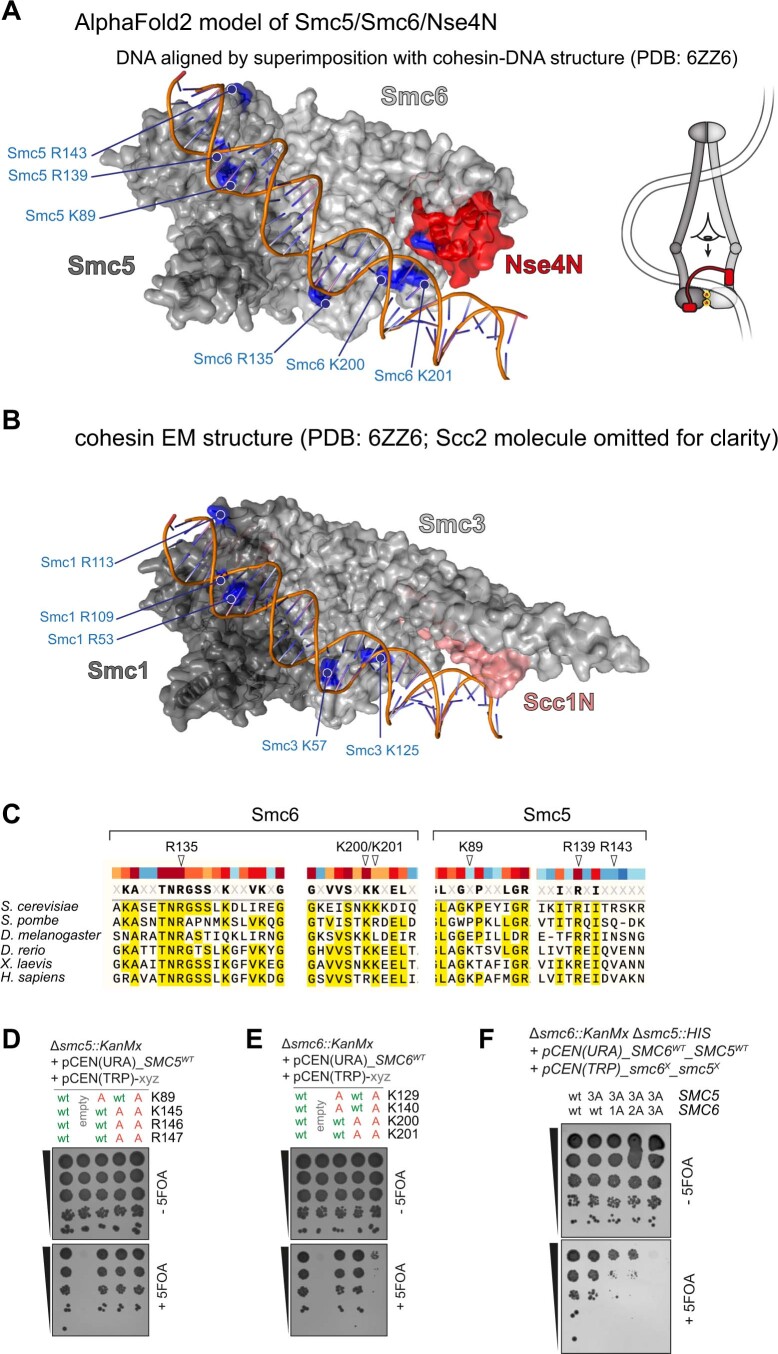
Extended Data Fig. 7 Identification of putative DNA binding residues on Smc6 and Smc5. (a) Model of ATP-engaged Smc6/Smc5 heads with the Nse4 N-terminus bound to the Smc6 neck. AlphaFold-Multimer models of Smc6/Nse4 and Smc5 were superimposed on their counterparts of the cohesin complex (PDB: 6ZZ6, see panel B). The DNA molecule from the cohesin cryo-EM structure contacts several putative DNA binding residues on Smc6 and Smc5. (b) Similar view on top of engaged heads in the cohesin cryo-EM structure (PDB: 6ZZ6) with DNA-interacting residues on Smc3 and Smc1 indicated. Note that the Scc2 molecule is not shown for simplicity. (c) Sequence alignment showing strong evolutionary conservation of examined residues in Smc6. Smc5 residues show weaker conservation consistent with results of functional assays shown in Fig. 4. (d) Positively charged residues on the Smc5 head were mutated to alanine (‘A’) and the mutant alleles were tested for function by plasmid shuffling. As in Fig. 4b but with residues selected based on a recent cryo-EM structure of Smc5/6 (Yu et al.43). (e) As in (D) but for residues on the Smc6 head. (f) Positively charged residues on Smc5 and Smc6 heads were mutated to alanines in combinations. As in Fig. 4b and c. Source data
Extended Data Fig. 7 Identification of putative DNA binding residues on Smc6 and Smc5. (a) Model of ATP-engaged Smc6/Smc5 heads with the Nse4 N-terminus bound to the Smc6 neck. AlphaFold-Multimer models of Smc6/Nse4 and Smc5 were superimposed on their counterparts of the cohesin complex (PDB: 6ZZ6, see panel B). The DNA molecule from the cohesin cryo-EM structure contacts several putative DNA binding residues on Smc6 and Smc5. (b) Similar view on top of engaged heads in the cohesin cryo-EM structure (PDB: 6ZZ6) with DNA-interacting residues on Smc3 and Smc1 indicated. Note that the Scc2 molecule is not shown for simplicity. (c) Sequence alignment showing strong evolutionary conservation of examined residues in Smc6. Smc5 residues show weaker conservation consistent with results of functional assays shown in Fig. 4. (d) Positively charged residues on the Smc5 head were mutated to alanine (‘A’) and the mutant alleles were tested for function by plasmid shuffling. As in Fig. 4b but with residues selected based on a recent cryo-EM structure of Smc5/6 (Yu et al.43). (e) As in (D) but for residues on the Smc6 head. (f) Positively charged residues on Smc5 and Smc6 heads were mutated to alanines in combinations. As in Fig. 4b and c. Source dataCC BY · PubMed Central
Extended Data Fig. 8 Entrapment assays involving Smc6-Nse4 fusion and BMOE crosslinking of interfaces. (a) Schematic representation of complexes used for experiments in Fig. 5 and Supplementary Fig. 8d. The ‘Nse4open’ complex has a split kleisin and thus has its SK ring permanently opened. The ‘Smc6-Nse4 fusion’ complex contains a peptide linker fusing the C-terminus of Smc6 with the N-terminus of Nse4. The linker contains a recognition site for the HRV 3C protease and can thus be opened by cleavage. (b) Schematic overview of entrapment in the absence and presence of the Smc6-Nse4 fusion. Without the fusion (top) during initial DNA segment capture the Smc6/Nse4 gate is opened by Nse5/6 (Smc5 and Smc6 arm distance is exaggerated for clarity). Upon gate closure and ATP hydrolysis, one of the loop strands is lost after escape between the disengaged heads, and the other strand becomes topologically entrapped in the SK ring. The green-filled area indicates the lumen of the ring compartment (SK) which is maintained even after protein denaturation due to crosslinking. (bottom) Scenarios for DNA entrapment by complexes with Smc6-Nse4 fusion (top row, middle panel) in combination with different cysteine pairs for crosslinking (other panels). Top row, left panel: Upon crosslinking of Smc5/Smc6 and Smc5/Nse4 interfaces an SK-plus-linker joint compartment becomes denaturation-resistant (orange-filled area) leading to DNA loop entrapment (not detected in this assay) rather than DNA entrapment. Top row, right panel: Crosslinking of only the Smc6/Nse4 interface leads to a denaturation-resistant linker compartment (blue-filled area) that topologically entraps a DNA strand. The SK compartment is also formed and entraps the other DNA strand, but it is sensitive to denaturation (green-dashed area). Bottom row: Crosslinking of all three interfaces entraps one DNA strand in a denaturation-resistant SK compartment (green-filled area) and another in the linker compartment (blue-filled area), only the latter of which can be released by incubation with 3C protease. Incompletely crosslinked rings lacking the Smc5/Smc6 or Smc5/Nse4 crosslinks (or both) entrap only the DNA strand in the linker compartment (in square bracket). (c) As in Fig. 5c but with cleavage of the linker prior to mixing of samples for DNA entrapment. (d) Salt-stable DNA binding of Smc5/6 complexes with a split Nse4 protein (‘Nse4 open’) or linked Smc6 and Nse4 proteins (‘Smc6-Nse4’ fusion protein). As in Fig. 3d. Pre-treatment with 3C protease cleaves the fusion linker peptide but does not alter DNA binding. Asterisks denote unspecific degradation products. (e) Insertion of a Spy-tag and Spy-Catcher into the Smc5 and Smc6 hinge-domains (see scheme on the left), respectively, leads to permanent, covalent fusion of the hinge domains as shown in the protein gel on the right. Source data
Extended Data Fig. 8 Entrapment assays involving Smc6-Nse4 fusion and BMOE crosslinking of interfaces. (a) Schematic representation of complexes used for experiments in Fig. 5 and Supplementary Fig. 8d. The ‘Nse4open’ complex has a split kleisin and thus has its SK ring permanently opened. The ‘Smc6-Nse4 fusion’ complex contains a peptide linker fusing the C-terminus of Smc6 with the N-terminus of Nse4. The linker contains a recognition site for the HRV 3C protease and can thus be opened by cleavage. (b) Schematic overview of entrapment in the absence and presence of the Smc6-Nse4 fusion. Without the fusion (top) during initial DNA segment capture the Smc6/Nse4 gate is opened by Nse5/6 (Smc5 and Smc6 arm distance is exaggerated for clarity). Upon gate closure and ATP hydrolysis, one of the loop strands is lost after escape between the disengaged heads, and the other strand becomes topologically entrapped in the SK ring. The green-filled area indicates the lumen of the ring compartment (SK) which is maintained even after protein denaturation due to crosslinking. (bottom) Scenarios for DNA entrapment by complexes with Smc6-Nse4 fusion (top row, middle panel) in combination with different cysteine pairs for crosslinking (other panels). Top row, left panel: Upon crosslinking of Smc5/Smc6 and Smc5/Nse4 interfaces an SK-plus-linker joint compartment becomes denaturation-resistant (orange-filled area) leading to DNA loop entrapment (not detected in this assay) rather than DNA entrapment. Top row, right panel: Crosslinking of only the Smc6/Nse4 interface leads to a denaturation-resistant linker compartment (blue-filled area) that topologically entraps a DNA strand. The SK compartment is also formed and entraps the other DNA strand, but it is sensitive to denaturation (green-dashed area). Bottom row: Crosslinking of all three interfaces entraps one DNA strand in a denaturation-resistant SK compartment (green-filled area) and another in the linker compartment (blue-filled area), only the latter of which can be released by incubation with 3C protease. Incompletely crosslinked rings lacking the Smc5/Smc6 or Smc5/Nse4 crosslinks (or both) entrap only the DNA strand in the linker compartment (in square bracket). (c) As in Fig. 5c but with cleavage of the linker prior to mixing of samples for DNA entrapment. (d) Salt-stable DNA binding of Smc5/6 complexes with a split Nse4 protein (‘Nse4 open’) or linked Smc6 and Nse4 proteins (‘Smc6-Nse4’ fusion protein). As in Fig. 3d. Pre-treatment with 3C protease cleaves the fusion linker peptide but does not alter DNA binding. Asterisks denote unspecific degradation products. (e) Insertion of a Spy-tag and Spy-Catcher into the Smc5 and Smc6 hinge-domains (see scheme on the left), respectively, leads to permanent, covalent fusion of the hinge domains as shown in the protein gel on the right. Source dataCC BY · PubMed Central

Verification

Claims the paper makes

The claims and statistics of this paper haven’t been checked yet.

Free: it reads the open-access text and checks the first 5 claims that cite a source we can check. Create a free account to check every claim.

A few free checks a day · papers already verified open instantly · results are public

Self-check

Percentages, sums and reported differences re-computed from the paper’s own numbers.

The self-check needs the paper’s full text.

Citations

Cites

Research works printed but not linked: 70

  1. 2019, Organization of Chromosomal DNA by SMC Complexes, Annu Rev Genet, 53, 445, 10.1146/annurev-genet-112618-043633 · DOI · Paper page
  2. 10.1126/science.aaz3418 · DOI · Paper page
  3. 10.1126/science.aar7831 · DOI · Paper page
  4. 10.1126/science.aaz4475 · DOI · Paper page
  5. Pradhan, B. et al. SMC complexes can traverse physical roadblocks bigger than their ring size. bioRxiv (2021). · DOI
  6. 2020, Structural insights into DNA loop extrusion by SMC protein complexes, Curr Opin Struct Biol, 65, 102, 10.1016/j.sbi.2020.06.009 · DOI
  7. 2022, SMC complexes: Lifting the lid on loop extrusion, Curr Opin Cell Biol, 74, 13, 10.1016/j.ceb.2021.12.003 · DOI
  8. 10.1146/annurev-genet-120417-031353 · DOI · Paper page
  9. 10.1074/jbc.M308828200 · DOI
  10. 10.1091/mbc.E04-05-0436 · DOI
  11. 10.1371/journal.pgen.1004071 · DOI
  12. 10.1371/journal.pgen.1004067 · DOI
  13. 10.1038/ncb1239 · DOI · Paper page
  14. 10.1038/ncb1619 · DOI · Paper page
  15. 10.1038/nature09791 · DOI
  16. 2020, Purified Smc5/6 Complex Exhibits DNA Substrate Recognition and Compaction, Mol Cell, 80, 1039, 10.1016/j.molcel.2020.11.012 · DOI · Paper page
  17. 2020, The Smc5/6 Core Complex Is a Structure-Specific DNA Binding and Compacting Machine, Mol Cell, 80, 1025, 10.1016/j.molcel.2020.11.011 · DOI · Paper page
  18. Yu, Y. et al. Integrative analysis reveals unique structural and functional features of the Smc5/6 complex. Proc Natl Acad Sci U S A 118(2021). · DOI · Paper page
  19. 2021, Nse5/6 is a negative regulator of the ATPase activity of the Smc5/6 complex, Nucleic Acids Res, 49, 4534, 10.1093/nar/gkab234 · DOI
  20. 2021, Nse5/6 inhibits the Smc5/6 ATPase and modulates DNA substrate binding, EMBO J, 40, e107807, 10.15252/embj.2021107807 · DOI · Paper page
  21. Hallett, S.T. et al. Cryo-EM structure of the Smc5/6 holo-complex. Nucleic Acids Res (2022). · DOI
  22. 10.1016/j.cub.2004.09.044 · DOI · Paper page
  23. 10.1002/bip.22843 · DOI · Paper page
  24. 10.1093/emboj/20.12.3238 · DOI
  25. 10.1038/nsmb.2488 · DOI
  26. 10.1126/science.1256917 · DOI · Paper page
  27. Wilhelm, L. et al. SMC condensin entraps chromosomal DNA by an ATP hydrolysis dependent loading mechanism in Bacillus subtilis. Elife 4(2015). · DOI · Paper page
  28. 10.1074/jbc.M608004200 · DOI
  29. 10.1016/j.molcel.2004.08.030 · DOI · Paper page
  30. 10.1016/j.str.2015.10.004 · DOI
  31. 10.1016/j.molcel.2009.06.032 · DOI
  32. Etheridge, T.J. et al. Live-cell single-molecule tracking highlights requirements for stable Smc5/6 chromatin association in vivo. Elife 10(2021). · DOI
  33. 10.1128/MCB.26.5.1617-1630.2006 · DOI
  34. Pradhan, B. et al. The Smc5/6 complex is a DNA loop extruding motor. bioRxiv (2022). · DOI
  35. Oravcová, M. et al. The Nse5/6-like SIMC1-SLF2 Complex Localizes SMC5/6 to Viral Replication Centers. bioRxiv (2022). · DOI
  36. 10.1016/j.molcel.2017.06.010 · DOI · Paper page
  37. 10.1016/j.molcel.2014.11.023 · DOI · Paper page
  38. 10.1016/j.molcel.2019.05.001 · DOI · Paper page
  39. 10.1016/j.molcel.2019.05.023 · DOI · Paper page
  40. 2020, Cryo-EM structures of holo condensin reveal a subunit flip-flop mechanism, Nat Struct Mol Biol, 27, 743, 10.1038/s41594-020-0457-x · DOI · Paper page
  41. Bürmann, F. , Funke, L.F.H. , Chin, J.W. & Löwe, J. Cryo-EM structure of MukBEF reveals DNA loop entrapment at chromosomal unloading sites . Molecular Cell (2021). · DOI
  42. 10.1016/j.celrep.2021.109051 · DOI · Paper page
  43. 10.1073/pnas.2202799119 · DOI · Paper page
  44. 10.15252/embj.201592934 · DOI · Paper page
  45. 10.1126/science.abb0981 · DOI · Paper page
  46. 10.15252/embj.201592462 · DOI · Paper page
  47. 10.1016/j.molcel.2020.07.013 · DOI · Paper page
  48. Collier, J.E. et al. Transport of DNA within cohesin involves clamping on top of engaged heads by Scc2 and entrapment within the ring by Scc3. Elife 9(2020). · DOI · Paper page
  49. Lee, B.-G. , Rhodes, J. & Löwe, J. Clamping of DNA shuts the condensin neck gate. bioRxiv (2021). · doi:10.1101/2021.10.29.466484
  50. 10.1126/science.abm4012 · DOI · Paper page
  51. Evans, R. et al. Protein complex prediction with AlphaFold-Multimer. bioRxiv (2021). · DOI · Paper page
  52. Collier, J.E. & Nasmyth, K.A. DNA passes through cohesin’s hinge as well as its Smc3-kleisin interface. Elife 11(2022). · DOI · Paper page
  53. 10.1093/nar/gkz497 · DOI · Paper page
  54. 10.1016/0076-6879(87)54076-9 · DOI · Paper page
  55. 10.1016/j.celrep.2015.07.048 · DOI
  56. 10.1038/nsmb.2087 · DOI · Paper page
  57. 10.1038/nature12867 · DOI · Paper page
  58. 10.1016/j.cell.2006.08.048 · DOI · Paper page
  59. 10.1016/j.cell.2012.07.028 · DOI · Paper page
  60. 10.1016/j.molcel.2016.01.025 · DOI · Paper page
  61. Ouyang, Z. & Yu, H. Releasing the cohesin ring: A rigid scaffold model for opening the DNA exit gate by Pds5 and Wapl. Bioessays 39(2017). · DOI
  62. 10.1038/emboj.2012.346 · DOI
  63. 10.1016/j.molcel.2016.01.026 · DOI · Paper page
  64. 10.1016/j.molcel.2019.03.037 · DOI · Paper page
  65. 10.1016/j.cell.2018.04.015 · DOI · Paper page
  66. 10.1038/s41594-020-0379-7 · DOI · Paper page
  67. 10.1016/j.cell.2015.11.030 · DOI · Paper page
  68. 2020, A role of the Nse4 kleisin and Nse1/Nse3 KITE subunits in the ATPase cycle of SMC5/6, Sci Rep, 10, 9694, 10.1038/s41598-020-66647-w · DOI
  69. Shaltiel, I.A. et al. A hold-and-feed mechanism drives directional DNA loop extrusion by condensin. bioRxiv, 2021.10.29.466147 (2021). · DOI · Paper page
  70. 10.1038/nmeth.2019 · DOI · Paper page

Metadata

Authors
Taschner, M., & Gruber, S.
Affiliations
University of Lausanne (CH)
Corresponding author
Gruber, Stephan
Venue
Nature Structural & Molecular Biology
Publisher
openRxiv
Published
2022-10-10
Volume
30
Issue
5
Pages
619-628
ISSN
1545-9985 (ISSN-L)
OpenAlex
W4362521393
Cited by
39
References
1
Citations by year
2026: 10 · 2025: 12 · 2024: 9 · 2023: 8
FWCI
3.48
Type
journal article
Language
en
Open access
Licence
cc-by
Locations
  • Nature Structural & Molecular Biology (publishedVersion, cc-by)
  • bioRxiv (Cold Spring Harbor Laboratory) (acceptedVersion, cc-by)
  • PubMed Central (publishedVersion, cc-by)
  • PubMed (publishedVersion)
  • SERVAL (Université de Lausanne) (submittedVersion, cc-by)
  • IRIS (submittedVersion, cc-by)
Funders
  • National Science Foundation
  • Schweizerischer Nationalfonds zur Förderung der Wissenschaftlichen Forschung (170242)
  • Université de Lausanne
  • H2020 European Research Council (724482)
Topics
  • Life Sciences › Biochemistry, Genetics and Molecular Biology › Molecular Biology › DNA Repair Mechanisms
  • Life Sciences › Biochemistry, Genetics and Molecular Biology › Molecular Biology › Genomics and Chromatin Dynamics
  • Life Sciences › Biochemistry, Genetics and Molecular Biology › Molecular Biology › Chromatin Remodeling and Cancer
Keywords
Smc5/6 complex, structural maintenance of chromosomes, DNA loop extrusion, DNA loading, ATP-dependent DNA binding, plasmid DNA, DNA repair, chromosome segregation
MeSH terms
Chromosomal Proteins, Non-Histone, Chromosomes, DNA, DNA Repair, Saccharomyces cerevisiae, Cell Cycle Proteins, Saccharomyces cerevisiae Proteins, Multiprotein Complexes

About these checks

Registry, retraction, Cites and statistics results are computed, not judged, with one exception: a printed reference that no index lists is read once by a model before it counts as a citation error. Claims are extracted by a model and kept only when their quote appears verbatim in the text; each cited source is then checked, with a model’s help, for whether it says what the claim relies on. Those verdicts can be wrong — read the claim’s quote and the cited source before relying on one.

Statistics and arithmetic are recomputed, not judged.

Confidence bands are shown only for fields calibrated on our annotated test set. None is calibrated yet, so no band is shown.

What counts as a claim ›How we measure confidence ›How we check this ›

Are you an author, or do you see a problem with this page? Request a review

Data sources

Bibliographic data: Semantic Scholar, OpenAlex, Crossref.

Contains information from the Semantic Scholar Academic Graph Datasets, made available under the ODC Attribution License (ODC-BY 1.0).

Semantic Scholar’s data is described in: Kinney, R., et al. (2023). The Semantic Scholar Open Data Platform. arXiv:2301.10140.