PreprintCited by 8Hearing, Cochlea, Tinnitus, GeneticsIon Channels and ReceptorsIon channel regulation and function

The DOI matches its Crossref record; 2 of the 2 references it cites were found in a registry or an index.

Complexes of vertebrate TMC1/2 and CIB2/3 proteins form hair-cell mechanotransduction cation channels

Open access

Preprint — published as 10.7554/elife.89719.3 · Paper page

Abstract

Calcium and integrin-binding protein 2 (CIB2) and CIB3 bind to transmembrane channel-like 1 (TMC1) and TMC2, the pore-forming subunits of the inner-ear mechano-electrical transduction (MET) apparatus. These interactions have been proposed to be functionally relevant across mechanosensory organs and vertebrate species. Here we show that both CIB2 and CIB3 can form heteromeric complexes with TMC1 and TMC2 and are integral for MET function in mouse cochlea and vestibular end organs as well as in zebrafish inner ear and lateral line. Our AlphaFold 2 models suggest that vertebrate CIB proteins can simultaneously interact with at least two cytoplasmic domains of TMC1 and TMC2 as validated using nuclear magnetic resonance spectroscopy of TMC1 fragments interacting with CIB2 and CIB3. Molecular dynamics simulations of TMC1/2 complexes with CIB2/3 predict that TMCs are structurally stabilized by CIB proteins to form cation channels. Overall, our work demonstrates that intact CIB2/3 and TMC1/2 complexes are integral to hair-cell MET function in vertebrate mechanosensory epithelia.

Overview

The claims and statistics of this paper haven’t been checked yet.

Run the check — free, no account needed. It reads the open-access text and checks the sources it cites.

A few free checks a day · papers already verified open instantly · results are public

Integrity signals

Registry

Crossref · 10.1101/2023.05.26.542533

Cites

2 of 2 references verified0 citation errors · 0 not checkable

Statistics

Figures

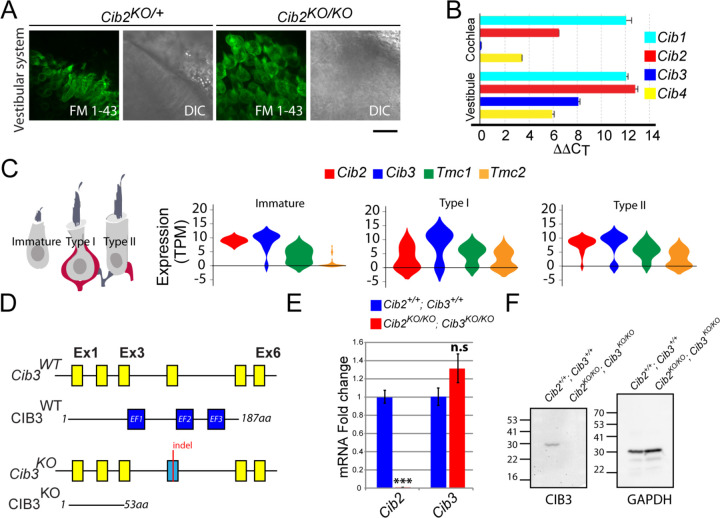
Figure 1. Expression of Cib genes in the mouse inner ear and generation of Cib3 knockout mice. (A) Maximum intensity projections of Z-stacks of confocal fluorescent images and corresponding DIC images of Cib2KO/+ and Cib2KO/KO cultured vestibular end organ explants imaged after exposure to 3 μM of FM 1–43 for 10 s. The samples were dissected at P5 and kept 2 days in vitro (P5 + 2div). Scale bar: 20 µm. (B) Real-time quantitative RT–PCR analysis of mRNA levels in the cochlear and vestibular sensory epithelia at P12 revealed different expression of Cib1, Cib2, Cib3, and Cib4 in the auditory and vestibular end organs. (C) Violin plots generated by gEAR portal showing the expression levels of Cib2, Cib3, Tmc1, and Tmc2 by scRNA-seq in immature, type 1, and type 2 utricular vestibular hair cells. TPM: transcript per million. (D) Gene structure of the wild-type and Cib3KO alleles. Exons are shown in yellow. Indel in exon 4 leads to a nonsense mutation and a premature stop at the protein level (p. Asp54*) (E) Real-time quantitative RT–PCR analysis using cDNA from brain tissue of Cib2;Cib3 mutant mice was used to analyze the expression of Cib2 and Cib3. Contrary to Cib2 mRNA, Cib3 mRNA appears to be stable in the Cib2KO/KO;Cib3KO/KO mice and does not go through nonsense mediated decay process. The relative expression of Cib2 and Cib3 members were normalized against hprt. Asterisks indicate statistical significance: ***, p<0.001 (Student’s t-test). n.s.: not significant. Error bars represent SEM. (F) Western Blot analysis on Cib2;Cib3 mouse heart tissue was performed using CIB3 antibody. No CIB3 protein was detected in the Cib2KO/KO;Cib3KO/KO mice, validating our mouse model. GAPDH detection was used as a loading control.
Figure 1. Expression of Cib genes in the mouse inner ear and generation of Cib3 knockout mice. (A) Maximum intensity projections of Z-stacks of confocal fluorescent images and corresponding DIC images of Cib2KO/+ and Cib2KO/KO cultured vestibular end organ explants imaged after exposure to 3 μM of FM 1–43 for 10 s. The samples were dissected at P5 and kept 2 days in vitro (P5 + 2div). Scale bar: 20 µm. (B) Real-time quantitative RT–PCR analysis of mRNA levels in the cochlear and vestibular sensory epithelia at P12 revealed different expression of Cib1, Cib2, Cib3, and Cib4 in the auditory and vestibular end organs. (C) Violin plots generated by gEAR portal showing the expression levels of Cib2, Cib3, Tmc1, and Tmc2 by scRNA-seq in immature, type 1, and type 2 utricular vestibular hair cells. TPM: transcript per million. (D) Gene structure of the wild-type and Cib3KO alleles. Exons are shown in yellow. Indel in exon 4 leads to a nonsense mutation and a premature stop at the protein level (p. Asp54*) (E) Real-time quantitative RT–PCR analysis using cDNA from brain tissue of Cib2;Cib3 mutant mice was used to analyze the expression of Cib2 and Cib3. Contrary to Cib2 mRNA, Cib3 mRNA appears to be stable in the Cib2KO/KO;Cib3KO/KO mice and does not go through nonsense mediated decay process. The relative expression of Cib2 and Cib3 members were normalized against hprt. Asterisks indicate statistical significance: ***, p<0.001 (Student’s t-test). n.s.: not significant. Error bars represent SEM. (F) Western Blot analysis on Cib2;Cib3 mouse heart tissue was performed using CIB3 antibody. No CIB3 protein was detected in the Cib2KO/KO;Cib3KO/KO mice, validating our mouse model. GAPDH detection was used as a loading control.CC BY-NC-ND · PubMed Central
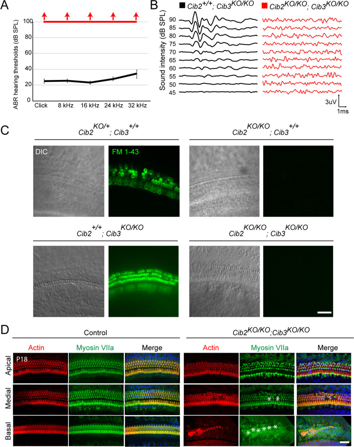
Figure 2. CIB2/3 double mutant mice have profound hearing loss. (A) ABR thresholds to broadband clicks and tone-pips with frequencies of 8 kHz, 16 kHz, 24 kHz and 32 kHz in Cib2+/+;Cib3KO/KO (black; n = 5) and Cib2KO/KO;Cib3KO/KO (red, n = 5) mice at P16. The same animals were tested with clicks and tone pips. (B) ABR pure tone traces of P32 Cib2+/+;Cib3KO/KO (black) and Cib2KO/KO;Cib3KO/KO (red) mice measured at 32 kHz. Hearing thresholds are in Decibels (dB SPL). (C) Maximum intensity projections of Z-stacks of confocal fluorescent images (right) and corresponding DIC images (left) of Cib2KO/+;Cib3+/+, Cib2KO/KO;Cib3+/+, Cib2+/+;Cib3KO/KO and Cib2KO/KO;Cib3KO/KO cultured organ of Corti explants imaged after exposure to 3 μM of FM 1–43 for 10 s. The samples were dissected at P5 and kept 2 days in vitro (P5 + 2div). Scale bar: 20 µm. (D) Maximum intensity projections of confocal Z-stacks of apical, medial and basal turns of organ of Corti of control and Cib2KO/KO;Cib3KO/KO mice immunostained with an anti-myosin VIIa antibody (green) and counterstained with phalloidin (red) at P18. Asterisks indicate hair cell loss. Scale bar: 10 µm.
Figure 2. CIB2/3 double mutant mice have profound hearing loss. (A) ABR thresholds to broadband clicks and tone-pips with frequencies of 8 kHz, 16 kHz, 24 kHz and 32 kHz in Cib2+/+;Cib3KO/KO (black; n = 5) and Cib2KO/KO;Cib3KO/KO (red, n = 5) mice at P16. The same animals were tested with clicks and tone pips. (B) ABR pure tone traces of P32 Cib2+/+;Cib3KO/KO (black) and Cib2KO/KO;Cib3KO/KO (red) mice measured at 32 kHz. Hearing thresholds are in Decibels (dB SPL). (C) Maximum intensity projections of Z-stacks of confocal fluorescent images (right) and corresponding DIC images (left) of Cib2KO/+;Cib3+/+, Cib2KO/KO;Cib3+/+, Cib2+/+;Cib3KO/KO and Cib2KO/KO;Cib3KO/KO cultured organ of Corti explants imaged after exposure to 3 μM of FM 1–43 for 10 s. The samples were dissected at P5 and kept 2 days in vitro (P5 + 2div). Scale bar: 20 µm. (D) Maximum intensity projections of confocal Z-stacks of apical, medial and basal turns of organ of Corti of control and Cib2KO/KO;Cib3KO/KO mice immunostained with an anti-myosin VIIa antibody (green) and counterstained with phalloidin (red) at P18. Asterisks indicate hair cell loss. Scale bar: 10 µm.CC BY-NC-ND · PubMed Central
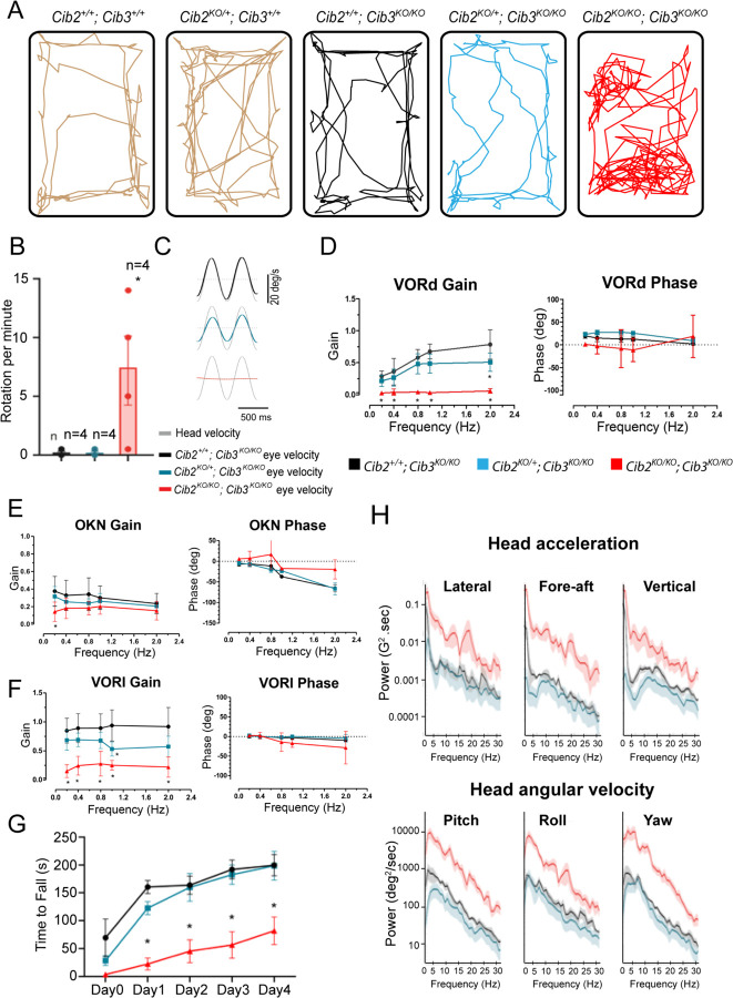
Figure 3. CIB2/3 double mutant mice have vestibular dysfunction. (A) Traces showing the open-field exploratory behavior of P60 Cib2+/+;Cib3+/+, Cib2KO/+;Cib3+/+, Cib2+/+;Cib3KO/KO, Cib2KO/+;Cib3KO/KO and Cib2KO/KO;Cib3KO/KO mutant mice. (B) Quantification of the number of rotations in 120 seconds (mean ± SEM), showing that, unlike Cib2+/+;Cib3KO/KO, and Cib2KO/+;Cib3KO/KO mice, Cib2KO/KO;Cib3KO/KO mutant mice display a circling behavior and a vestibular defect (t‐test) (n = 4 for each genotype). (C) Examples of head-velocity (grey) and resultant eye-velocity traces evoked during VORd testing. Note, the eye movement is compensatory, and the trace has been inverted to facilitate comparison with head velocity. (D) VORd Gain and phase (mean ± SD) plotted as a function of frequency for Cib2+/+;Cib3KO/KO (n = 4), Cib2KO/+;Cib3KO/KO (n = 5), Cib2KO/KO;Cib3KO/KO (n = 4). (E) OKN gain and phase (mean ± SD) plotted as a function of frequency for Cib2+/+; Cib3KO/KO (n = 4), Cib2KO/+;Cib3KO/KO (n = 5), Cib2KO/KO;Cib3KO/KO (n = 4). (F) VORl gain and phase (mean ± SD) plotted as a function of frequency for Cib2+/+;Cib3KO/KO (n = 4), Cib2KO/+;Cib3KO/KO (n = 5), Cib2KO/KO;Cib3KO/KO (n = 4). Comparisons made with two-way ANOVA followed by Bonferroni post-hoc test for (A-D); *p < 0.05. (G) Quantification of the time mice remained on the rotating rod with increasing acceleration (mean ± SEM). Comparisons made with two-way ANOVA followed by Bonferroni post-hoc test for (F-K); *P<0.002. (Cib2+/+;Cib3KO/KO (n = 6), Cib2KO/+;Cib3KO/KO (n = 6), Cib2KO/KO;Cib3KO/KO (n = 6)). (H) Comparison of power spectra density of head movements in translational axes (top) and rotational axes (bottom) between wild type (Cib2+/+;Cib3KO/KO, black), heterozygous mutants (Cib2KO/+;Cib3KO/KO, blue), and homozygous mutants (Cib2KO/KO;Cib3KO/KO, red). The double knockout Cib2KO/KO;Cib3KO/KO exhibit significantly higher power than Cib2+/+;Cib3KO/KO and Cib2KO/+;Cib3KO/KO across all frequencies (0–30 Hz), in all six translational and rotational axes.
Figure 3. CIB2/3 double mutant mice have vestibular dysfunction. (A) Traces showing the open-field exploratory behavior of P60 Cib2+/+;Cib3+/+, Cib2KO/+;Cib3+/+, Cib2+/+;Cib3KO/KO, Cib2KO/+;Cib3KO/KO and Cib2KO/KO;Cib3KO/KO mutant mice. (B) Quantification of the number of rotations in 120 seconds (mean ± SEM), showing that, unlike Cib2+/+;Cib3KO/KO, and Cib2KO/+;Cib3KO/KO mice, Cib2KO/KO;Cib3KO/KO mutant mice display a circling behavior and a vestibular defect (t‐test) (n = 4 for each genotype). (C) Examples of head-velocity (grey) and resultant eye-velocity traces evoked during VORd testing. Note, the eye movement is compensatory, and the trace has been inverted to facilitate comparison with head velocity. (D) VORd Gain and phase (mean ± SD) plotted as a function of frequency for Cib2+/+;Cib3KO/KO (n = 4), Cib2KO/+;Cib3KO/KO (n = 5), Cib2KO/KO;Cib3KO/KO (n = 4). (E) OKN gain and phase (mean ± SD) plotted as a function of frequency for Cib2+/+; Cib3KO/KO (n = 4), Cib2KO/+;Cib3KO/KO (n = 5), Cib2KO/KO;Cib3KO/KO (n = 4). (F) VORl gain and phase (mean ± SD) plotted as a function of frequency for Cib2+/+;Cib3KO/KO (n = 4), Cib2KO/+;Cib3KO/KO (n = 5), Cib2KO/KO;Cib3KO/KO (n = 4). Comparisons made with two-way ANOVA followed by Bonferroni post-hoc test for (A-D); *p < 0.05. (G) Quantification of the time mice remained on the rotating rod with increasing acceleration (mean ± SEM). Comparisons made with two-way ANOVA followed by Bonferroni post-hoc test for (F-K); *P<0.002. (Cib2+/+;Cib3KO/KO (n = 6), Cib2KO/+;Cib3KO/KO (n = 6), Cib2KO/KO;Cib3KO/KO (n = 6)). (H) Comparison of power spectra density of head movements in translational axes (top) and rotational axes (bottom) between wild type (Cib2+/+;Cib3KO/KO, black), heterozygous mutants (Cib2KO/+;Cib3KO/KO, blue), and homozygous mutants (Cib2KO/KO;Cib3KO/KO, red). The double knockout Cib2KO/KO;Cib3KO/KO exhibit significantly higher power than Cib2+/+;Cib3KO/KO and Cib2KO/+;Cib3KO/KO across all frequencies (0–30 Hz), in all six translational and rotational axes.CC BY-NC-ND · PubMed Central
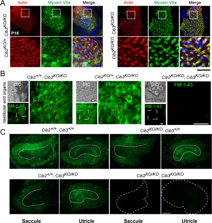
Figure 4. Vestibular hair cells do not have functional MET channels at rest in CIB2/3 double mutant mice. (A) Maximum intensity projections of confocal Z-stacks of the vestibular end organs of P18 Cib2KO/+;Cib3KO/KO and Cib2KO/KO;Cib3KO/KO mutant mice immunostained with myosin VIIa antibody (green) and counterstained with phalloidin (red) and DAPI (blue). Scale bar = 50 µm. (B) Adult vestibular end organs of Cib2+/+;Cib3KO/KO, Cib2KO/+;Cib3KO/KO and Cib2KO/KO;Cib3KO/KO mutant mice imaged after exposure to 3 μM of FM 1–43 for 10 s. Vestibular hair cell bodies (green doted lines), and stereocilia bundles (red arrows) are shown. Scale bar: 100 µm. (C) FM 1-43FX (fixable form) labeling of saccules and utricles by IP injection at P60. Obvious reduction in signal in Cib2KO/KO;Cib3KO/KO, and subtle extrastriolar reduction in Cib2+/+;Cib3KO/KO mice was observed. Extrastriolar regions are delimited with white lines. Scale bar = 100 µm.
Figure 4. Vestibular hair cells do not have functional MET channels at rest in CIB2/3 double mutant mice. (A) Maximum intensity projections of confocal Z-stacks of the vestibular end organs of P18 Cib2KO/+;Cib3KO/KO and Cib2KO/KO;Cib3KO/KO mutant mice immunostained with myosin VIIa antibody (green) and counterstained with phalloidin (red) and DAPI (blue). Scale bar = 50 µm. (B) Adult vestibular end organs of Cib2+/+;Cib3KO/KO, Cib2KO/+;Cib3KO/KO and Cib2KO/KO;Cib3KO/KO mutant mice imaged after exposure to 3 μM of FM 1–43 for 10 s. Vestibular hair cell bodies (green doted lines), and stereocilia bundles (red arrows) are shown. Scale bar: 100 µm. (C) FM 1-43FX (fixable form) labeling of saccules and utricles by IP injection at P60. Obvious reduction in signal in Cib2KO/KO;Cib3KO/KO, and subtle extrastriolar reduction in Cib2+/+;Cib3KO/KO mice was observed. Extrastriolar regions are delimited with white lines. Scale bar = 100 µm.CC BY-NC-ND · PubMed Central
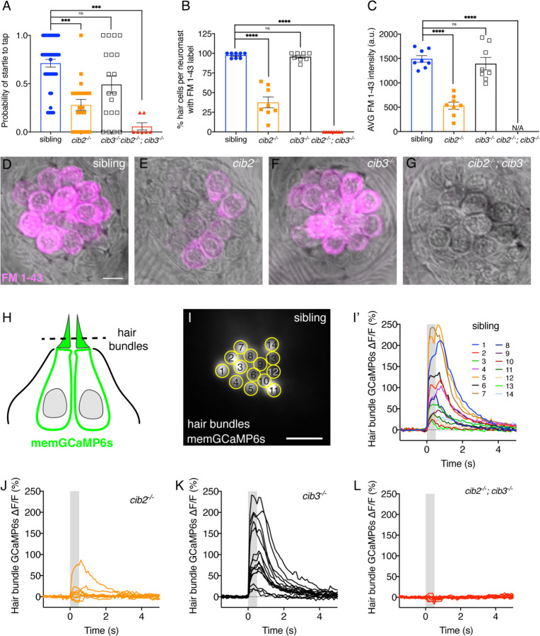
Figure 5. Both Cib2 and Cib3 are required in zebrafish for acoustic startle and MET function. (A) Compared to controls (siblings) cib3 mutants have a normal acoustic startle response. Cib2;cib3 double mutants completely lack an acoustic startle response, while cib2 mutants have a reduced probability to startle compared to controls (n = 43 sibling, 20 cib2, 19 cib3 and 7 cib2;cib3 animals). (B) Compared to controls cib3 mutants have a normal % of hair cells that label with FM 1–43 per neuromast. In cib2;cib3 mutants no hair cells label with FM 1–43, and in cib2 mutants a significantly reduced % of hair cells label with FM 1–43 per neuromast. (C) The average intensity of FM 1–43 labeling in cib3 mutant hair cells is similar to controls. In contrast, in cib2 mutants, hair cells that label with FM 1–43, have a significantly reduced intensity compared to controls (n = 8 neuromasts per genotype in B and C). (D-G) Representative neuromasts labeled with FM 1–43 for each genotype. FM 1–43 label is overlaid onto laser scanning DIC images. (H) Schematic showing the localization of membrane-localized GCaMP6s (memGCaMP6s); the indicator used to measure Ca2+ influx into lateral-line hair bundles. The plane used for Ca2+ imaging and to determine if a hair cell is mechanosensitive is indicated by the dashed line. (I-I’) Representative example of a hair bundle imaging plane (I), along with the resulting Ca2+ responses (I’) from hair bundles from a control neuromast. The ROIs used to measure Ca2+ signals in each hair bundle are indicated in (I). (J-L) Representative examples of Ca2+ responses from neuromasts in cib2 (J), cib3 (K) and cib2;cib3 mutants (L). FM 1–43 imaging and behavior were performed at 5 dpf. Ca2+ imaging was performed at 5 or 6 dpf. A Kruskal-Wallis test was used in A; A one-way ANOVA was used in B and C. The scale bar in D and I = 5 µm. The online version of this article includes source data and the following figure supplement(s) for Figure 5.
Figure 5. Both Cib2 and Cib3 are required in zebrafish for acoustic startle and MET function. (A) Compared to controls (siblings) cib3 mutants have a normal acoustic startle response. Cib2;cib3 double mutants completely lack an acoustic startle response, while cib2 mutants have a reduced probability to startle compared to controls (n = 43 sibling, 20 cib2, 19 cib3 and 7 cib2;cib3 animals). (B) Compared to controls cib3 mutants have a normal % of hair cells that label with FM 1–43 per neuromast. In cib2;cib3 mutants no hair cells label with FM 1–43, and in cib2 mutants a significantly reduced % of hair cells label with FM 1–43 per neuromast. (C) The average intensity of FM 1–43 labeling in cib3 mutant hair cells is similar to controls. In contrast, in cib2 mutants, hair cells that label with FM 1–43, have a significantly reduced intensity compared to controls (n = 8 neuromasts per genotype in B and C). (D-G) Representative neuromasts labeled with FM 1–43 for each genotype. FM 1–43 label is overlaid onto laser scanning DIC images. (H) Schematic showing the localization of membrane-localized GCaMP6s (memGCaMP6s); the indicator used to measure Ca2+ influx into lateral-line hair bundles. The plane used for Ca2+ imaging and to determine if a hair cell is mechanosensitive is indicated by the dashed line. (I-I’) Representative example of a hair bundle imaging plane (I), along with the resulting Ca2+ responses (I’) from hair bundles from a control neuromast. The ROIs used to measure Ca2+ signals in each hair bundle are indicated in (I). (J-L) Representative examples of Ca2+ responses from neuromasts in cib2 (J), cib3 (K) and cib2;cib3 mutants (L). FM 1–43 imaging and behavior were performed at 5 dpf. Ca2+ imaging was performed at 5 or 6 dpf. A Kruskal-Wallis test was used in A; A one-way ANOVA was used in B and C. The scale bar in D and I = 5 µm. The online version of this article includes source data and the following figure supplement(s) for Figure 5.CC BY-NC-ND · PubMed Central
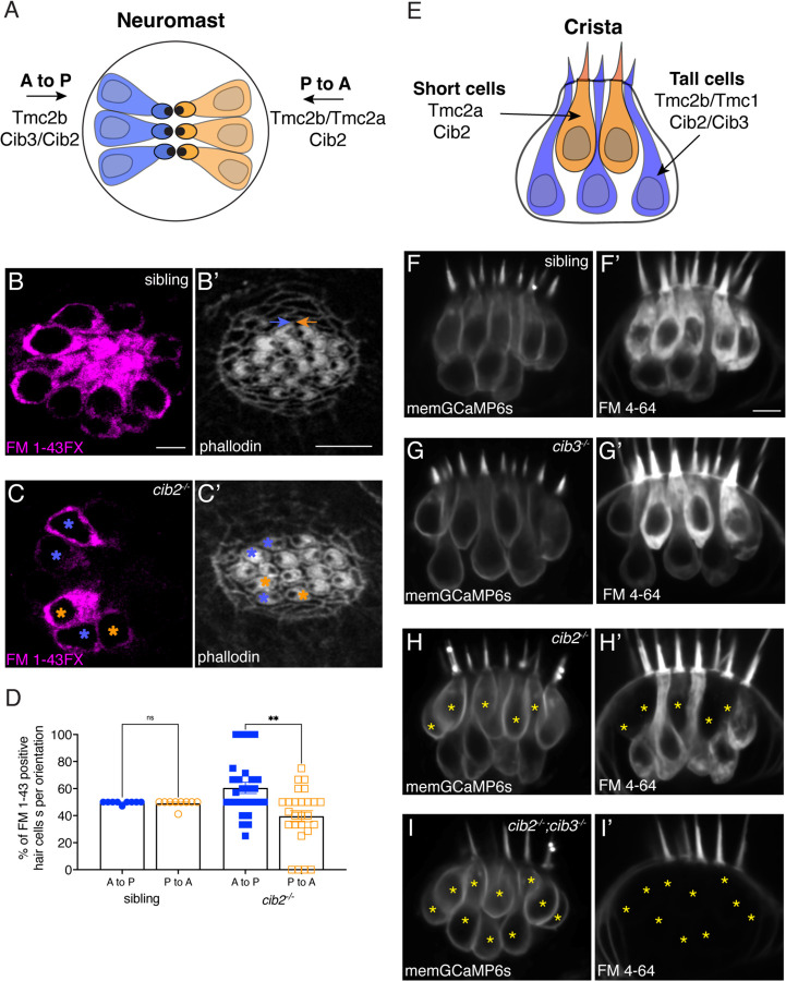
Figure 6. Cib2 is required in specific subsets of hair cells in the zebrafish pLL and inner ear. (A) Overview of Tmc requirements in pLL hair cells. Hair cells that sense posterior flow (orange) rely on Tmc2b, while hair cells that sense anterior flow (blue) can rely on Tmc2b and/or Tmc2a for mechanosensitive function. (B-C’) FM 1-43FX labeling in pLL neuromasts from sibling control (B) and cib2 mutants (C). The residual cells in cib2 mutants rely on Cib3 for mechanosensitive function. Phalloidin label can be used to link FM 1-43FX label to the orientation of pLL hair cells (C,C’; see colored asterisks). In B’, orange and blue arrows rest above hair cells that are oriented to respond to anterior and posterior flow, respectively. (D) Quantification reveals that a similar percentage of anterior and posterior responsive pLL hair cells label with FM 1-43FX in sibling controls (posterior flow: 49.7 %, anterior flow: 49.2 %, n = 9 neuromasts). In contrast, in cib2 mutants, a significantly higher percentage of posterior responsive pLL hair cells label with FM 1-43FX in cib2 mutants (posterior flow: 60.5 %, anterior flow: 39.5 %, n = 26 neuromasts). (E) Overview of Tmc and Cib requirements in the hair cells of zebrafish cristae. Short teardrop-shaped cells rely primarily on Tmc2a, while tall gourd-shaped cells rely primarily on Tmc2b and Tmc1. (F-I’) Medial cristae for each genotype. MemGCaMP6s labels all hair cells, while FM 4–64 labels hair cells with intact mechanosensitive function. In siblings and cib3 mutants, both teardrop and gourd cells label with FM 4–64 (F-F’, G-G’). In cib2;cib3 double mutants, no hair cells label with FM 4–64 (asterisks) (I-I’). In cib2 mutants many short cells fail to label with FM 4–64 (asterisks) (H-H’), (n = 13 double het sibling, 7 cib3, 14 cib2 and 4 double mutant crista). All images were acquired at 5 dpf. A Kruskal-Wallis test was used in D; P > 0.01. The scale bars in B, B’, and F’ = 5 µm.
Figure 6. Cib2 is required in specific subsets of hair cells in the zebrafish pLL and inner ear. (A) Overview of Tmc requirements in pLL hair cells. Hair cells that sense posterior flow (orange) rely on Tmc2b, while hair cells that sense anterior flow (blue) can rely on Tmc2b and/or Tmc2a for mechanosensitive function. (B-C’) FM 1-43FX labeling in pLL neuromasts from sibling control (B) and cib2 mutants (C). The residual cells in cib2 mutants rely on Cib3 for mechanosensitive function. Phalloidin label can be used to link FM 1-43FX label to the orientation of pLL hair cells (C,C’; see colored asterisks). In B’, orange and blue arrows rest above hair cells that are oriented to respond to anterior and posterior flow, respectively. (D) Quantification reveals that a similar percentage of anterior and posterior responsive pLL hair cells label with FM 1-43FX in sibling controls (posterior flow: 49.7 %, anterior flow: 49.2 %, n = 9 neuromasts). In contrast, in cib2 mutants, a significantly higher percentage of posterior responsive pLL hair cells label with FM 1-43FX in cib2 mutants (posterior flow: 60.5 %, anterior flow: 39.5 %, n = 26 neuromasts). (E) Overview of Tmc and Cib requirements in the hair cells of zebrafish cristae. Short teardrop-shaped cells rely primarily on Tmc2a, while tall gourd-shaped cells rely primarily on Tmc2b and Tmc1. (F-I’) Medial cristae for each genotype. MemGCaMP6s labels all hair cells, while FM 4–64 labels hair cells with intact mechanosensitive function. In siblings and cib3 mutants, both teardrop and gourd cells label with FM 4–64 (F-F’, G-G’). In cib2;cib3 double mutants, no hair cells label with FM 4–64 (asterisks) (I-I’). In cib2 mutants many short cells fail to label with FM 4–64 (asterisks) (H-H’), (n = 13 double het sibling, 7 cib3, 14 cib2 and 4 double mutant crista). All images were acquired at 5 dpf. A Kruskal-Wallis test was used in D; P > 0.01. The scale bars in B, B’, and F’ = 5 µm.CC BY-NC-ND · PubMed Central
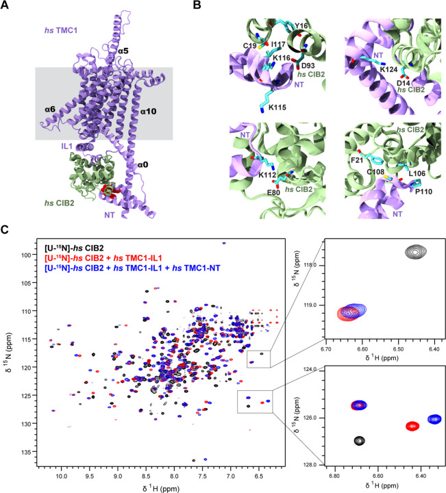
Figure 7. AF2 predictions and NMR data support a clamp-like model for the human TMC1 and CIB2 complex. (A) AF2 model of hs TMC1 in complex with hs CIB2. Side view of monomeric hs TMC1 (light purple) in complex with hs CIB2 (light sea green). N-terminal residues of TMC1 known to interact with CIB2 are in red. (B) Top left panel shows residues at the hs TMC1-NT fragment (115–117) shown to weaken interaction with CIB2 upon mutation (Liang et al., 2021) at the interface with CIB2. Additional panels show details of contacts between TMC1 and CIB2 with formation of K124:D14 and K112:E80 salt bridges, as well as hydrophobic interactions. (C) Overlay of 1H-15N TROSY-HSQC spectra of hs [U-15N]-CIB2 (black) alone and bound to either only hs TMC1-IL (red) or both hs TMC1-IL and hs TMC1-NT (blue). NMR data were obtained in the presence of 3 mM CaCl2. These data are also shown in Figure 7-figure supplement 7.
Figure 7. AF2 predictions and NMR data support a clamp-like model for the human TMC1 and CIB2 complex. (A) AF2 model of hs TMC1 in complex with hs CIB2. Side view of monomeric hs TMC1 (light purple) in complex with hs CIB2 (light sea green). N-terminal residues of TMC1 known to interact with CIB2 are in red. (B) Top left panel shows residues at the hs TMC1-NT fragment (115–117) shown to weaken interaction with CIB2 upon mutation (Liang et al., 2021) at the interface with CIB2. Additional panels show details of contacts between TMC1 and CIB2 with formation of K124:D14 and K112:E80 salt bridges, as well as hydrophobic interactions. (C) Overlay of 1H-15N TROSY-HSQC spectra of hs [U-15N]-CIB2 (black) alone and bound to either only hs TMC1-IL (red) or both hs TMC1-IL and hs TMC1-NT (blue). NMR data were obtained in the presence of 3 mM CaCl2. These data are also shown in Figure 7-figure supplement 7.CC BY-NC-ND · PubMed Central
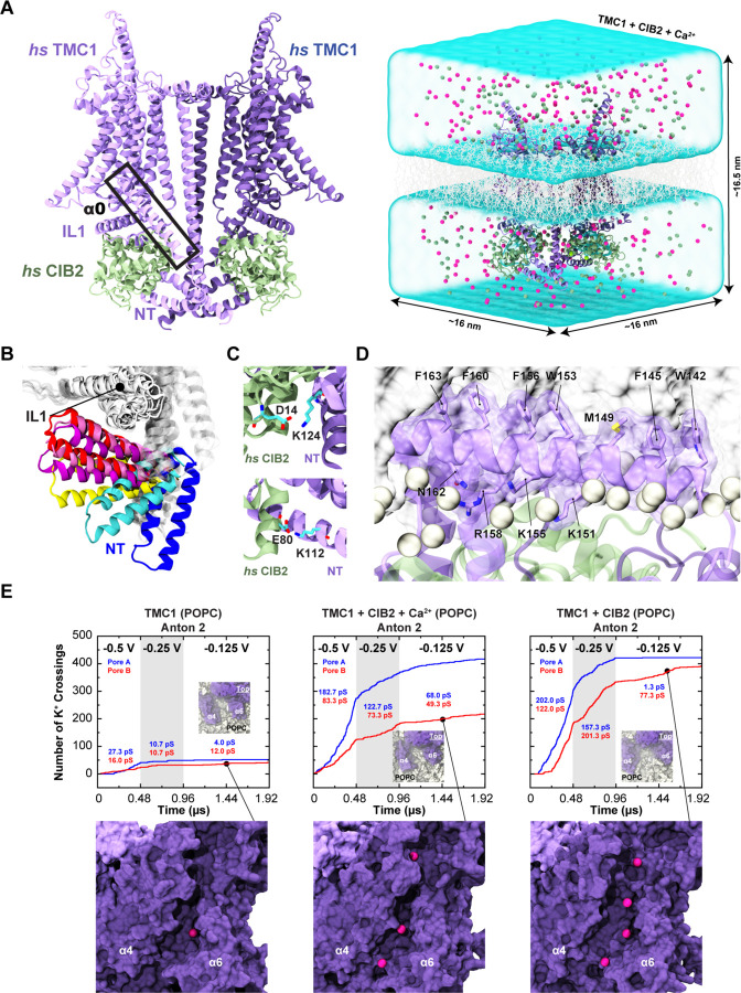
Figure 8. Simulations of AF2 predictions show that human TMC1 and CIB2 complexes form cation channels. (A) Left: AF2 model of the dimeric hs TMC1 + hs CIB2 complex. Right: A complete simulation system of hs TMC1 in complex with hs CIB2-Ca2+ is shown in surface representation. hs TMC1 is shown in light (monomer A) and dark (monomer B) purple, hs CIB2 in light sea green, water in transparent light blue, lipid membrane in gray lines, and K+ and Cl- ions are shown as pink and green spheres, respectively. (B) The hs TMC1-NT domain explores diverse conformational space in the absence of CIB during equilibrium simulations (S3a). (C) Salt bridges K124:D14 and K112:E80 remain throughout equilibrium simulations. (D) Snapshot of amphipathic helix α0 inserting into the lipid bilayer (S4c). (E) Number of K+ crossings as a function of time for pores A and B of hs TMC1, hs TMC1 + hs CIB2 + Ca2+, and hs TMC1 + hs CIB2 in a POPC bilayer at −0.5 V, −0.25 V, and −0.125 V. Insets show top views of pore region with TMC1 (dark purple) and lipids (light gray) in surface representation (protein and lipids partially excluded for clarity). Representative images of K+ permeating the pore region between α4 and α6 of hs TMC1 are shown with protein in surface representation and ions as pink spheres. Conductance values calculated for K+ crossings.
Figure 8. Simulations of AF2 predictions show that human TMC1 and CIB2 complexes form cation channels. (A) Left: AF2 model of the dimeric hs TMC1 + hs CIB2 complex. Right: A complete simulation system of hs TMC1 in complex with hs CIB2-Ca2+ is shown in surface representation. hs TMC1 is shown in light (monomer A) and dark (monomer B) purple, hs CIB2 in light sea green, water in transparent light blue, lipid membrane in gray lines, and K+ and Cl- ions are shown as pink and green spheres, respectively. (B) The hs TMC1-NT domain explores diverse conformational space in the absence of CIB during equilibrium simulations (S3a). (C) Salt bridges K124:D14 and K112:E80 remain throughout equilibrium simulations. (D) Snapshot of amphipathic helix α0 inserting into the lipid bilayer (S4c). (E) Number of K+ crossings as a function of time for pores A and B of hs TMC1, hs TMC1 + hs CIB2 + Ca2+, and hs TMC1 + hs CIB2 in a POPC bilayer at −0.5 V, −0.25 V, and −0.125 V. Insets show top views of pore region with TMC1 (dark purple) and lipids (light gray) in surface representation (protein and lipids partially excluded for clarity). Representative images of K+ permeating the pore region between α4 and α6 of hs TMC1 are shown with protein in surface representation and ions as pink spheres. Conductance values calculated for K+ crossings.CC BY-NC-ND · PubMed Central

Verification

Claims the paper makes

The claims and statistics of this paper haven’t been checked yet.

Run the check — free, no account needed. It reads the open-access text and checks the sources it cites.

A few free checks a day · papers already verified open instantly · results are public

Self-check

Percentages, sums and reported differences re-computed from the paper’s own numbers.

The self-check needs the paper’s full text.

Citations

Cites

Research works printed but not linked: 94

  1. 10.1523/JNEUROSCI.1163-06.2006 · DOI
  2. 10.1126/sciadv.abo1126 · DOI
  3. 10.1016/j.devcel.2022.03.001 · DOI
  4. 10.7554/eLife.38433 · DOI
  5. 10.1152/jn.00590.2007 · DOI
  6. 10.1152/jn.90319.2008 · DOI
  7. 10.1021/ct300400x · DOI
  8. 10.1038/s41467-018-04589-8 · DOI
  9. 10.1038/nn.2295 · DOI
  10. 10.1085/jgp.202112959 · DOI
  11. 10.1016/j.abb.2009.05.002 · DOI
  12. 10.1111/cge.13170 · DOI
  13. 10.1038/273377a0 · DOI
  14. 10.1093/HMG/DDAA045 · DOI
  15. 10.1038/s41467-017-01604-2 · DOI
  16. 10.1038/s41583-019-0153-1 · DOI
  17. 10.1016/j.neuron.2020.03.033 · DOI
  18. 10.1038/s41598-019-51573-3 · DOI
  19. 10.1523/JNEUROSCI.1351-17.2017 · DOI
  20. 10.7554/eLife.28474 · DOI
  21. 10.3389/fnmol.2019.00320 · DOI
  22. 10.1093/hmg/9.14.2189 · DOI
  23. 10.1101/2021.10.04.463034 · DOI · Paper page
  24. 10.1002/cphy.c160049 · DOI
  25. 10.1073/pnas.2210849119 · DOI
  26. 10.1038/nrg1377 · DOI
  27. 10.1038/s41467-017-00061-1 · DOI
  28. 10.1016/j.cell.2009.09.010 · DOI
  29. 10.1073/pnas.0911632106 · DOI
  30. 10.1523/JNEUROSCI.1096-15.2015 · DOI
  31. 10.1016/j.bbamem.2011.09.030 · DOI
  32. 10.1139/o2012-021 · DOI
  33. 10.1002/jcc.23354.CHARMM36 · DOI
  34. 10.1016/0263-7855(96)00018-5 · DOI · Paper page
  35. 10.1038/s41586-022-05314-8 · DOI
  36. 10.7554/eLife.20010 · DOI
  37. 10.1371/journal.pone.0000880 · DOI
  38. 10.1002/jcc.20945 · DOI · Paper page
  39. 10.1016/j.bpj.2009.04.013 · DOI
  40. 10.7554/eLife.41845 · DOI
  41. 10.1038/347631a0 · DOI
  42. 10.1172/JCI60405 · DOI
  43. 10.1007/s00424-014-1582-3 · DOI
  44. 10.1038/nature06091 · DOI
  45. 10.1093/bioinformatics/bts199 · DOI
  46. 10.1085/jgp.201311068 · DOI
  47. 10.1016/j.cub.2023.02.033 · DOI
  48. 10.1021/acs.jctc.8b01066 · DOI
  49. 10.1016/j.neuron.2021.05.007 · DOI
  50. 10.1063/1.4825247 · DOI
  51. 10.1038/s41467-018-07564-5 · DOI
  52. 10.1073/pnas.0608536103 · DOI
  53. 10.3791/58794 · DOI
  54. 10.1073/pnas.1402152111 · DOI
  55. 10.1523/JNEUROSCI.2216-16.2017 · DOI
  56. 10.1242/bio.028795 · DOI
  57. 10.1523/JNEUROSCI.23-10-04054.2003 · DOI
  58. 10.15252/emmm.201708087 · DOI
  59. 10.1086/342193 · DOI
  60. 10.1080/01677063.2017.1342246 · DOI
  61. 10.1016/S0896-6273(00)80455-9 · DOI
  62. 10.1007/s10858-004-8195-7 · DOI
  63. 10.1007/s10858-016-0049-6 · DOI
  64. 10.1371/journal.pgen.1007635 · DOI
  65. 10.1016/j.neuron.2018.07.033 · DOI
  66. 10.1016/j.neuron.2013.06.019 · DOI
  67. 10.1242/dmm.006429 · DOI
  68. 10.1002/jcc.20289 · DOI · Paper page
  69. 10.3389/fncir.2013.00001 · DOI
  70. 10.1016/j.celrep.2023.112245 · DOI
  71. 10.1038/ng.2426 · DOI
  72. 10.1038/ejhg.2015.157 · DOI
  73. 10.1016/j.ydbio.2004.05.004 · DOI
  74. 10.1109/SC.2014.9 · DOI
  75. 10.1523/JNEUROSCI.2878-16.2017 · DOI
  76. 10.7554/eLife.82978 · DOI
  77. 10.1038/nature02483 · DOI
  78. 10.1523/JNEUROSCI.0163-20.2020 · DOI
  79. 10.3389/fnmol.2023.1274822 · DOI
  80. 10.1038/nature02484 · DOI
  81. 10.1038/nature11590 · DOI
  82. 10.1126/stke.2192004re4 · DOI
  83. 10.3389/fnmol.2018.00274 · DOI
  84. 10.1016/j.bbagen.2014.08.004 · DOI
  85. 10.1038/nprot.2016.141 · DOI
  86. 10.1101/2021.09.17.460860 · DOI
  87. 10.1523/JNEUROSCI.1807-22.2023 · DOI
  88. 10.3389/fnmol.2017.00401 · DOI
  89. 10.1093/bioinformatics/btp033 · DOI
  90. 10.1016/j.cell.2012.10.041 · DOI
  91. 10.1038/s41594-018-0117-6 · DOI
  92. 10.1038/s41467-018-02974-x · DOI · Paper page
  93. 10.1523/JNEUROSCI.6184-11.2012 · DOI
  94. 10.3389/fcell.2020.570486 · DOI

Metadata

Authors
Giese, A. P. J., Weng, W.-H., Kindt, K. S., Chang, H. H. V., Montgomery, J. S., Ratzan, E. M., Beirl, A. J., Rivera, R. A., Lotthammer, J. M., Walujkar, S., Foster, M. P., Zobeiri, O. A., Holt, J. R., Riazuddin, S., Cullen, K. E., Sotomayor, M., & Ahmed, Z. M.
Affiliations
  • University of Maryland, Baltimore (US)
  • The Ohio State University (US)
  • National Institutes of Health (US)
  • McGill University (CA)
  • Boston Children's Hospital (US)
  • Johns Hopkins University (US)
Corresponding author
  • Sotomayor, Marcos
  • Ahmed, Zubair M.
Venue
eLife
Publisher
openRxiv
Published
2023-05-30
Volume
12
ISSN
2050-084X (ISSN-L)
OpenAlex
W4378837212
Cited by
8
References
2
Citations by year
2025: 3 · 2024: 3 · 2023: 2
FWCI
1.90
Type
journal article
Language
en
Open access
Licence
cc-by
Locations
  • eLife (publishedVersion, cc-by)
  • bioRxiv (Cold Spring Harbor Laboratory) (acceptedVersion, cc-by-nc-nd)
  • PubMed (publishedVersion)
  • PubMed Central (submittedVersion, cc-by-nc-nd)
Funders
  • Ohio State University
  • Ohio Supercomputer Center (PAS1037, PAA0217)
  • National Institutes of Health (1ziadc000085-01, 1uf1ns111695-01)
  • National Institute on Deafness and Other Communication Disorders (1ZIADC000085-01, R01DC018304, R01DC015271, R01DC012564, R01DC018061, R01DC002390)
  • National Institute of General Medical Sciences (T32GM144293)
  • Eunice Kennedy Shriver National Institute of Child Health and Human Development (P50HD105351)
Topics
  • Life Sciences › Neuroscience › Sensory Systems › Hearing, Cochlea, Tinnitus, Genetics
  • Life Sciences › Neuroscience › Sensory Systems › Ion Channels and Receptors
  • Life Sciences › Biochemistry, Genetics and Molecular Biology › Molecular Biology › Ion channel regulation and function
Keywords
TMC-1, TMC2, CIB2, mechanoelectrical transduction, hair cell mechanotransduction, cation channels, heteromeric protein complexes, cochlea, vestibular organs, lateral line, zebrafish, mice, protein-protein interaction

About these checks

Registry, retraction, Cites and statistics results are computed, not judged, with one exception: a printed reference that no index lists is read once by a model before it counts as a citation error. Claims are extracted by a model and kept only when their quote appears verbatim in the text; each cited source is then checked, with a model’s help, for whether it says what the claim relies on. Those verdicts can be wrong — read the claim’s quote and the cited source before relying on one.

Statistics and arithmetic are recomputed, not judged.

Confidence bands are shown only for fields calibrated on our annotated test set. None is calibrated yet, so no band is shown.

What counts as a claim ›How we measure confidence ›How we check this ›

Are you an author, or do you see a problem with this page? Request a review