PreprintCited by 1Cellular transport and secretionPlant Molecular Biology ResearchPhotosynthetic Processes and Mechanisms

The DOI matches its Crossref record; its reference list is not available.

Regulation of Vacuole Fusion in Stomata by Dephosphorylation of the HOPS subunit VPS39

Open access
Pullen, Anne-Marie, Billings, Grant, Hodgens, Charles, White, Gisele, Akpa, Belinda S., Rojas-Pierce, Marcela·2025·The Plant Journal

Preprint — published as 10.1111/tpj.70640 · Paper page

Abstract

Understanding how plants regulate water loss is important for improving crop productivity. Tight control of stomatal opening and closing is essential for the uptake of CO 2 while mitigating water vapor loss. The opening of stomata is regulated in part by homotypic vacuole fusion, which is mediated by conserved ho motypic vacuole p rotein s orting (HOPS) and vacuolar SNARE (soluble N-ethylmaleimide sensitive factor attachment protein receptors) complexes. HOPS tethers apposing vacuole membranes and promotes the formation of trans -SNARE complexes to mediate fusion. In yeast, HOPS dissociates from the assembled SNARE complex to complete vacuole fusion, but little is known about this process in plants. HOPS-specific subunits VACUOLE PROTEIN SORTING39 (VPS39) and VPS41 are required for homotypic plant vacuole fusion, and a computational model predicted that post-translational modifications of HOPS may be needed for plant stomatal vacuole fusion. Here, we characterized a viable T-DNA insertion allele of VPS39 which demonstrated a critical role of VPS39 in stomatal vacuole fusion. We found that VPS39 has increased levels of phosphorylation when stomata are closed versus open, and that VPS39 function in stomata and embryonic development requires dynamic changes in phosphorylation. Our data are consistent with VPS39 phosphorylation altering vacuole dynamics in response to environmental cues, similar to well-established phosphorylation cascades that regulate ion transport during stomatal opening. SIGNIFICANCE STATEMENT: Vacuole fusion is important for stomata opening but how it is regulated in response of stomata opening signals is not characterized. This research demonstrated the role of the HOPS complex in vacuole fusion in stomata, and it identified phosphorylation sites in the HOPS subunit VPS39 that are critical for vacuole fusion. One Ser residue was enriched in closed stomata and represents a putative site for control of vacuole fusion downstream of stomata opening signals.

Overview

The claims and statistics of this paper haven’t been checked yet.

Run the check — free, no account needed. It reads the open-access text and checks the sources it cites.

A few free checks a day · papers already verified open instantly · results are public

Integrity signals

Registry

Crossref · 10.1101/2025.10.02.680005

Cites

Statistics

Figures

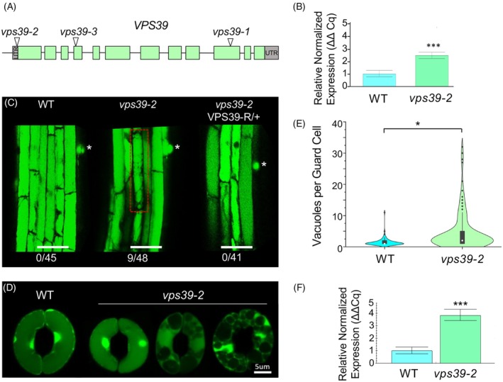
Figure 1 The HOPS subunit VPS39 regulates vacuole fusion in Arabidopsis. (A) Map of VPS39 with the location of T‐DNA insertions in vps39 alleles. Exons are represented in blocks, and green represents CDS. T‐DNA is located 2 bp upstream of the translation start site in vps39‐2. (B) Relative gene expression levels of VPS39 in 14‐day‐old seedlings by RT‐qPCR. Data are presented as relative normalized expression ±95% confidence interval from n = 30 seedlings per genotype. Gene expression was normalized to PEX4 (UBC21) and expressed relative to WT. Significant differences by one‐way ANOVA are indicated; P = 6.73 × 10⁻⁷ (***). (C) Mild vacuole fragmentation phenotype in vps39‐2 roots. Asterisk represents the location of root hair primordia, and a cell with fragmented vacuoles is highlighted in red. The number of seedlings with at least one cell with fragmented vacuoles is shown. Roots were stained with BCECF. Scale bar: 50 μm. (D) Vacuole phenotype of vps39‐2 stomata under opening conditions. Leaf epidermal peels of WT and vps39‐2 were exposed to stomatal opening conditions and stained with BCECF. Images of WT and vps39‐2 stomata are shown, with vps39‐2 exhibiting variable vacuole numbers. (E) Vacuole quantification from stomatal opening assays as in (D). A violin plot shows the distribution of the data along with a box and whisker plot with the median noted as a white dot and outliers as black diamonds. Statistical analysis was performed using a generalized linear mixed model comparing vacuoles per guard cell. The model containing genotype was favored (P = 0.01). n = 97–139 guard cells per genotype from 5 to 8 plants per genotype. (F) RT‐qPCR relative normalized expression of VPS39 in guard cell‐enriched tissue from 5‐week‐old WT or vps39‐2 plants. n = 12 plants per genotype. Data are represented as relative normalized expression ±95% confidence interval. Gene expression was normalized to PEX4. Statistical analysis was performed using the Mann–Whitney U test; P = 4.66 × 10⁻⁵ (***).
Figure 1 The HOPS subunit VPS39 regulates vacuole fusion in Arabidopsis. (A) Map of VPS39 with the location of T‐DNA insertions in vps39 alleles. Exons are represented in blocks, and green represents CDS. T‐DNA is located 2 bp upstream of the translation start site in vps39‐2. (B) Relative gene expression levels of VPS39 in 14‐day‐old seedlings by RT‐qPCR. Data are presented as relative normalized expression ±95% confidence interval from n = 30 seedlings per genotype. Gene expression was normalized to PEX4 (UBC21) and expressed relative to WT. Significant differences by one‐way ANOVA are indicated; P = 6.73 × 10⁻⁷ (***). (C) Mild vacuole fragmentation phenotype in vps39‐2 roots. Asterisk represents the location of root hair primordia, and a cell with fragmented vacuoles is highlighted in red. The number of seedlings with at least one cell with fragmented vacuoles is shown. Roots were stained with BCECF. Scale bar: 50 μm. (D) Vacuole phenotype of vps39‐2 stomata under opening conditions. Leaf epidermal peels of WT and vps39‐2 were exposed to stomatal opening conditions and stained with BCECF. Images of WT and vps39‐2 stomata are shown, with vps39‐2 exhibiting variable vacuole numbers. (E) Vacuole quantification from stomatal opening assays as in (D). A violin plot shows the distribution of the data along with a box and whisker plot with the median noted as a white dot and outliers as black diamonds. Statistical analysis was performed using a generalized linear mixed model comparing vacuoles per guard cell. The model containing genotype was favored (P = 0.01). n = 97–139 guard cells per genotype from 5 to 8 plants per genotype. (F) RT‐qPCR relative normalized expression of VPS39 in guard cell‐enriched tissue from 5‐week‐old WT or vps39‐2 plants. n = 12 plants per genotype. Data are represented as relative normalized expression ±95% confidence interval. Gene expression was normalized to PEX4. Statistical analysis was performed using the Mann–Whitney U test; P = 4.66 × 10⁻⁵ (***).CC BY · PubMed Central
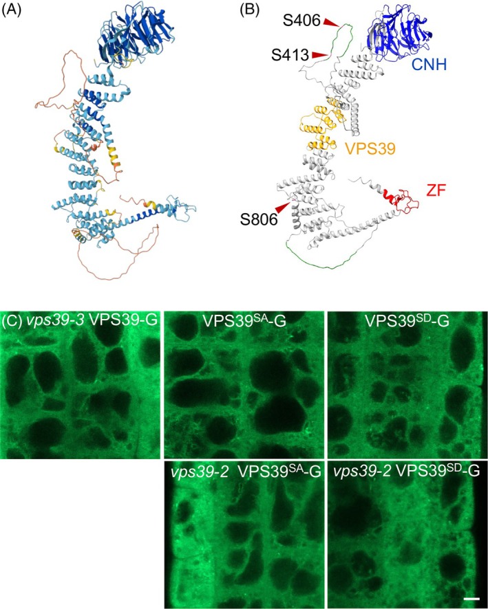
Figure 2 VPS39‐GFP phosphorylation mutants do not display altered protein localization. (A) Predicted Arabidopsis VPS39 structure from Alphafold 2.0. (B) Domain structure of VPS39. A Conserved Citron Homology (CNH, blue), a VPS39/TGFβR association domain 1 (orange) and VPS39‐associated zinc finger (red) are predicted by Uniprot. Two predicted intrinsically disordered regions (green) are indicated as predicted by MobiDB. The positions of S406, S413, S806 (pink) used for phosphomutant analysis are indicated with arrowheads. (C) Localization of intact or phosphomutant versions of VPS39‐GFP in the roots of complemented vps39‐3, WT, or vps39‐2. Images are representative from 16 to 23 seedlings per genotype. Scale bar corresponds to 5 μm.
Figure 2 VPS39‐GFP phosphorylation mutants do not display altered protein localization. (A) Predicted Arabidopsis VPS39 structure from Alphafold 2.0. (B) Domain structure of VPS39. A Conserved Citron Homology (CNH, blue), a VPS39/TGFβR association domain 1 (orange) and VPS39‐associated zinc finger (red) are predicted by Uniprot. Two predicted intrinsically disordered regions (green) are indicated as predicted by MobiDB. The positions of S406, S413, S806 (pink) used for phosphomutant analysis are indicated with arrowheads. (C) Localization of intact or phosphomutant versions of VPS39‐GFP in the roots of complemented vps39‐3, WT, or vps39‐2. Images are representative from 16 to 23 seedlings per genotype. Scale bar corresponds to 5 μm.CC BY · PubMed Central
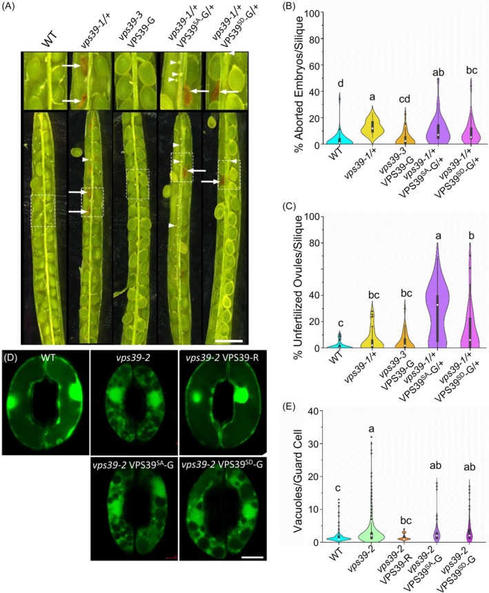
Figure 3 Phosphomutant versions of VPS39‐GFP at S406, 413, and 806 are not functional. (A) Embryo abortion phenotype in siliques from WT, complemented vps39‐3 VPS39‐GFP, vps39‐1/+ heterozygotes, and vps39‐1/+ carrying the VPS39SD‐GFP or VPS39SA‐GFP constructs. Arrows denote aborted embryos. Arrowheads denote unfertilized ovules or very early aborted embryos. Scale bar: 1 mm. (B) Quantification of aborted embryos from (A). Statistical analysis was performed with a Kruskal–Wallis test followed by post‐hoc Dunn's test with Šidák correction. Significant differences are shown using compact letter display (P < 0.0051). n = 30–42 siliques/genotype from 6 to 11 plants/genotype. (C) Quantification of the apparent unfertilized ovules of phosphomutant lines from (A). Kruskal–Wallis followed by post‐hoc Dunn's test with Šidák correction for multiple comparisons P < 0.0051. n = 30–42 siliques per genotype from 6 to 11 plants per genotype. (D) Vacuole fusion phenotypes of vps39‐2 mutants expressing phosphomutant VPS39‐GFP in stomata. Epidermal peels from WT, vps39‐2, and vps39‐2 expressing VPS39SD‐GFP or VPS39SA‐GFP were exposed to opening conditions for 2 h, stained with BCECF, and imaged by confocal microscopy. Examples of highly fragmented vacuoles of phosphomutant lines are shown. Scale bar: 5 μm. (E) Quantification of the number of vacuoles per guard cell as seen in (D). Statistical analysis was performed using a generalized linear mixed model comparing vacuoles per guard cell followed by bootstrap analysis. Significant difference found in the bootstrap analysis is denoted by compact letter display. The model containing genotype was favored (P = 1.30 × 10⁻⁷). n = 97–436 guard cells from 9 to 26 plants per genotype. This model includes stomata data from Figure 1E. Data distributions in (B), (C), and (E) are plotted as violin plots along with box and whisker plots with the median noted as a white dot and outliers as black dots.
Figure 3 Phosphomutant versions of VPS39‐GFP at S406, 413, and 806 are not functional. (A) Embryo abortion phenotype in siliques from WT, complemented vps39‐3 VPS39‐GFP, vps39‐1/+ heterozygotes, and vps39‐1/+ carrying the VPS39SD‐GFP or VPS39SA‐GFP constructs. Arrows denote aborted embryos. Arrowheads denote unfertilized ovules or very early aborted embryos. Scale bar: 1 mm. (B) Quantification of aborted embryos from (A). Statistical analysis was performed with a Kruskal–Wallis test followed by post‐hoc Dunn's test with Šidák correction. Significant differences are shown using compact letter display (P < 0.0051). n = 30–42 siliques/genotype from 6 to 11 plants/genotype. (C) Quantification of the apparent unfertilized ovules of phosphomutant lines from (A). Kruskal–Wallis followed by post‐hoc Dunn's test with Šidák correction for multiple comparisons P < 0.0051. n = 30–42 siliques per genotype from 6 to 11 plants per genotype. (D) Vacuole fusion phenotypes of vps39‐2 mutants expressing phosphomutant VPS39‐GFP in stomata. Epidermal peels from WT, vps39‐2, and vps39‐2 expressing VPS39SD‐GFP or VPS39SA‐GFP were exposed to opening conditions for 2 h, stained with BCECF, and imaged by confocal microscopy. Examples of highly fragmented vacuoles of phosphomutant lines are shown. Scale bar: 5 μm. (E) Quantification of the number of vacuoles per guard cell as seen in (D). Statistical analysis was performed using a generalized linear mixed model comparing vacuoles per guard cell followed by bootstrap analysis. Significant difference found in the bootstrap analysis is denoted by compact letter display. The model containing genotype was favored (P = 1.30 × 10⁻⁷). n = 97–436 guard cells from 9 to 26 plants per genotype. This model includes stomata data from Figure 1E. Data distributions in (B), (C), and (E) are plotted as violin plots along with box and whisker plots with the median noted as a white dot and outliers as black dots.CC BY · PubMed Central
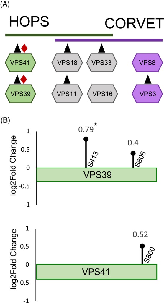
Figure 4 Phosphoproteomics analysis of guard cell‐enriched tissue reveals VPS39 phosphorylation changes between open and closed stomata. (A) Depiction of HOPS‐specific (green), CORVET‐specific (purple), and shared core (gray) subunits detected in the proteome (▲) or the phosphoproteome (♦) from guard cell‐enriched tissue. (B) Lollipop representation of phosphorylation sites mapped onto the VPS39 and VPS41 primary sequences. Each lollipop denotes a modified residue. The X‐axis represents the residue position along each protein sequence, and the Y‐axis represents Log2FC of phosphorylated residues between closed and open samples. Increased phosphorylation was detected in the closed condition for both proteins (Log2FC >0.5), but only S413 on VPS39 was significant (*, Absolute Log2FC >0.5 and P < 0.05).
Figure 4 Phosphoproteomics analysis of guard cell‐enriched tissue reveals VPS39 phosphorylation changes between open and closed stomata. (A) Depiction of HOPS‐specific (green), CORVET‐specific (purple), and shared core (gray) subunits detected in the proteome (▲) or the phosphoproteome (♦) from guard cell‐enriched tissue. (B) Lollipop representation of phosphorylation sites mapped onto the VPS39 and VPS41 primary sequences. Each lollipop denotes a modified residue. The X‐axis represents the residue position along each protein sequence, and the Y‐axis represents Log2FC of phosphorylated residues between closed and open samples. Increased phosphorylation was detected in the closed condition for both proteins (Log2FC >0.5), but only S413 on VPS39 was significant (*, Absolute Log2FC >0.5 and P < 0.05).CC BY · PubMed Central
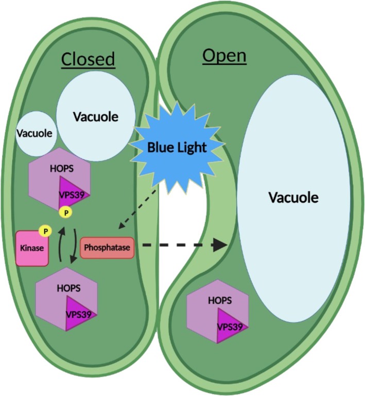
Figure 5 Proposed model for regulation of stomatal vacuole fusion by phosphorylation of HOPS. We predict that the HOPS subunit VPS39 accumulates in its phosphorylated form in closed stomata, which may promote HOPS association with the vacuolar SNAREs at the tonoplast. Dissociation of HOPS is necessary to complete fusion, and this could be triggered by VPS39 dephosphorylation in response to stomatal opening signals such as blue light. Kinases and phosphatases involved in this process are unknown. Image generated in BioRender (Biorender.com).
Figure 5 Proposed model for regulation of stomatal vacuole fusion by phosphorylation of HOPS. We predict that the HOPS subunit VPS39 accumulates in its phosphorylated form in closed stomata, which may promote HOPS association with the vacuolar SNAREs at the tonoplast. Dissociation of HOPS is necessary to complete fusion, and this could be triggered by VPS39 dephosphorylation in response to stomatal opening signals such as blue light. Kinases and phosphatases involved in this process are unknown. Image generated in BioRender (Biorender.com).CC BY · PubMed Central

Verification

Claims the paper makes

The claims and statistics of this paper haven’t been checked yet.

Run the check — free, no account needed. It reads the open-access text and checks the sources it cites.

A few free checks a day · papers already verified open instantly · results are public

Self-check

Percentages, sums and reported differences re-computed from the paper’s own numbers.

The self-check needs the paper’s full text.

Citations

No citation lists for this paper

No citation lists for this paper yet — checked again daily.

References 108 · Cited by 1

Research works printed but not linked: 102

  1. 10.1073/pnas.1320421111 · DOI
  2. 10.1242/jcs.107805 · DOI
  3. 10.18637/jss.v067.i01 · DOI · Paper page
  4. 10.1101/pdb.prot072983 · DOI
  5. 10.1073/pnas.1807763115 · DOI
  6. 10.1073/pnas.1117797109 · DOI
  7. 10.32614/Rj-2017-066 · DOI
  8. 10.1083/jcb.201004092 · DOI
  9. 10.1104/pp.16.01848 · DOI
  10. 10.1111/nph.17135 · DOI
  11. 10.1080/19420889.2017.1395993 · DOI
  12. 10.1126/science.8108733 · DOI
  13. 10.1093/plphys/kiac028 · DOI
  14. 10.1038/nplants.2016.179 · DOI
  15. 10.1016/j.cub.2022.04.040 · DOI
  16. 10.1016/0092-8674(90)90482-T · DOI
  17. 10.1046/j.1365-313x.1998.00343.x · DOI
  18. 10.1016/j.tplants.2020.01.008 · DOI
  19. Davison AC , Hinkley DV (1997) Bootstrap methods and their application. Cambridge University Press, · DOI · Paper page
  20. 10.1016/j.cub.2024.06.001 · DOI
  21. 10.1038/ncb2270 · DOI
  22. 10.1105/tpc.107.057711 · DOI
  23. 10.1111/j.1365-313X.2011.04786.x · DOI
  24. 10.1073/pnas.95.26.15781 · DOI
  25. 10.3791/55969 · DOI
  26. 10.1093/pcp/pcu038 · DOI
  27. 10.1104/pp.105.067520 · DOI
  28. 10.1111/j.1365-3040.2009.01993.x · DOI
  29. 10.1073/pnas.0912021106 · DOI
  30. 10.1111/nph.16168 · DOI
  31. 10.1038/s41467-024-45248-5 · DOI
  32. 10.1074/jbc.M109.000737 · DOI
  33. 10.1038/s41467-017-01237-5 · DOI
  34. 10.1093/insilicoplants/diae015 · DOI
  35. 10.1016/j.devcel.2014.06.006 · DOI
  36. 10.1074/mcp.RA120.002105 · DOI
  37. 10.21769/BioProtoc.2447 · DOI
  38. 10.1093/pcp/pcv076 · DOI
  39. 10.1105/tpc.010215 · DOI
  40. 10.1146/annurev-arplant-042809-112226 · DOI
  41. 10.1126/science.adk0850 · DOI · Paper page
  42. 10.1016/j.mcpro.2022.100477 · DOI
  43. 10.1016/j.semcdb.2017.07.008 · DOI
  44. 10.1111/nph.17208 · DOI
  45. 10.1126/science.283.5401.537 · DOI
  46. 10.1111/j.1365-3040.2012.02592.x · DOI
  47. 10.1105/tpc.7.4.473 · DOI
  48. 10.1007/BF02773350 · DOI
  49. 10.1038/343186a0 · DOI
  50. 10.18637/jss.v112.i01 · DOI
  51. 10.1038/s41586-020-2094-2 · DOI
  52. 10.1038/s41596-018-0006-9 · DOI
  53. 10.1038/s41598-023-34273-x · DOI
  54. 10.1111/j.1365-3040.1995.tb00188.x · DOI
  55. 10.1105/tpc.007906 · DOI
  56. 10.1093/bioinformatics/btx015 · DOI
  57. 10.1016/j.ceb.2009.05.007 · DOI
  58. 10.1126/science.1173041 · DOI
  59. 10.1073/pnas.2220270120 · DOI
  60. 10.1105/tpc.9.3.409 · DOI
  61. 10.1093/nar/gkae969 · DOI
  62. 10.1002/pro.4466 · DOI · Paper page
  63. 10.1093/nar/gkx1071 · DOI
  64. 10.1074/mcp.M114.040352 · DOI
  65. 10.1016/S1534-5807(01)00024-7 · DOI
  66. 10.1091/mbc.12.12.3733 · DOI
  67. 10.1093/plcell/koae243 · DOI
  68. 10.1146/annurev.arplant.52.1.627 · DOI
  69. 10.1073/pnas.84.12.4108 · DOI
  70. 10.7554/eLife.27396 · DOI
  71. 10.1073/pnas.97.17.9402 · DOI
  72. 10.2307/2684063 · DOI
  73. 10.7554/eLife.80901 · DOI · Paper page
  74. 10.1111/plb.13382 · DOI
  75. 10.1093/mp/ssm029 · DOI
  76. 10.1016/0092-8674(93)90376-2 · DOI
  77. 10.7554/eLife.67578 · DOI
  78. 10.1073/pnas.0903801106 · DOI
  79. 10.1038/msb.2008.32 · DOI
  80. 10.1093/mp/ssm017 · DOI · Paper page
  81. 10.1038/s41467-021-26333-5 · DOI · Paper page
  82. 10.1111/nph.15593 · DOI
  83. 10.1105/tpc.105.038950 · DOI
  84. 10.1038/26412 · DOI · Paper page
  85. 10.1038/ncomms3094 · DOI · Paper page
  86. 10.1073/pnas.1717839115 · DOI
  87. 10.1093/nar/gkae1010 · DOI · Paper page
  88. 10.1247/csf.29.49 · DOI
  89. 10.1126/scisignal.2003509 · DOI
  90. 10.1105/tpc.113.114827 · DOI
  91. 10.1083/jcb.200308071 · DOI
  92. 10.1111/jipb.13536 · DOI
  93. 10.1016/j.molcel.2017.12.002 · DOI
  94. 2019, Welcome to the Tidyverse, Journal of open source software, 4, 1686, 10.21105/joss.01686 · DOI · Paper page
  95. 10.1146/annurev-cellbio-100109-104131 · DOI
  96. 10.1038/nsmb.1451 · DOI
  97. 10.1111/tpj.14345 · DOI
  98. 10.1093/jxb/erae270 · DOI
  99. 10.1007/s10265-006-0048-x · DOI
  100. 10.1083/jcb.151.3.551 · DOI · Paper page
  101. 10.1093/plcell/koae073 · DOI
  102. 10.1093/mp/ssu019 · DOI

Metadata

Authors
Pullen, A.-M., Billings, G., Hodgens, C., White, G., Akpa, B. S., & Rojas-Pierce, M.
Affiliations
  • North Carolina State University (US)
  • University of Tennessee at Knoxville (US)
  • University of Tennessee System (US)
Venue
The Plant Journal
Publisher
openRxiv
Published
2025-10-02
Volume
124
Issue
6
Pages
e70640-e70640
ISSN
0960-7412 (ISSN-L)
OpenAlex
W4417514813
Cited by
1
References
108
Citations by year
2026: 1
FWCI
0.50
Type
journal article
Language
en
Open access
Locations
  • The Plant Journal (publishedVersion, cc-by)
  • bioRxiv (Cold Spring Harbor Laboratory) (acceptedVersion, cc-by)
  • PubMed (publishedVersion)
  • PubMed Central (publishedVersion, cc-by)
Funders
  • National Science Foundation (1918746)
  • U.S. Department of Agriculture
  • North Carolina State University
  • National Institute of Food and Agriculture (HATCH 7005574)
  • Agricultural Research Service
  • National Institute for Basic Biology
Topics
  • Life Sciences › Biochemistry, Genetics and Molecular Biology › Cell Biology › Cellular transport and secretion
  • Life Sciences › Agricultural and Biological Sciences › Plant Science › Plant Molecular Biology Research
  • Life Sciences › Biochemistry, Genetics and Molecular Biology › Molecular Biology › Photosynthetic Processes and Mechanisms
Keywords
HOPS complex, VPS39, VPS41, phosphorylation, trans-SNARE complex, stomatal opening, water vapor loss, post-translational modification
MeSH terms
Membrane Fusion, Phosphorylation, Vacuoles, Arabidopsis, Arabidopsis Proteins, Vesicular Transport Proteins, SNARE Proteins, Plant Stomata

About these checks

Registry, retraction, Cites and statistics results are computed, not judged, with one exception: a printed reference that no index lists is read once by a model before it counts as a citation error. Claims are extracted by a model and kept only when their quote appears verbatim in the text; each cited source is then checked, with a model’s help, for whether it says what the claim relies on. Those verdicts can be wrong — read the claim’s quote and the cited source before relying on one.

Statistics and arithmetic are recomputed, not judged.

Confidence bands are shown only for fields calibrated on our annotated test set. None is calibrated yet, so no band is shown.

What counts as a claim ›How we measure confidence ›How we check this ›

Are you an author, or do you see a problem with this page? Request a review

How to cite

Cite CoChat for the checks on this page, and the paper for its own findings.

This page
CoChat. (2026). Citation check: Regulation of Vacuole Fusion in Stomata by Dephosphorylation of the HOPS subunit VPS39. https://app.cochat.ai/papers/10.1101/2025.10.02.680005
The paper
Pullen, A.-M., Billings, G., Hodgens, C., White, G., Akpa, B. S., & Rojas-Pierce, M. (2025). Regulation of Vacuole Fusion in Stomata by Dephosphorylation of the HOPS subunit VPS39. The Plant Journal. https://doi.org/10.1101/2025.10.02.680005