

The DOI matches its Crossref record; 1 of the 1 references it cites were found in a registry or an index.
Visualizing the mechanism of quinol oxidation and inhibition of a bd -type oxidase using cryo-EM
Open accessPreprint — published as 10.1126/sciadv.aec9946 · Paper page
Abstract
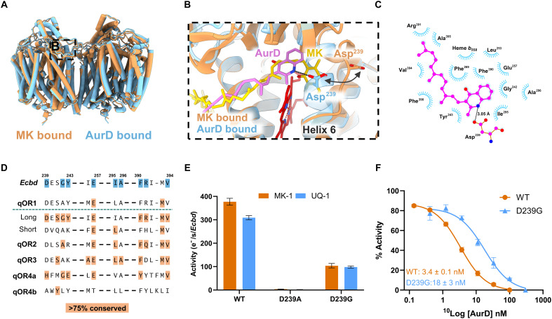
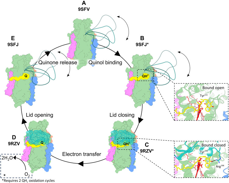
Cytochrome bd is a prokaryotic terminal oxidase recognized as an antibiotic target against various pathogens. Despite its critical role in respiration, failure to capture the mechanism of enzyme catalysis and inhibition prohibits structure guided drug discovery. Here, we present cryo-electron microscopy structures of Escherichia coli cytochrome bd -I in monomeric and dimeric forms, along all stages of quinol turn-over and in an inhibitor-bound state. We identify a dynamic Q-loop lid that undergoes a disorder-to-order transition upon substrate binding to the dimer, completing the active site and enabling catalysis. Structure-guided mutagenesis confirms Tyr243 and Arg298 as essential catalytic residues unique to long Q-loop oxidases, highlighting evolutionary divergence from short Q-loop variants. Inhibition by Aurachin D triggers refolding of the active site, occluding substrate access via a conserved Asp239-mediated mechanism. The structural and mechanistic insights presented here establish a comprehensive framework, opening new ways for drug discovery.
Overview
The claims and statistics of this paper haven’t been checked yet.
Run the check — free, no account needed. It reads the open-access text and checks the sources it cites.
Integrity signals
Registry
Cites
Statistics
Figures
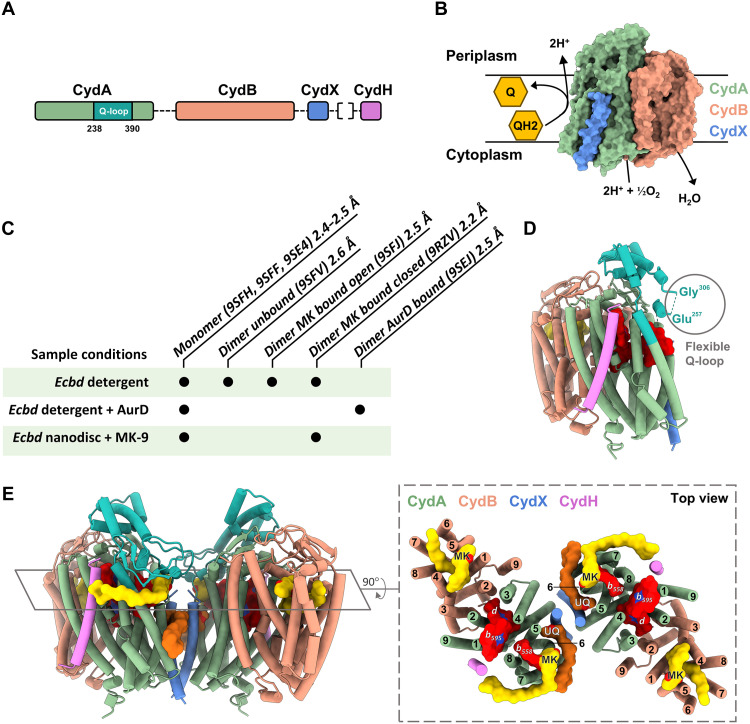
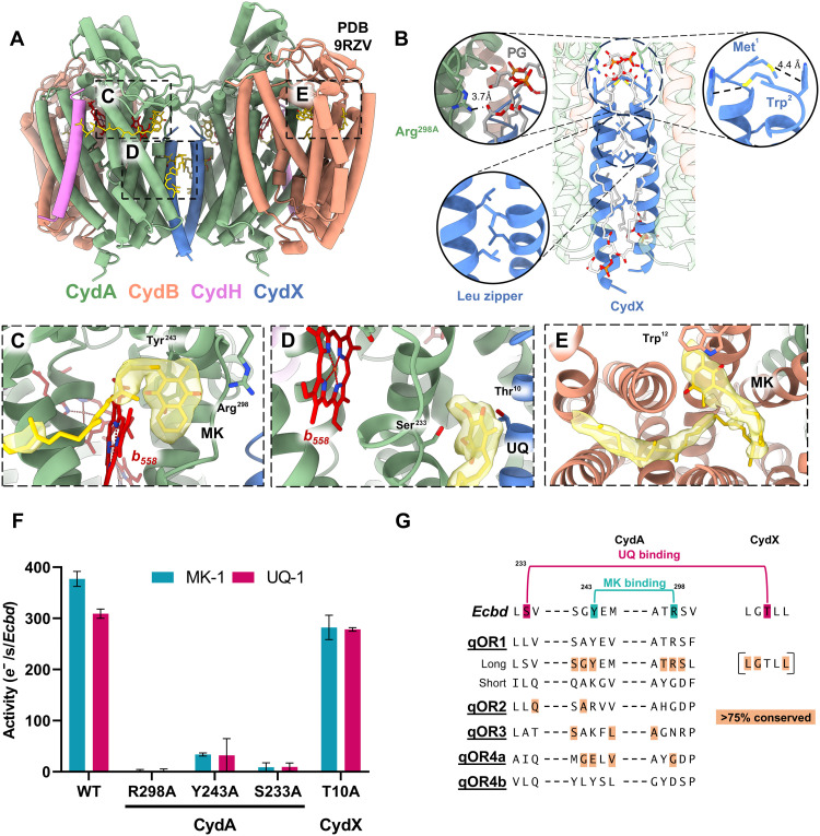
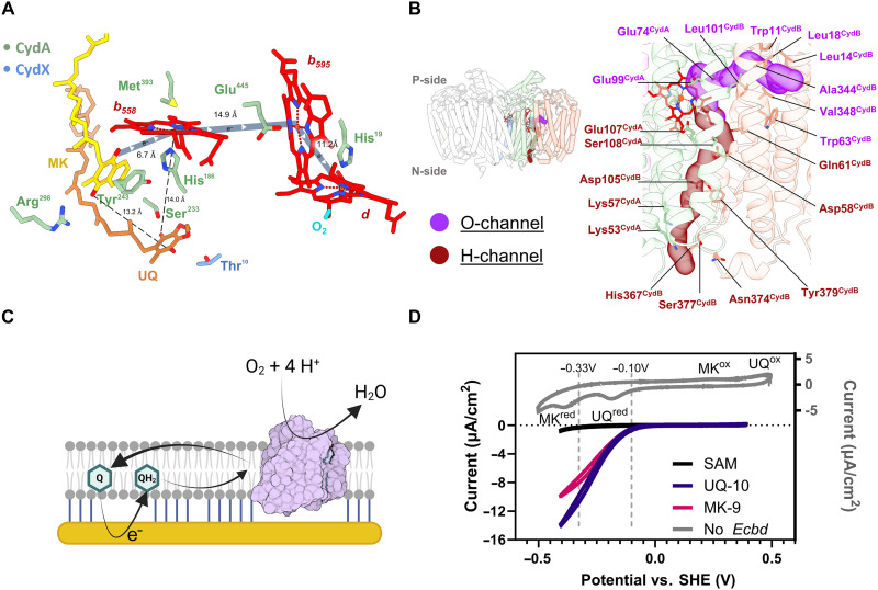
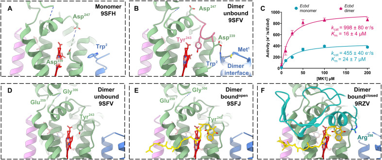
Verification
Claims the paper makes
The claims and statistics of this paper haven’t been checked yet.
Run the check — free, no account needed. It reads the open-access text and checks the sources it cites.
Self-check
Percentages, sums and reported differences re-computed from the paper’s own numbers.
The self-check needs the paper’s full text.
Citations
Cites
Highly accurate protein structure prediction with AlphaFold
Jumper, J., Evans, R., Pritzel, A., Green, T., Figurnov, M., Ronneberger, O., Tunyasuvunakool, K., Bates, R., Žídek, A., Potapenko, A., et al. (2021) · Nature · doi:10.1038/s41586-021-03819-2 · Cited by 43,764
Research works printed but not linked: 52
- 2022, Recent Advances in Structural Studies of Cytochrome bd and Its Potential Application as a Drug Target, Int J Mol Sci, 23, 3166, 10.3390/ijms23063166 · DOI
- 10.1056/NEJMp1314385 · DOI
- 10.1099/00221287-142-4-755 · DOI · Paper page
- 10.1021/bi700862u · DOI
- 10.1038/nchembio.135 · DOI
- X. Xia , S. Wu , L. Li , B. Xu , G. Wang , The cytochrome bd complex is essential for chromate and sulfide resistance and is regulated by a GbsR-type regulator, CydE, in Alishewanella Sp. WH16-1. Front Microbiol 9 (2018). · DOI
- C. J. Beebout , L. A. Sominsky , A. R. Eberly , G. T. Van Horn , M. Hadjifrangiskou , Cytochrome bd promotes Escherichia coli biofilm antibiotic tolerance by regulating accumulation of noxious chemicals. Biofilms and Microbiomes 7 (2021). · DOI
- 10.1016/j.pbiomolbio.2019.11.002 · DOI
- 10.15252/emmm.202114123 · DOI
- J. Jones-Carson , M. Husain , L. Liu , D. J. Orlicky , A. Vázquez-Torres , Cytochrome bd-dependent bioenergetics and antinitrosative defenses in Salmonella pathogenesis. mBio 7 (2016). · DOI
- M. Shepherd , M. E. S. Achard , A. Idris , M. Totsika , M. D. Phan , K. M. Peters , S. Sarkar , C. A. Ribeiro , L. V. Holyoake , D. Ladakis , G. C. Ulett , M. J. Sweet , R. K. Poole , A. G. McEwan , M. A. Schembri , The cytochrome bd-I respiratory oxidase augments survival of multidrug-resistant Escherichia coli during infection. Sci Rep 6 (2016). · DOI
- 2024, Steroid Drugs Inhibit Bacterial Respiratory Oxidases and Are Lethal Toward Methicillin-Resistant Staphylococcus aureus, Journal of Infectious Diseases, 230, e149, 10.1093/infdis/jiad540 · DOI
- 10.1126/science.aay0967 · DOI
- 10.1126/science.aaf2477 · DOI
- H. G. Goojani , J. Konings , H. Hakvoort , S. Hong , R. B. Gennis , J. Sakamoto , H. Lill , D. Bald , The carboxy-terminal insert in the Q-loop is needed for functionality of Escherichia coli cytochrome bd-I. Biochim Biophys Acta Bioenerg 1861 (2020). · DOI
- 2020, The long Q-loop of Escherichia coli cytochrome bd oxidase is required for assembly and structural integrity, FEBS Lett, 594, 1577, 10.1002/1873-3468.13749 · DOI
- 10.1016/S0005-2728(97)00034-0 · DOI · Paper page
- A. Theßeling , T. Rasmussen , S. Burschel , D. Wohlwend , J. Kägi , R. Müller , B. Böttcher , T. Friedrich , Homologous bd oxidases share the same architecture but differ in mechanism. Nat Commun 10 (2019). · DOI
- T. N. Grund , M. Radloff , D. Wu , H. G. Goojani , L. F. Witte , W. Jösting , S. Buschmann , H. Müller, Elamri , S. Welsch , H. Schwalbe , H. Michel , D. Bald , S. Safarian , Mechanistic and structural diversity between cytochrome bd isoforms of Escherichia coli. Proc Natl Acad Sci U S A 118 (2021). · DOI
- A. Grauel , J. Kägi , T. Rasmussen , I. Makarchuk , S. Oppermann , A. F. A. Moumbock , D. Wohlwend , R. Müller , F. Melin , S. Günther , P. Hellwig , B. Böttcher , T. Friedrich , Structure of Escherichia coli cytochrome bd-II type oxidase with bound aurachin D. Nat Commun 12 (2021). · DOI
- R. Timkovich , M. S. Cork , R. B. Gennis , P. Y. Johnson , Proposed structure of heme d, a prosthetic group of bacterial terminal oxidases. J Am Chem Soc 107 (1985). · DOI
- 2025, Menaquinone-specific turnover by Mycobacterium tuberculosis cytochrome bd is redox regulated by the Q-loop disulfide bond, J Biol Chem, 301, 108094, 10.1016/j.jbc.2024.108094 · DOI
- 10.1021/la047732f · DOI
- 10.1021/jacs.9b10254 · DOI
- 2010, A study of cytochrome bo3 in a tethered bilayer lipid membrane, Biochim Biophys Acta Bioenerg, 1797, 1917, 10.1016/j.bbabio.2010.01.012 · DOI
- 2023, Rapid and Highly Stable Membrane Reconstitution by LAiR Enables the Study of Physiological Integral Membrane Protein Functions, ACS Cent Sci, 9, 494, 10.1021/acscentsci.2c01170 · DOI
- 10.1021/ja056972u · DOI
- 10.1093/nar/gky309 · DOI
- M. Janczak , J. Vilhjálmsdóttir , P. Ädelroth , Proton transfer in cytochrome bd-I from E. coli involves Asp-105 in CydB. Biochim Biophys Acta Bioenerg 1865 (2024). · DOI
- S. Safarian , H. K. Opel-Reading , D. Wu , A. R. Mehdipour , K. Hards , L. K. Harold , M. Radloff , I. Stewart , S. Welsch , G. Hummer , G. M. Cook , K. L. Krause , H. Michel , The cryo-EM structure of the bd oxidase from M. tuberculosis reveals a unique structural framework and enables rational drug design to combat TB. Nat Commun 12 (2021). · DOI
- M. Radloff , I. Elamri , T. N. Grund , L. F. Witte , K. F. Hohmann , S. Nakagaki , H. G. Goojani , H. Nasiri , Hideto Miyoshi , D. Bald , H. Xie , J. Sakamoto , H. Schwalbe , S. Safarian , Short-chain aurachin D derivatives are selective inhibitors of E. coli cytochrome bd-I and bd-II oxidases. Sci Rep 11 (2021). · DOI
- 10.1021/bi061946+ · DOI
- R. J. Allen , E. P. Brenner , C. E. VanOrsdel , J. J. Hobson , D. J. Hearn , M. R. Hemm , Conservation analysis of the CydX protein yields insights into small protein identification and evolution. BMC Genomics 15 (2014). · DOI
- 10.1016/j.febslet.2014.03.036 · DOI
- 10.1128/JB.00324-13 · DOI
- 10.1021/bi060192w · DOI
- 10.1021/jacs.8b10836 · DOI
- 10.1021/jacs.8b10836 · DOI
- W. Wang , Y. Gao , Y. Tang , X. Zhou , Y. Lai , S. Zhou , Y. Zhang , X. Yang , F. Liu , L. W. Guddat , Q. Wang , Z. Rao , H. Gong , Cryo-EM structure of mycobacterial cytochrome bd reveals two oxygen access channels. Nat Commun 12 (2021). · DOI
- T. N. Grund , Y. Kabashima , T. Kusumoto , D. Wu , S. Welsch , J. Sakamoto , H. Michel , S. Safarian , The cryoEM structure of cytochrome bd from C. glutamicum provides novel insights into structural properties of actinobacterial terminal oxidases. Front Chem 10 (2023). · DOI
- 10.1128/JB.00562-09 · DOI
- 10.1128/JB.02176-14 · DOI
- 2024, The effect of lipid composition on the thermal stability of nanodiscs, Biochim Biophys Acta Biomembr, 1866, 184239, 10.1016/j.bbamem.2023.184239 · DOI
- 10.1016/S0040-4020(97)00698-4 · DOI
- 2007, Phase separation in mixed self-assembled monolayers and its effect on biomimetic membranes, Sensors and Actuators, B, 124, 501, 10.1016/j.snb.2007.01.014 · DOI
- 10.1038/nmeth.4169 · DOI · Paper page
- 10.1038/s41592-020-00990-8 · DOI
- 10.1016/j.jsb.2015.08.007 · DOI · Paper page
- 10.1107/S0907444910007493 · DOI · Paper page
- P. D. Adams , R. W. Grosse-Kunstleve , L.-W. Hung , T. R. Ioerger , A. J. Mccoy , N. W. Moriarty , R. J. Read , J. C. Sacchettini , N. K. Sauter , T. C. Terwilliger , Biological Crystallography PHENIX: building new software for automated crystallographic structure determination. Acta Crystallographica Section D 58 (2002). · DOI · Paper page
- 10.1002/PRO.3943 · DOI · Paper page
- 10.1021/ci200227u · DOI
Metadata
- Authors
- van der Velden, T. T., Kayastha, K., Pelser, F., Brünle, S., & Jeuken, L. J. C.
- Affiliations
- Leiden University (NL)
- Corresponding author
- Jeuken, Lars J. C.
- Venue
- Science Advances
- Publisher
- openRxiv
- Published
- 2025-10-10
- Volume
- 12
- Issue
- 21
- Pages
- eaec9946-eaec9946
- ISSN
- 2375-2548 (ISSN-L)
- PMC
- PMC13189123
- OpenAlex
- W7161809970
- Cited by
- 2
- References
- 1
- Citations by year
- 2026: 2
- FWCI
- 2.70
- Type
- journal article
- Language
- en
- Open access
- gold
- doi.org
- Licence
- Locations
- Science Advances (publishedVersion, cc-by)
- bioRxiv (Cold Spring Harbor Laboratory) (acceptedVersion, cc-by)
- PubMed (publishedVersion)
- PubMed Central (publishedVersion, cc-by)
- Leiden Repository (Leiden University) (submittedVersion, cc-by)
- Leiden Repository (Leiden University) (submittedVersion)
- Funders
- Oncode accalerator (NGFOP2201)
- Netherlands Electron Microscopy Infrastructure (184.034.014)
- Topics
- Life Sciences › Biochemistry, Genetics and Molecular Biology › Structural Biology › Enzyme Structure and Function
- Life Sciences › Biochemistry, Genetics and Molecular Biology › Structural Biology › Advanced Electron Microscopy Techniques and Applications
- Physical Sciences › Chemistry › Inorganic Chemistry › Metal-Catalyzed Oxygenation Mechanisms
- Keywords
- cytochrome bd oxidase, quinol oxidation, disorder-to-order transition, catalytic residues, terminal oxidase, antibiotic targets, structure-based drug design, protein dimerization
- MeSH terms
- Cytochrome b Group, Enzyme Inhibitors, Escherichia coli, Hydroquinones, Models, Molecular, Oxidation-Reduction, Oxidoreductases, Protein Conformation, Benzoquinones, Catalytic Domain, Cryoelectron Microscopy, Escherichia coli Proteins, Electron Transport Chain Complex Proteins
About these checks
Registry, retraction, Cites and statistics results are computed, not judged, with one exception: a printed reference that no index lists is read once by a model before it counts as a citation error. Claims are extracted by a model and kept only when their quote appears verbatim in the text; each cited source is then checked, with a model’s help, for whether it says what the claim relies on. Those verdicts can be wrong — read the claim’s quote and the cited source before relying on one.
Statistics and arithmetic are recomputed, not judged.
Confidence bands are shown only for fields calibrated on our annotated test set. None is calibrated yet, so no band is shown.
What counts as a claim ›How we measure confidence ›How we check this ›
Are you an author, or do you see a problem with this page? Request a review
How to cite
Cite CoChat for the checks on this page, and the paper for its own findings.
- This page
- CoChat. (2026). Citation check: Visualizing the mechanism of quinol oxidation and inhibition of a bd -type oxidase using cryo-EM. https://app.cochat.ai/papers/10.1101/2025.10.10.681541
- The paper
- van der Velden, T. T., Kayastha, K., Pelser, F., Brünle, S., & Jeuken, L. J. C. (2025). Visualizing the mechanism of quinol oxidation and inhibition of a bd -type oxidase using cryo-EM. Science Advances. https://doi.org/10.1101/2025.10.10.681541
Data sources
Bibliographic data: Semantic Scholar, OpenAlex, Crossref.
Contains information from the Semantic Scholar Academic Graph Datasets, made available under the ODC Attribution License (ODC-BY 1.0).
Semantic Scholar’s data is described in: Kinney, R., et al. (2023). The Semantic Scholar Open Data Platform. arXiv:2301.10140.
