

The DOI matches its Crossref record; 10 of the 10 references it cites were found in a registry or an index.
Automated Reconstruction of a Serial-Section EM Drosophila Brain with Flood-Filling Networks and Local Realignment
Abstract
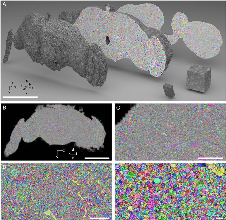
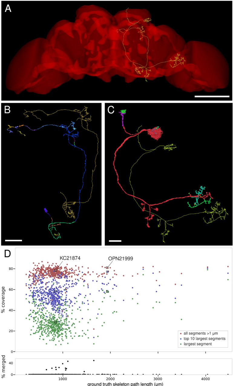
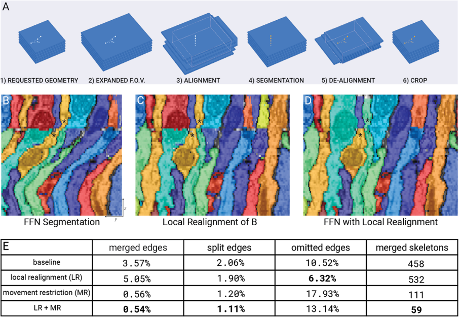
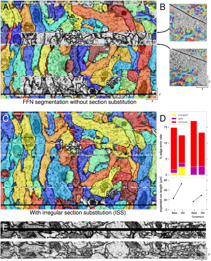
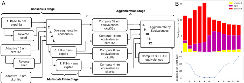
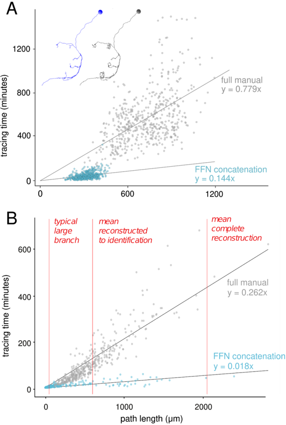
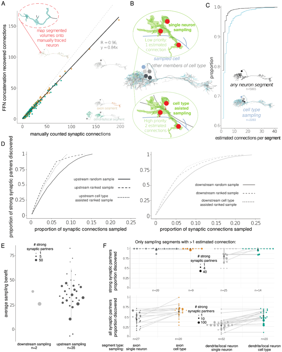
Reconstruction of neural circuitry at single-synapse resolution is a key target for improving understanding of the nervous system in health and disease. Serial section transmission electron microscopy (ssTEM) is among the most prolific imaging methods employed in pursuit of such reconstructions. We demonstrate how Flood-Filling Networks (FFNs) can be used to computationally segment a forty-teravoxel whole-brain Drosophila ssTEM volume. To compensate for data irregularities and imperfect global alignment, FFNs were combined with procedures that locally re-align serial sections as well as dynamically adjust and synthesize image content. The proposed approach produced a largely merger-free segmentation of the entire ssTEM Drosophila brain, which we make freely available. As compared to manual tracing using an efficient skeletonization strategy, the segmentation enabled circuit reconstruction and analysis workflows that were an order of magnitude faster.
Overview
The claims and statistics of this paper haven’t been checked yet.
Run the check — free, no account needed. It reads the open-access text and checks the sources it cites.
Integrity signals
Registry
Cites
Statistics
Figures
Verification
Claims the paper makes
The claims and statistics of this paper haven’t been checked yet.
Run the check — free, no account needed. It reads the open-access text and checks the sources it cites.
Claims other papers make about this one
Each is checked against this paper’s own text: does it say what the citing paper says it does?
The cited source says what the claim attributes to it.
In the citing paper
“Since manual skeletonization is laborious, the walled garden community is starting to migrate to semi-automated reconstruction 17 based on combining automatically generated skeletons 18.”
From FlyWire: online community for whole-brain connectomics (2022) · cites this paper as [18]
Self-check
Percentages, sums and reported differences re-computed from the paper’s own numbers.
The self-check needs the paper’s full text.
Citations
Cites
The structure of the nervous system of the nematode Caenorhabditis elegans
White, J. G., Southgate, E., Thomson, J. N., & Brenner, S. (1986) · Philosophical Transactions of the Royal Society of London. B, Biological Sciences · doi:10.1098/rstb.1986.0056 · Cited by 5,589 · cited 1×
“elegans (Varshney et al., 2011; White et al., 1986), larval Drosophila (Ohyama et al.”
Connectomic reconstruction of the inner plexiform layer in the mouse retina
Helmstaedter, M., Briggman, K. L., Turaga, S. C., Jain, V., Seung, H. S., & Denk, W. (2013) · Nature · doi:10.1038/nature12346 · Cited by 997
Self-citationStructural Properties of the Caenorhabditis elegans Neuronal Network
Varshney, L. R., Chen, B. L., Paniagua, E., Hall, D. H., & Chklovskii, D. B. (2011) · PLoS Computational Biology · doi:10.1371/journal.pcbi.1001066 · Cited by 867
Dendritic Spine Pathology: Cause or Consequence of Neurological Disorders?
Fiala, J. C., Spacek, J., & Harris, K. M. (2002) · Brain Research Reviews · doi:10.1016/s0165-0173(02)00158-3 · Cited by 811
A visual motion detection circuit suggested by Drosophila connectomics
Takemura, S.-Y., Bharioke, A., Lu, Z., Nern, A., Vitaladevuni, S., Rivlin, P. K., Katz, W. T., Olbris, D. J., Plaza, S. M., Winston, P., et al. (2013) · Nature · doi:10.1038/nature12450 · Cited by 686
A multilevel multimodal circuit enhances action selection in drosophila
Ohyama, T., Schneider-Mizell, C. M., Fetter, R. D., Aleman, J. V., Franconville, R., Rivera-Alba, M., Mensh, B. D., Branson, K. M., Simpson, J. H., Truman, J. W., et al. (2015) · Nature · doi:10.1038/nature14297 · Cited by 502
A connectome of a learning and memory center in the adult Drosophila brain
Takemura, S.-Y., Aso, Y., Hige, T., Wong, A., Lu, Z., Xu, C. S., Rivlin, P. K., Hess, H., Zhao, T., Parag, T., et al. (2017) · eLife · doi:10.7554/elife.26975 · Cited by 367
Quantitative neuroanatomy for connectomics in Drosophila
Schneider-Mizell, C. M., Gerhard, S., Longair, M., Kazimiers, T., Li, F., Zwart, M. F., Champion, A., Midgley, F. M., Fetter, R. D., Saalfeld, S., et al. (2016) · eLife · doi:10.7554/elife.12059 · Cited by 332
Self-citationFlyWire: online community for whole-brain connectomics
Dorkenwald, S., McKellar, C. E., Macrina, T., Kemnitz, N., Lee, K., Lu, R., Wu, J., Popovych, S., Mitchell, E., Nehoran, B., et al. (2022) · Nature Methods · doi:10.1038/s41592-021-01330-0 · Cited by 289
Functional and anatomical specificity in a higher olfactory centre
Frechter, S., Bates, A. S., Tootoonian, S., Dolan, M.-J., Manton, J., Jamasb, A. R., Kohl, J., Bock, D., & Jefferis, G. (2019) · eLife · doi:10.7554/elife.44590 · Cited by 105
Self-citation
Research works printed but not linked: 41
- 10.3390/molecules17010355 · DOI
- 10.1016/j.neuron.2015.09.003 · DOI
- 10.1038/nature09802 · DOI
- Buhmann, Julia , Renate Krause , Rodrigo Ceballos Lentini , Nils Eckstein , Matthew Cook , Srinivas Turaga , and Jan Funke . 2018. “Synaptic Partner Prediction from Point Annotations in Insect Brains.” arXiv [cs.CV]. arXiv. http://arxiv.org/abs/1806.08205. · DOI · Paper page
- Buhmann, Julia , Arlo Sheridan , Stephan Gerhard , Renate Krause , Tri Nguyen , Larissa Heinrich , Philipp Schlegel , et al. 2019. “Automatic Detection of Synaptic Partners in a Whole-Brain Drosophila EM Dataset.” bioRxiv. https://doi.org/10.1101/2019.12.12.874172. · DOI · Paper page
- 2019, Neural Arbors Are Pareto Optimal, Proceedings of the Royal Society B: Biological Sciences, 286, 20182727, 10.1098/rspb.2018.2727 · DOI
- 10.1016/j.neuron.2016.06.012 · DOI · Paper page
- 2018, Communication from Learned to Innate Olfactory Processing Centers Is Required for Memory Retrieval in Drosophila, Neuron, 100, 651, 10.1016/j.neuron.2018.08.037 · DOI · Paper page
- Heinrich, Larissa , Jan Funke , Constantin Pape , Juan Nunez-Iglesias , and Stephan Saalfeld . 2018. “Synaptic Cleft Segmentation in Non-Isotropic Volume Electron Microscopy of the Complete Drosophila Brain.” arXiv [cs.CV]. arXiv. http://arxiv.org/abs/1805.02718. · DOI · Paper page
- He, Kaiming , Xiangyu Zhang , Shaoqing Ren , and Jian Sun . 2016. “Deep Residual Learning for Image Recognition.” In Proceedings of the IEEE Conference on Computer Vision and Pattern Recognition, 770–78.
- 2016, Organization of Descending Neurons in Drosophila Melanogaster, Scientific Reports, 6, 20259, 10.1038/srep20259 · DOI
- 2018, Fully-Automatic Synapse Prediction and Validation on a Large Data Set, Frontiers in Neural Circuits, 12, 87, 10.3389/fncir.2018.00087 · DOI · Paper page
- Huoviala, Paavo , Michael-John Dolan , Fiona Love , Shahar Frechter , Ruairi J. V. Roberts , Zane Mitrevica , Philipp Schlegel , et al. 2018. “Neural Circuit Basis of Aversive Odour Processing inDrosophilafrom Sensory Input to Descending Output.” https://doi.org/10.1101/394403. · DOI · Paper page
- Januszewski, Michal , and Viren Jain . 2019. “Segmentation-Enhanced CycleGAN.” bioRxiv. https://doi.org/10.1101/548081. · DOI
- 2018, High-Precision Automated Reconstruction of Neurons with Flood-Filling Networks, Nature Methods, 15, 605, 10.1038/s41592-018-0049-4 · DOI · Paper page
- 2018, A Topological Representation of Branching Neuronal Morphologies, Neuroinformatics, 16, 3, 10.1007/s12021-017-9341-1 · DOI
- 10.1016/j.cell.2015.06.054 · DOI · Paper page
- Khairy, Khaled , Gennady Denisov , and Stephan Saalfeld . 2018. “Joint Deformable Registration of Large EM Image Volumes: A Matrix Solver Approach.” arXiv [cs.CV]. arXiv. http://arxiv.org/abs/1804.10019. · arXiv
- 10.1038/nature13240 · DOI · Paper page
- Kornfeld, Jörgen , Sam E. Benezra , Rajeevan T. Narayanan , Fabian Svara , Robert Egger , Marcel Oberlaender , Winfried Denk , and Michael A. Long . 2017. “EM Connectomics Reveals Axonal Target Variation in a Sequence-Generating Network.” eLife 6 (March). https://doi.org/10.7554/eLife.24364. · DOI
- 10.1016/j.conb.2018.04.030 · DOI
- Kreshuk, Anna , Jan Funke , Albert Cardona , and Fred A. Hamprecht . 2015. “Who Is Talking to Whom: Synaptic Partner Detection in Anisotropic Volumes of Insect Brain.” In Medical Image Computing and Computer-Assisted Intervention -- MICCAI 2015, 661–68. Springer International Publishing. · DOI · Paper page
- Lee, Kisuk , Jonathan Zung , Peter Li , Viren Jain , and H. Sebastian Seung . 2017. “Superhuman Accuracy on the SNEMI3D Connectomics Challenge.” arXiv [cs.CV]. arXiv. http://arxiv.org/abs/1706.00120. · arXiv
- 10.1038/nature17192 · DOI
- Lewis, J. P. 1995. “Fast Template Matching.” Vision Interface 95 (May). http://dx.doi.org/.
- Rangamani, Padmini , Miriam Bell , Andrea Cugno , Donya Ohadi , Thomas M. Bartol , Ravi Iyengar , and Terrence J. Sejnowski . 2019. “Dendritic Spine Geometry and Ultrastructure Dictate the Spatiotemporal Dynamics of Second Messengers.” Biophysical Journal. https://doi.org/10.1016/j.bpj.2018.11.1651. · DOI
- 10.1093/bioinformatics/btp266 · DOI · Paper page
- 2012, Elastic Volume Reconstruction from Series of Ultra-Thin Microscopy Sections, Nature Methods, 9, 717, 10.1038/nmeth.2072 · DOI · Paper page
- Sato, M. , I. Bitter , M. A. Bender , A. E. Kaufman , and M. Nakajima . 2000. “TEASAR: Tree-Structure Extraction Algorithm for Accurate and Robust Skeletons.” In Proceedings the Eighth Pacific Conference on Computer Graphics and Applications, 281–449. ieeexplore.ieee.org.
- 10.1038/nature24005 · DOI · Paper page
- Shan Xu, C. , Michal Januszewski , Zhiyuan Lu , Shin-Ya Takemura , Kenneth J. Hayworth , Gary Huang , Kazunori Shinomiya , et al. 2020. “A Connectome of the Adult Drosophila Central Brain.” bioRxiv. https://doi.org/10.1101/2020.01.21.911859. · DOI
- Shepherd, Gordon M. , and Sten Grillner . 2018. Handbook of Brain Microcircuits. Oxford University Press. · DOI
- 2014, A Genetic and Computational Approach to Structurally Classify Neuronal Types, Nature Communications, 5, 3512, 10.1038/ncomms4512 · DOI
- Takemura, Shin-Ya , Aljoscha Nern , Dmitri B. Chklovskii , Louis K. Scheffer , Gerald M. Rubin , and Ian A. Meinertzhagen . 2017. “The Comprehensive Connectome of a Neural Substrate for ‘ON’ Motion Detection in Drosophila.” eLife 6 (April). https://doi.org/10.7554/eLife.24394. · DOI · Paper page
- 10.1073/pnas.1509820112 · DOI · Paper page
- 10.1038/nn.4290 · DOI
- Wetzel, A. W. , J. Bakal , M. Dittrich , D. G. C. Hildebrand , J. L. Morgan , and J. W. Lichtman . 2016. “Registering Large Volume Serial-Section Electron Microscopy Image Sets for Neural Circuit Reconstruction Using FFT Signal Whitening.” In 2016 IEEE Applied Imagery Pattern Recognition Workshop (AIPR), 1–10.
- Zhao, Ting , and Stephen M. Plaza . 2014. “Automatic Neuron Type Identification by Neurite Localization in the Drosophila Medulla.” arXiv [q-bio.NC]. arXiv. http://arxiv.org/abs/1409.1892. · arXiv
- 10.1016/j.cell.2018.06.019 · DOI · Paper page
- Zheng, Zhihao , Feng Li , Corey Fisher , Iqbal J. Ali , Nadiya Sharifi , Steven Calle-Schuler , Joseph Hsu , et al. 2020. “Structured Sampling of Olfactory Input by the Fly Mushroom Body.” bioRxiv. https://doi.org/10.1101/2020.04.17.047167. · DOI · Paper page
- Zuiderveld, Karel . 1994. “Contrast Limited Adaptive Histogram Equalization.” In Graphics Gems IV, edited by Paul S. Heckbert , 474–85. San Diego, CA, USA: Academic Press Professional, Inc. · DOI · Paper page
Cited by
Sorted by citations
Deep learning for cellular image analysis
Moen, E., Bannon, D., Kudo, T., Graf, W., Covert, M., & Van Valen, D. (2019) · Nature Methods · doi:10.1038/s41592-019-0403-1 · Cited by 1,225
A connectome and analysis of the adult Drosophila central brain
Scheffer, L. K., Xu, C. S., Januszewski, M., Lu, Z., Takemura, S.-Y., Hayworth, K. J., Huang, G. B., Shinomiya, K., Maitlin-Shepard, J., Berg, S., et al. (2020) · eLife · doi:10.7554/elife.57443 · Cited by 1,128
Self-citationHulse, B. K., Haberkern, H., Franconville, R., Turner-Evans, D., Takemura, S.-Y., Wolff, T., Noorman, M., Dreher, M., Dan, C., Parekh, R., et al. (2021) · eLife · doi:10.7554/elife.66039 · Cited by 392
FlyWire: online community for whole-brain connectomics
Dorkenwald, S., McKellar, C. E., Macrina, T., Kemnitz, N., Lee, K., Lu, R., Wu, J., Popovych, S., Mitchell, E., Nehoran, B., et al. (2022) · Nature Methods · doi:10.1038/s41592-021-01330-0 · Cited by 289
Eckstein, N., Bates, A. S., Champion, A., Du, M., Yin, Y., Schlegel, P., Lu, A. K.-Y., Rymer, T., Finley-May, S., Paterson, T., et al. (2024) · Cell · doi:10.1016/j.cell.2024.03.016 · Cited by 277
Self-citationPhelps, J. S., Hildebrand, D. G. C., Graham, B. J., Kuan, A. T., Thomas, L. A., Nguyen, T. M., Buhmann, J., Azevedo, A. W., Sustar, A., Agrawal, S., et al. (2021) · Cell · doi:10.1016/j.cell.2020.12.013 · Cited by 265
The natverse, a versatile toolbox for combining and analysing neuroanatomical data
Bates, A. S., Manton, J. D., Jagannathan, S. R., Costa, M., Schlegel, P., Rohlfing, T., & Jefferis, G. S. (2020) · eLife · doi:10.7554/elife.53350 · Cited by 215
Self-citationInformation flow, cell types and stereotypy in a full olfactory connectome
Schlegel, P., Bates, A. S., Stürner, T., Jagannathan, S. R., Drummond, N., Hsu, J., Serratosa Capdevila, L., Javier, A., Marin, E. C., Barth-Maron, A., et al. (2021) · eLife · doi:10.7554/elife.66018 · Cited by 178
Self-citationFunctional connectomics spanning multiple areas of mouse visual cortex
The MICrONS Consortium, Bae, J. A., Baptiste, M., Bishop, C. A., Bodor, A. L., Brittain, D., Buchanan, J., Bumbarger, D. J., Castro, M. A., Celii, B., et al. (2021) · doi:10.1101/2021.07.28.454025 · Cited by 214
Eckstein, N., Bates, A. S., Champion, A., Du, M., Yin, Y., Schlegel, P., Lu, A. K.-Y., Rymer, T., Finley-May, S., Paterson, T., et al. (2020) · doi:10.1101/2020.06.12.148775 · Cited by 115
Self-citationMarin, E. C., Büld, L., Theiss, M., Sarkissian, T., Roberts, R. J. V., Turnbull, R., Tamimi, I. F. M., Pleijzier, M. W., Laursen, W. J., Drummond, N., et al. (2020) · Current Biology · doi:10.1016/j.cub.2020.06.028 · Cited by 121
Self-citationGenerating parallel representations of position and identity in the olfactory system
Taisz, I., Donà, E., Münch, D., Bailey, S. N., Morris, B. J., Meechan, K. I., Stevens, K. M., Varela-Martínez, I., Gkantia, M., Schlegel, P., et al. (2023) · Cell · doi:10.1016/j.cell.2023.04.038 · Cited by 79
Self-citationStructured sampling of olfactory input by the fly mushroom body
Zheng, Z., Li, F., Fisher, C., Ali, I. J., Sharifi, N., Calle-Schuler, S., Hsu, J., Masoodpanah, N., Kmecova, L., Kazimiers, T., et al. (2022) · Current Biology · doi:10.1016/j.cub.2022.06.031 · Cited by 75
Self-citationTaste quality and hunger interactions in a feeding sensorimotor circuit
Shiu, P. K., Sterne, G. R., Engert, S., Dickson, B. J., & Scott, K. (2022) · eLife · doi:10.7554/elife.79887 · Cited by 56
Binary and analog variation of synapses between cortical pyramidal neurons
Dorkenwald, S., Turner, N. L., Macrina, T., Lee, K., Lu, R., Wu, J., Bodor, A. L., Bleckert, A. A., Brittain, D., Kemnitz, N., et al. (2019) · doi:10.1101/2019.12.29.890319 · Cited by 130
“Other methods for handling ssEM image defects are being proposed (Li et al. 2019), and we can look forward to further gains in automated reconstruction accuracy in the future.”
MethodPetascale neural circuit reconstruction: automated methods
Macrina, T., Lee, K., Lu, R., Turner, N. L., Wu, J., Popovych, S., Silversmith, W., Kemnitz, N., Bae, J. A., Castro, M. A., et al. (2021) · doi:10.1101/2021.08.04.455162 · Cited by 55
Hulse, B. K., Haberkern, H., Franconville, R., Turner-Evans, D. B., Takemura, S., Wolff, T., Noorman, M., Dreher, M., Dan, C., Parekh, R., et al. (2020) · doi:10.1101/2020.12.08.413955 · Cited by 392
“…and PFL neurons, in the FAFB volume as well (Rayshubskiy et al., 2020; Turner-Evans et al., 2020), and more complete EM reconstructions of that volume (Dorkenwald et al., 2020; Li et al., 2019) should be able to clarify whether these variations exactly match what is seen in the hemibrain volume.”
BackgroundMethodWiring patterns from auditory sensory neurons to the escape and song-relay pathways in fruit flies
Kim, H., Horigome, M., Ishikawa, Y., Li, F., Lauritzen, J. S., Card, G., Bock, D. D., & Kamikouchi, A. (2020) · Journal of Comparative Neurology · doi:10.1002/cne.24877 · Cited by 27
Self-citationEye structure shapes neuron function in Drosophila motion vision
Zhao, A., Gruntman, E., Nern, A., Iyer, N. A., Rogers, E. M., Koskela, S., Siwanowicz, I., Dreher, M., Flynn, M. A., Laughland, C. W., et al. (2022) · doi:10.1101/2022.12.14.520178 · Cited by 28
Self-citationAutomatic Detection of Synaptic Partners in a Whole-Brain Drosophila EM Dataset
Buhmann, J., Sheridan, A., Gerhard, S., Krause, R., Nguyen, T., Heinrich, L., Schlegel, P., Lee, W.-C. A., Wilson, R., Saalfeld, S., et al. (2019) · doi:10.1101/2019.12.12.874172 · Cited by 21
Self-citationZhao, A., Nern, A., Koskela, S., Dreher, M., Erginkaya, M., Laughland, C. W., Ludwigh, H., Thomson, A., Hoeller, J., Parekh, R., et al. (2023) · doi:10.1101/2023.10.16.562634 · Cited by 15
Self-citation
Metadata
- Authors
- Li, P. H., Lindsey, L. F., Januszewski, M., Zheng, Z., Bates, A. S., Taisz, I., Tyka, M., Nichols, M., Li, F., Perlman, E., Maitin-Shepard, J., Blakely, T., Leavitt, L., Jefferis, G. S. X. E., Bock, D., & Jain, V.
- Publisher
- openRxiv
- Published
- 2019-04-11
- Cited by
- 55
- References
- 10
- Type
- Preprint
About these checks
Registry, retraction, Cites and statistics results are computed, not judged, with one exception: a printed reference that no index lists is read once by a model before it counts as a citation error. Claims are extracted by a model and kept only when their quote appears verbatim in the text; each cited source is then checked, with a model’s help, for whether it says what the claim relies on. Those verdicts can be wrong — read the claim’s quote and the cited source before relying on one.
Statistics and arithmetic are recomputed, not judged.
Confidence bands are shown only for fields calibrated on our annotated test set. None is calibrated yet, so no band is shown.
What counts as a claim ›How we measure confidence ›How we check this ›
Are you an author, or do you see a problem with this page? Request a review
