Cited by 58

The DOI matches its Crossref record; 1 of the 1 references it cites were found in a registry or an index.

Micro RNA‐132/212 family enhances arteriogenesis after hindlimb ischaemia through modulation of the Ras‐MAPK pathway

Lei, Zhiyong, van Mil, Alain, Brandt, Maarten M., Grundmann, Sebastian, Hoefer, Imo, Smits, Michiel, el Azzouzi, Hamid, Fukao, Taro, Cheng, Caroline, Doevendans, Pieter A., Sluijter, Joost P.G.·2015·Journal of Cellular and Molecular Medicine

Abstract

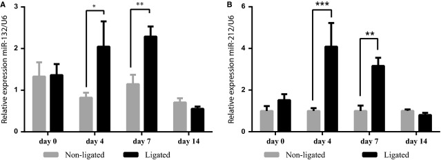
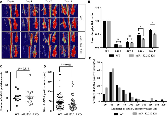
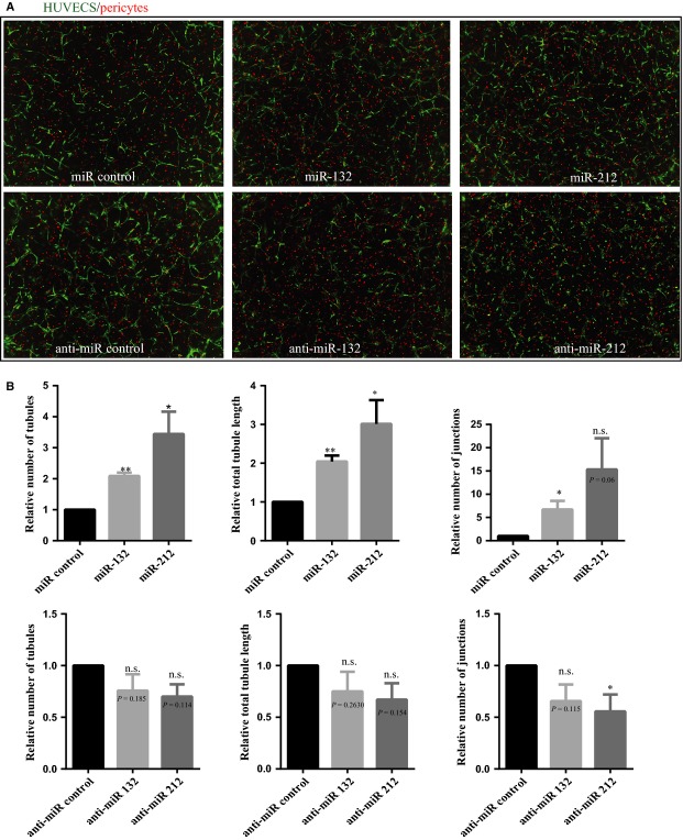
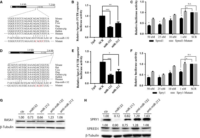
Arteriogenesis is a complicated process induced by increased local shear‐and radial wall‐stress, leading to an increase in arterial diameter. This process is enhanced by growth factors secreted by both inflammatory and endothelial cells in response to physical stress. Although therapeutic promotion of arteriogenesis is of great interest for ischaemic diseases, little is known about the modulation of the signalling cascades via micro RNA s. We observed that miR‐132/212 expression was significantly upregulated after occlusion of the femoral artery. miR‐132/212 knockout (KO) mice display a slower perfusion recovery after hind‐limb ischaemia compared to wildtype (WT) mice. Immunohistochemical analysis demonstrates a clear trend towards smaller collateral arteries in KO mice. Although Ex vivo aortic ring assays score similar number of branches in miR‐132/212 KO mice compared to WT, it can be stimulated with exogenous miR‐132, a dominant member of the miR‐132/212 family. Moreover, in in vitro pericyte‐endothelial co‐culture cell assays, overexpression of miR‐132 and mir‐212 in endothelial cells results in enhanced vascularization, as shown by an increase in tubular structures and junctions. Our results suggested that miR‐132/212 may exert their effects by enhancing the Ras‐Mitogen‐activated protein kinases MAPK signalling pathway through direct inhibition of Rasa1, and Spred1. The miR‐132/212 cluster promotes arteriogenesis by modulating Ras‐MAPK signalling via direct targeting of its inhibitors Rasa1 and Spred1.

Overview

The claims and statistics of this paper haven’t been checked yet.

Run the check — free, no account needed. It reads the open-access text and checks the sources it cites.

A few free checks a day · papers already verified open instantly · results are public

Integrity signals

Registry

Crossref · 10.1111/jcmm.12586

Cites

1 of 1 references verified0 citation errors · 0 not checkable

Statistics

Figures

Figure 1 miR-132/212 expression after hind-limb ischaemia. (A) miR-132 expression as measured by qPCR assays. N = 5, values in the graph are shown as mean ± SEM, *P < 0.05; **P < 0.01. (B) miR-212 expression as measured by qPCR assays. N = 5, values in the graph are shown as mean ± SEM, **P < 0.01; ***P < 0.001.
Figure 1 miR-132/212 expression after hind-limb ischaemia. (A) miR-132 expression as measured by qPCR assays. N = 5, values in the graph are shown as mean ± SEM, *P < 0.05; **P < 0.01. (B) miR-212 expression as measured by qPCR assays. N = 5, values in the graph are shown as mean ± SEM, **P < 0.01; ***P < 0.001.CC BY · PubMed Central
Figure 2 miR-132/212 knockout mice show slower blood flow recovery rate after hindlimb ischaemia. (A) Laser Doppler images of WT and miR-132/212 KO at 0, 4, 7 and 14 days after ligation of femoral artery. Low or no perfusion is displayed as dark blue, whereas the highest degree of perfusion is displayed as red. (B) Quantification of laser Doppler image (Ratio R/L) as shown in A (N = 10 for WT and 13 for KO. Values in the graph are shown as mean ± SEM, *P < 0.05. (C) Quantification of number of αSMA positive vessels as determined by αSMA staining on adductor muscle on day 14. (N = 10 for WT and 13 for KO. Values in the graph are shown as mean ± SEM). (D) Quantification of the diameter of αSMA positive vessels as determined by αSMA staining on adductor muscle on day 14 (N = 10 for WT and 13 for KO. Values in the graph are shown as mean ± SEM). (E) Quantitative analysis the percentage of arteries in different size range. Note the higher percentage in the small vessels (≦400 a.u.) but lower in the larger vessel in the miR-132/212 KO mice (N = 10 for WT and 13 for KO.
Figure 2 miR-132/212 knockout mice show slower blood flow recovery rate after hindlimb ischaemia. (A) Laser Doppler images of WT and miR-132/212 KO at 0, 4, 7 and 14 days after ligation of femoral artery. Low or no perfusion is displayed as dark blue, whereas the highest degree of perfusion is displayed as red. (B) Quantification of laser Doppler image (Ratio R/L) as shown in A (N = 10 for WT and 13 for KO. Values in the graph are shown as mean ± SEM, *P < 0.05. (C) Quantification of number of αSMA positive vessels as determined by αSMA staining on adductor muscle on day 14. (N = 10 for WT and 13 for KO. Values in the graph are shown as mean ± SEM). (D) Quantification of the diameter of αSMA positive vessels as determined by αSMA staining on adductor muscle on day 14 (N = 10 for WT and 13 for KO. Values in the graph are shown as mean ± SEM). (E) Quantitative analysis the percentage of arteries in different size range. Note the higher percentage in the small vessels (≦400 a.u.) but lower in the larger vessel in the miR-132/212 KO mice (N = 10 for WT and 13 for KO.CC BY · PubMed Central
Figure 3 Effect of miR132 and miR212 in HUVECs angiogenesis in co-culture with pericytes. (A) Representative image from HUVECs and pericytes co-culture assay with miR-132 and 212 transfection. HUVECs labelled in green with GFP, pericytes in labelled with PKH26 in red. (B) Quantification of the HUVECs and pericytes co-culture assay with anti-miR-132 and 212 transfection (N = 3, values in the graph are shown as mean ± SEM, *P < 0.05; **P < 0.01).
Figure 3 Effect of miR132 and miR212 in HUVECs angiogenesis in co-culture with pericytes. (A) Representative image from HUVECs and pericytes co-culture assay with miR-132 and 212 transfection. HUVECs labelled in green with GFP, pericytes in labelled with PKH26 in red. (B) Quantification of the HUVECs and pericytes co-culture assay with anti-miR-132 and 212 transfection (N = 3, values in the graph are shown as mean ± SEM, *P < 0.05; **P < 0.01).CC BY · PubMed Central
Figure 4 Identification ofSpred1 and Spry1 as direct miR-132/212 targets by luciferase assay and in cultured HUVECs. (A) The position of predicted miR-132 and miR212 targets of Spred1 by Targetscan and mutant form of 3′UTR as indicated in red. (B) Luciferase assay of Spred1-3′UTR reporter in response to transfection with indicated scramble or microRNA mimics at final concentration of 25 nM. N ≥ 3, values in the graph are shown as mean ± SEM, **P < 0.01; ***P < 0.001. (C) Luciferase activity of wildtype and mutant Spred1-3′UTR in response to different dose of miR-132, N = 6 for WT; N = 3 for mutant. Values in the graph are shown as mean ± SEM *P < 0.05. (D) The position of predicted miR-132 and miR212 targets of Spred1 by Targetscan and mutant form of 3′UTR as indicated in red. (E) Luciferase assay of Spred1-3′UTR reporter in response to transfection with indicated scramble or microRNA mimics at final concentration of 25 nM. N ≥ 3, values in the graph are shown as mean ± SEM; **P < 0.05; ***P < 0.001. (F) Luciferase activity of wildtype and mutant Spred1-3′UTR in response to different final concentration of miR-132 mimics transfection, N = 6 for WT; N = 3 for mutants. Values in the graph are shown as mean ± SEM, *P < 0.05. (G) RASA1 expression after miR-132, miR212 overexpression and inhibition in HUVECs. The number above indicates the relative expression compared with sham normalized by β-Tubulin expression. (H) SPRED1 and SPRY1 expressions after miR-132, miR212 overexpression and inhibition in HUVECs. The number above indicates the relative expression compared with sham normalized by β-Tubulin expression.
Figure 4 Identification ofSpred1 and Spry1 as direct miR-132/212 targets by luciferase assay and in cultured HUVECs. (A) The position of predicted miR-132 and miR212 targets of Spred1 by Targetscan and mutant form of 3′UTR as indicated in red. (B) Luciferase assay of Spred1-3′UTR reporter in response to transfection with indicated scramble or microRNA mimics at final concentration of 25 nM. N ≥ 3, values in the graph are shown as mean ± SEM, **P < 0.01; ***P < 0.001. (C) Luciferase activity of wildtype and mutant Spred1-3′UTR in response to different dose of miR-132, N = 6 for WT; N = 3 for mutant. Values in the graph are shown as mean ± SEM *P < 0.05. (D) The position of predicted miR-132 and miR212 targets of Spred1 by Targetscan and mutant form of 3′UTR as indicated in red. (E) Luciferase assay of Spred1-3′UTR reporter in response to transfection with indicated scramble or microRNA mimics at final concentration of 25 nM. N ≥ 3, values in the graph are shown as mean ± SEM; **P < 0.05; ***P < 0.001. (F) Luciferase activity of wildtype and mutant Spred1-3′UTR in response to different final concentration of miR-132 mimics transfection, N = 6 for WT; N = 3 for mutants. Values in the graph are shown as mean ± SEM, *P < 0.05. (G) RASA1 expression after miR-132, miR212 overexpression and inhibition in HUVECs. The number above indicates the relative expression compared with sham normalized by β-Tubulin expression. (H) SPRED1 and SPRY1 expressions after miR-132, miR212 overexpression and inhibition in HUVECs. The number above indicates the relative expression compared with sham normalized by β-Tubulin expression.CC BY · PubMed Central
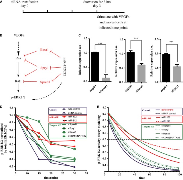
Figure 5 miR-132/212 modulate Ras-MAPK signalling by suppressing Rasa1, Spred1 and Spry1 in HUVECs. (A) Experimental setting for quantitative measure of active ERK1/2 using Bio-plex phosopho-ERK1/2 assay. (B) A working model for miR-132/212 in modulation of Ras-MAPK pathway. (C) Quantification of Spred1, Spry1 or Rasa1 expression level after siRNA transfection against Spred1, Spry1 or Rasa1 in HUVECs, Values in the graph are shown as mean ± SEM, *** P < 0.001 n = 3. (D) Quantification of phosophorylated ERK1/2 level by Bio-plex pro phosopho-ERK1/2 set. Note the sustain ERK1/2 phosphorylation is prolonged after miR-132, 212 transfection or siRNA against Spred1, Spry1, Rasa1 or combinations of the three. (E) Modelling the decay of phosophorylated ERK1/2 level from D.
Figure 5 miR-132/212 modulate Ras-MAPK signalling by suppressing Rasa1, Spred1 and Spry1 in HUVECs. (A) Experimental setting for quantitative measure of active ERK1/2 using Bio-plex phosopho-ERK1/2 assay. (B) A working model for miR-132/212 in modulation of Ras-MAPK pathway. (C) Quantification of Spred1, Spry1 or Rasa1 expression level after siRNA transfection against Spred1, Spry1 or Rasa1 in HUVECs, Values in the graph are shown as mean ± SEM, *** P < 0.001 n = 3. (D) Quantification of phosophorylated ERK1/2 level by Bio-plex pro phosopho-ERK1/2 set. Note the sustain ERK1/2 phosphorylation is prolonged after miR-132, 212 transfection or siRNA against Spred1, Spry1, Rasa1 or combinations of the three. (E) Modelling the decay of phosophorylated ERK1/2 level from D.CC BY · PubMed Central
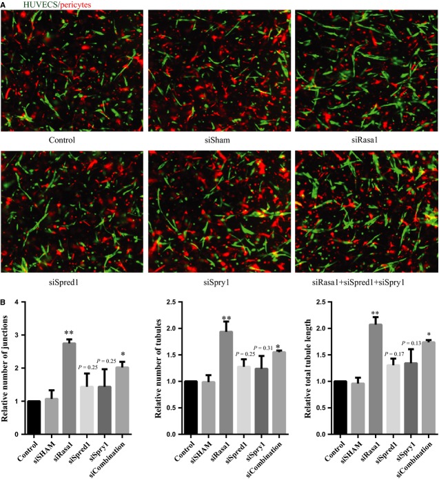
Figure 6 Knockdown targets of miR132 and miR212 Rasa1, Spred1 and Spry1 mimics effect of miR-132 and miR-212 in HUVECs pericytes neovascularization assay. (A) Representative image from HUVECs and pericytes co-culture assay after transfection with siRNAs against Spred1, Spry1, Rasa1 and combination of these three siRNA. (B) Quantification of the number of tubules, junctions and total tubule length in the HUVECs and pericytes co-culture assay after transfection with siRNAs against Spred1, Spry1 and Rasa1, Values in the graph are shown as mean ± SEM, *P < 0.05; **P < 0.01; n = 3.
Figure 6 Knockdown targets of miR132 and miR212 Rasa1, Spred1 and Spry1 mimics effect of miR-132 and miR-212 in HUVECs pericytes neovascularization assay. (A) Representative image from HUVECs and pericytes co-culture assay after transfection with siRNAs against Spred1, Spry1, Rasa1 and combination of these three siRNA. (B) Quantification of the number of tubules, junctions and total tubule length in the HUVECs and pericytes co-culture assay after transfection with siRNAs against Spred1, Spry1 and Rasa1, Values in the graph are shown as mean ± SEM, *P < 0.05; **P < 0.01; n = 3.CC BY · PubMed Central
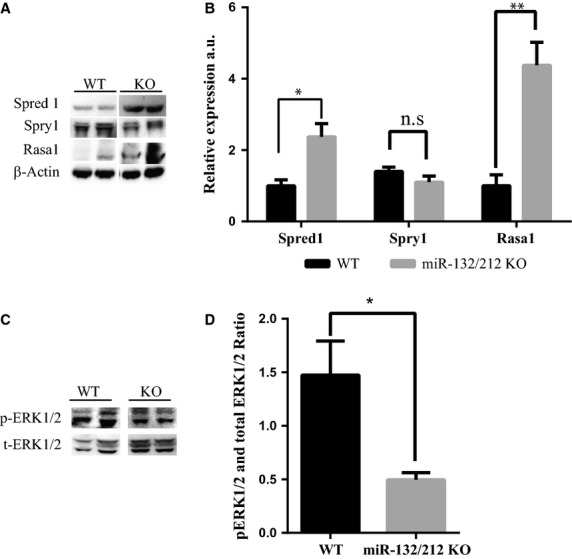
Figure 7 Expression of miR-132/212 targets and phosophorylated ERK1/2 in ischaemia limb. (A) Spred1, Rasa1 and Spry1 expression as determined by Western blot, normalized by β-Actin. (B) Quantification Spred1, Rasa1 and Spry1 expression as determined by Western blot in B. (C) Phosophorylated ERK1/2 expression in the thigh WT and KO mice on 14 days after hindlimb ischaemia as determined by Western blot on day 14. (D) Quantification of phosophorylated ERK1/2 expression in the thigh WT and KO mice on 14 days after hindlimb ischaemia as determined by Western blot, normalized by β-Actin.
Figure 7 Expression of miR-132/212 targets and phosophorylated ERK1/2 in ischaemia limb. (A) Spred1, Rasa1 and Spry1 expression as determined by Western blot, normalized by β-Actin. (B) Quantification Spred1, Rasa1 and Spry1 expression as determined by Western blot in B. (C) Phosophorylated ERK1/2 expression in the thigh WT and KO mice on 14 days after hindlimb ischaemia as determined by Western blot on day 14. (D) Quantification of phosophorylated ERK1/2 expression in the thigh WT and KO mice on 14 days after hindlimb ischaemia as determined by Western blot, normalized by β-Actin.CC BY · PubMed Central

Verification

Claims the paper makes

The claims and statistics of this paper haven’t been checked yet.

Run the check — free, no account needed. It reads the open-access text and checks the sources it cites.

A few free checks a day · papers already verified open instantly · results are public

Claims other papers make about this one

Each is checked against this paper’s own text: does it say what the citing paper says it does?

Self-check

Percentages, sums and reported differences re-computed from the paper’s own numbers.

The self-check needs the paper’s full text.

Citations

Cites

Show all 1 references

Research works printed but not linked: 40

  1. 10.1038/nature10144 · DOI · Paper page
  2. 10.1002/humu.22431 · DOI
  3. 10.1111/j.1582-4934.2006.tb00290.x · DOI
  4. 10.1002/dmrr.2232 · DOI
  5. 10.1016/S0008-6363(01)00546-6 · DOI
  6. 10.1161/01.CIR.0000145142.85645.EA · DOI
  7. 10.1371/journal.pone.0005467 · DOI
  8. 10.1007/s00109-013-1017-1 · DOI
  9. 10.1073/pnas.1105254108 · DOI · Paper page
  10. 10.1038/nm.2186 · DOI · Paper page
  11. 10.1016/j.devcel.2008.07.008 · DOI · Paper page
  12. 10.1161/CIRCULATIONAHA.111.039008 · DOI
  13. 10.1126/science.1174381 · DOI
  14. 10.1161/CIRCRESAHA.107.153916 · DOI
  15. 10.1161/CIRCRESAHA.111.251546 · DOI · Paper page
  16. 10.1074/jbc.M111.322669 · DOI
  17. 10.1096/fj.13-238527 · DOI · Paper page
  18. 10.1172/JCI70230 · DOI
  19. 10.1016/j.ajpath.2012.04.014 · DOI
  20. 10.1038/ng.2990 · DOI
  21. 10.1161/CIRCULATIONAHA.110.000323 · DOI
  22. 10.1161/ATVBAHA.113.303050 · DOI
  23. 10.1161/ATVBAHA.109.197434 · DOI
  24. 10.1093/cvr/cvs003 · DOI
  25. 10.1038/nprot.2011.435 · DOI
  26. 10.1182/blood-2009-05-222364 · DOI
  27. 10.1038/nature08170 · DOI · Paper page
  28. 10.1016/j.ymeth.2012.08.006 · DOI
  29. 10.1038/nprot.2014.043 · DOI
  30. 10.1016/j.biochi.2008.10.010 · DOI
  31. 10.1038/ncb867 · DOI · Paper page
  32. 10.1038/35088082 · DOI
  33. 10.1016/j.devcel.2013.03.019 · DOI
  34. 10.1182/blood-2012-12-474601 · DOI
  35. 10.1007/s00395-008-0760-x · DOI
  36. 10.1016/j.tcb.2005.11.004 · DOI
  37. 10.1002/bies.20632 · DOI
  38. 10.1016/j.molcel.2007.06.017 · DOI · Paper page
  39. 10.1093/eurheartj/ehu344 · DOI
  40. 10.1042/CS20140005 · DOI

Cited by

Sorted by citations

See all 58

Metadata

Authors
Lei, Z., van Mil, A., Brandt, M. M., Grundmann, S., Hoefer, I., Smits, M., el Azzouzi, H., Fukao, T., Cheng, C., Doevendans, P. A., & Sluijter, J. P. G.
Affiliations
  • Division Heart and Lungs Department of Cardiology University Medical Center Utrecht Utrecht The Netherlands
  • Experimental Cardiology Erasmus Medical Center Rotterdam The Netherlands
  • Department of Cardiology and Angiology I University Heart Center Freiburg ‐ Bad Krozingen Freiburg Germany
  • Max Planck Institute of Immunobiology and Epigenetics Freiburg Germany
Venue
Journal of Cellular and Molecular Medicine
Publisher
Wiley
Published
2015-05-06
Volume
19
Issue
8
Pages
1994-2005
ISSN
  • 1582-1838
  • 1582-4934
Cited by
58
References
1
Language
en
Funders
  • Netherlands CardioVascular Research Initiative
  • Dutch Heart Foundation
  • Dutch Federation of University Medical Centers
  • Netherlands Organization for Health Research and Development
  • Royal Netherlands Academy of Sciences

About these checks

Registry, retraction, Cites and statistics results are computed, not judged, with one exception: a printed reference that no index lists is read once by a model before it counts as a citation error. Claims are extracted by a model and kept only when their quote appears verbatim in the text; each cited source is then checked, with a model’s help, for whether it says what the claim relies on. Those verdicts can be wrong — read the claim’s quote and the cited source before relying on one.

Statistics and arithmetic are recomputed, not judged.

Confidence bands are shown only for fields calibrated on our annotated test set. None is calibrated yet, so no band is shown.

What counts as a claim ›How we measure confidence ›How we check this ›

Are you an author, or do you see a problem with this page? Request a review

How to cite

Cite CoChat for the checks on this page, and the paper for its own findings.

This page
CoChat. (2026). Citation check: Micro RNA‐132/212 family enhances arteriogenesis after hindlimb ischaemia through modulation of the Ras‐MAPK pathway. https://app.cochat.ai/papers/10.1111/jcmm.12586
The paper
Lei, Z., van Mil, A., Brandt, M. M., Grundmann, S., Hoefer, I., Smits, M., el Azzouzi, H., Fukao, T., Cheng, C., Doevendans, P. A., & Sluijter, J. P. G. (2015). Micro RNA‐132/212 family enhances arteriogenesis after hindlimb ischaemia through modulation of the Ras‐MAPK pathway. Journal of Cellular and Molecular Medicine. https://doi.org/10.1111/jcmm.12586