

Programmable and contractile materials through cell encapsulation in fibrous hydrogel assemblies
Open accessPublished version of a preprint
The DOI matches its Crossref record; 77 of the 77 references it cites were found in a registry or an index.
Abstract
Synthetic fibrous materials were engineered to contract under cellular forces similar to natural extracellular matrix.
Overview
The claims and statistics of this paper haven’t been checked yet.
Free: it reads the open-access text and checks the first 5 claims that cite a source we can check. Create a free account to check every claim.
Integrity signals
Registry
Cites
Statistics
Figures
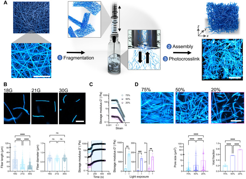

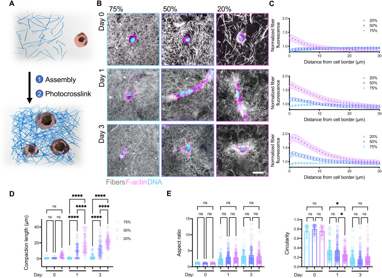


Verification
Claims the paper makes
The claims and statistics of this paper haven’t been checked yet.
Free: it reads the open-access text and checks the first 5 claims that cite a source we can check. Create a free account to check every claim.
Claims other papers make about this one
Each is checked against this paper’s own text: does it say what the citing paper says it does?
The cited source doesn’t support the claim as written.
In the citing paper
“Similar phenomena were observed in a study reported by Burdick et al., (2021) with higher fiber densities yielding higher strain [23].”
From Development of silk microfiber-reinforced bioink for muscle tissue engineering and in situ printing by a handheld 3D printer (2025) · cites this paper as [23]
Checked by a reasoning model — may be wrong.
Experimental — accuracy not yet measuredThe source backs part of the claim, not all of it — or backs it for a different population, condition or comparison.
In the citing paper
“Fiber-reinforced composites show enhanced mechanical strength and fiber-induced cell alignment in various cell studies[23, 26–30].”
From Development of silk microfiber-reinforced bioink for muscle tissue engineering and in situ printing by a handheld 3D printer (2025) · cites this paper as [23]
Experimental — accuracy not yet measuredThe source backs part of the claim, not all of it — or backs it for a different population, condition or comparison.
In the citing paper
“This finding is consistent with previous studies that have demonstrated similar outcomes, including fiber-oriented cell alignment [23, 27].”
From Development of silk microfiber-reinforced bioink for muscle tissue engineering and in situ printing by a handheld 3D printer (2025) · cites this paper as [23]
Experimental — accuracy not yet measuredThe source backs part of the claim, not all of it — or backs it for a different population, condition or comparison.
In the citing paper
“Literature suggests that higher fiber densities can impediment cell recruitment due to increased mechanical resistance, resulting in reduced cell spreading and increased cell circularity [23].”
From Development of silk microfiber-reinforced bioink for muscle tissue engineering and in situ printing by a handheld 3D printer (2025) · cites this paper as [23]
Experimental — accuracy not yet measuredThe cited source says what the claim attributes to it.
In the citing paper
“Fiber-reinforced hydrogel composites recently gained attention with their unique fibrous structure mimicking native tissue architecture [23–25].”
From Development of silk microfiber-reinforced bioink for muscle tissue engineering and in situ printing by a handheld 3D printer (2025) · cites this paper as [23]
Self-check
Percentages, sums and reported differences re-computed from the paper’s own numbers.
The self-check needs the paper’s full text.
Citations
Cites
Myofibroblasts and mechano-regulation of connective tissue remodelling
Tomasek, J. J., Gabbiani, G., Hinz, B., Chaponnier, C., & Brown, R. A. (2002) · Nature Reviews Molecular Cell Biology · doi:10.1038/nrm809 · Cited by 4,290 · cited 1×
“For example, cell contractility is responsible for many self-assembly or morphogenic processes in vivo, such as shaping tissues in embryonic development (8) and closing wounds during healing (9).”
BackgroundA practical guide to hydrogels for cell culture
Caliari, S. R., & Burdick, J. A. (2016) · Nature Methods · doi:10.1038/nmeth.3839 · Cited by 2,075
Self-citationNo evidence that carotenoid pigments boost either immune or antioxidant defenses in a songbird
Koch, R. E., Kavazis, A. N., Hasselquist, D., Hood, W. R., Zhang, Y., Toomey, M. B., & Hill, G. E. (2018) · Nature Communications · doi:10.1038/s41467-018-02974-x · Cited by 1,924
3D bioprinting of collagen to rebuild components of the human heart
Lee, A., Hudson, A. R., Shiwarski, D. J., Tashman, J. W., Hinton, T. J., Yerneni, S., Bliley, J. M., Campbell, P. G., & Feinberg, A. W. (2019) · Science · doi:10.1126/science.aav9051 · Cited by 1,782
Nonlinear elasticity in biological gels
Storm, C., Pastore, J. J., MacKintosh, F. C., Lubensky, T. C., & Janmey, P. A. (2005) · Nature · doi:10.1038/nature03521 · Cited by 1,703
Self-citationSLIM: Simultaneous Logic-in-Memory Computing Exploiting Bilayer Analog OxRAM Devices
Kingra, S. K., Parmar, V., Chang, C.-C., Hudec, B., Hou, T.-H., & Suri, M. (2020) · Scientific Reports · doi:10.1038/s41598-020-59121-0 · Cited by 1,439
Advanced maturation of human cardiac tissue grown from pluripotent stem cells
Ronaldson-Bouchard, K., Ma, S. P., Yeager, K., Chen, T., Song, L., Sirabella, D., Morikawa, K., Teles, D., Yazawa, M., & Vunjak-Novakovic, G. (2018) · Nature · doi:10.1038/s41586-018-0016-3 · Cited by 1,272
Yang, B.-J., Han, X.-X., Yin, L.-L., Xing, M.-Q., Xu, Z.-H., & Xue, H.-W. (2016) · Nature Communications · doi:10.1038/ncomms11388 · Cited by 1,202
Synthetic alternatives to Matrigel
Aisenbrey, E. A., & Murphy, W. L. (2020) · Nature Reviews Materials · doi:10.1038/s41578-020-0199-8 · Cited by 1,016 · cited 1×
“Furthermore, other ECM-derived matrices, such as Matrigel and decellularized ECM, are widely used in cell culture, in tissue engineering, and even as clinical therapies; however, their batch variability and animal sourcing are limiting (13).”
BackgroundBiomanufacturing of organ-specific tissues with high cellular density and embedded vascular channels
Skylar-Scott, M. A., Uzel, S. G. M., Nam, L. L., Ahrens, J. H., Truby, R. L., Damaraju, S., & Lewis, J. A. (2019) · Science Advances · doi:10.1126/sciadv.aaw2459 · Cited by 943
Cited by
Zhang, Y. S., Haghiashtiani, G., Hübscher, T., Kelly, D. J., Lee, J. M., Lutolf, M., McAlpine, M. C., Yeong, W. Y., Zenobi-Wong, M., & Malda, J. (2021) · Nature Reviews Methods Primers · doi:10.1038/s43586-021-00073-8 · Cited by 391
Engineered hydrogels for mechanobiology
Blache, U., Ford, E. M., Ha, B., Rijns, L., Chaudhuri, O., Dankers, P. Y. W., Kloxin, A. M., Snedeker, J. G., & Gentleman, E. (2022) · Nature Reviews Methods Primers · doi:10.1038/s43586-022-00179-7 · Cited by 227
Light-based vat-polymerization bioprinting
Levato, R., Dudaryeva, O., Garciamendez-Mijares, C. E., Kirkpatrick, B. E., Rizzo, R., Schimelman, J., Anseth, K. S., Chen, S., Zenobi-Wong, M., & Zhang, Y. S. (2023) · Nature Reviews Methods Primers · doi:10.1038/s43586-023-00231-0 · Cited by 176
Ding, A., Tang, F., & Alsberg, E. (2025) · Chemical Reviews · doi:10.1021/acs.chemrev.4c00070 · Cited by 165
Granular Hydrogels in Biofabrication: Recent Advances and Future Perspectives
Daly, A. C. (2023) · Advanced Healthcare Materials · doi:10.1002/adhm.202301388 · Cited by 153
Biomedical applications of stimuli-responsive “smart” interpenetrating polymer network hydrogels
Wu, J., Xue, W., Yun, Z., Liu, Q., & Sun, X. (2024) · Materials Today Bio · doi:10.1016/j.mtbio.2024.100998 · Cited by 178
“They have found application in diverse fields, including food application [2], drug delivery [3 – 7], gene editing [8], biosensors [9,10], cell encapsulation [11,12], and biomedical implants [13], garnering significant attention and widespread utilization.”
BackgroundLithography-based 3D printing of hydrogels
Dhand, A. P., Davidson, M. D., & Burdick, J. A. (2024) · Nature Reviews Bioengineering · doi:10.1038/s44222-024-00251-9 · Cited by 136
“…also be seeded within 3D-printed hydrogel structures, for example, seeding collagen gels loaded with fibroblasts or mesenchymal stem cells within DLP-printed micropillar hydrogel moulds has been used to create engineered microtissues that are useful for disease modelling or drug discovery 47,144 .”
Self-citationProgramming hydrogels to probe spatiotemporal cell biology
Qazi, T. H., Blatchley, M. R., Davidson, M. D., Yavitt, F. M., Cooke, M. E., Anseth, K. S., & Burdick, J. A. (2022) · Cell stem cell · doi:10.1016/j.stem.2022.03.013 · Cited by 105
Self-citationAdvancing Synthetic Hydrogels through Nature‐Inspired Materials Chemistry
Soliman, B. G., Nguyen, A. K., Gooding, J. J., & Kilian, K. A. (2024) · Advanced Materials · doi:10.1002/adma.202404235 · Cited by 111
Magnetically‐Assisted 3D Bioprinting of Anisotropic Tissue‐Mimetic Constructs
Pardo, A., Bakht, S. M., Gómez‐Florit, M., Rial, R., Monteiro, R. F., Teixeira, S. P. B., Taboada, P., Reis, R. L., Domingues, R. M. A., & Gomes, M. E. (2022) · Advanced Functional Materials · doi:10.1002/adfm.202208940 · Cited by 93
Li, S., Li, X., Xu, Y., Fan, C., Li, Z. A., Zheng, L., Luo, B., Li, Z., Lin, B., Zha, Z., et al. (2023) · Bioactive Materials · doi:10.1016/j.bioactmat.2023.09.012 · Cited by 86
Middle-out methods for spatiotemporal tissue engineering of organoids
Blatchley, M. R., & Anseth, K. S. (2023) · Nature Reviews Bioengineering · doi:10.1038/s44222-023-00039-3 · Cited by 94
3D Printing of Self‐Assembling Nanofibrous Multidomain Peptide Hydrogels
Farsheed, A. C., Thomas, A. J., Pogostin, B. H., & Hartgerink, J. D. (2023) · Advanced Materials · doi:10.1002/adma.202210378 · Cited by 84
Miksch, C. E., Skillin, N. P., Kirkpatrick, B. E., Hach, G. K., Rao, V. V., White, T. J., & Anseth, K. S. (2022) · Small · doi:10.1002/smll.202200951 · Cited by 74
Lose the Stress: Viscoelastic Materials for Cell Engineering
Carvalho, E. M., & Kumar, S. (2021) · Acta Biomaterialia · doi:10.1016/j.actbio.2022.03.058 · Cited by 63
“A relatively new fabrication strategy based on fragmenting and reassembling electrospun fibers offers an additional degree of tunability [102, 103].”
Pramanick, A., Hayes, T., Sergis, V., McEvoy, E., Pandit, A. S., & Daly, A. C. (2024) · Advanced Functional Materials · doi:10.1002/adfm.202414559 · Cited by 65
Loebel, C., Saleh, A. M., Jacobson, K. R., Daniels, R., Mauck, R. L., Calve, S., & Burdick, J. A. (2022) · Nature Protocols · doi:10.1038/s41596-021-00652-9 · Cited by 57
Self-citation4D printing for biomedical applications
Mandal, A., & Chatterjee, K. (2024) · Journal of Materials Chemistry B · doi:10.1039/d4tb00006d · Cited by 62
Precision Hydrogels for the Study of Cancer Cell Mechanobiology
Sievers, J., Mahajan, V., Welzel, P. B., Werner, C., & Taubenberger, A. V. (2023) · Advanced Healthcare Materials · doi:10.1002/adhm.202202514 · Cited by 53
Microinterfaces in biopolymer-based bicontinuous hydrogels guide rapid 3D cell migration
Xu, K. L., Caprio, N. D., Fallahi, H., Dehghany, M., Davidson, M. D., Laforest, L. G., Cheung, B. C. H., Zhang, Y., Wu, M., Shenoy, V. B., et al. (2024) · Nature Communications · doi:10.1038/s41467-024-46774-y · Cited by 50
Self-citationSynergistic Influence of Fibrous Pattern Orientation and Modulus on Cellular Mechanoresponse
Sun, Q., Pan, X., Wang, P., & Wei, Q. (2024) · Nano Letters · doi:10.1021/acs.nanolett.4c01352 · Cited by 45
Recent advances in collagen-based hydrogels: Materials, preparation and applications
She, J., Liu, J., Mu, Y., Lv, S., Tong, J., Liu, L., He, T., Wang, J., & Wei, D. (2024) · Reactive and Functional Polymers · doi:10.1016/j.reactfunctpolym.2024.106136 · Cited by 55
New dimensions of electrospun nanofiber material designs for biotechnological uses
Kamaraj, M., Moghimi, N., Chen, J., Morales, R., Chen, S., Khademhosseini, A., & John, J. V. (2024) · Trends in Biotechnology · doi:10.1016/j.tibtech.2023.11.008 · Cited by 44
Fibrous hydrogels by electrospinning: Novel platforms for biomedical applications
Lee, J. W., & Song, K. H. (2023) · Journal of Tissue Engineering · doi:10.1177/20417314231191881 · Cited by 48
Skillin, N. P., Kirkpatrick, B. E., Herbert, K. M., Nelson, B. R., Hach, G. K., Günay, K. A., Khan, R. M., DelRio, F. W., White, T. J., & Anseth, K. S. (2024) · Science Advances · doi:10.1126/sciadv.adn0235 · Cited by 40
Stoecker, L., Cedillo‐Servín, G., König, N. F., de Graaf, F. V., García‐Jiménez, M., Hofmann, S., Ito, K., Wentzel, A. S., & Castilho, M. D. (2025) · Advanced Materials · doi:10.1002/adma.202410292 · Cited by 45
Hierarchical Design of Tissue‐Mimetic Fibrillar Hydrogel Scaffolds
Pardo, A., Gómez‐Florit, M., Davidson, M. D., Öztürk-Öncel, M. Ö., Domingues, R. M. A., Burdick, J. A., & Gomes, M. E. (2024) · Advanced Healthcare Materials · doi:10.1002/adhm.202303167 · Cited by 37
Self-citationTunable Mesoscopic Collagen Island Architectures Modulate Stem Cell Behavior
Nguyen, R. Y., Cabral, A. T., Rossello‐Martinez, A., Zulli, A., Gong, X., Zhang, Q., Yan, J. Q., & Mak, M. (2023) · Advanced Materials · doi:10.1002/adma.202207882 · Cited by 44
Ohnsorg, M. L., Mash, K. M., Khang, A., Rao, V. V., Kirkpatrick, B. E., Bera, K., & Anseth, K. S. (2024) · Advanced Materials · doi:10.1002/adma.202403198 · Cited by 33
Yang, P., Boer, G. E., Snow, F., Williamson, A., Cheeseman, S., Samarasinghe, R. M., Rifai, A., Priyam, A., Elnathan, R., Guijt, R. M., et al. (2025) · Chemical Engineering Journal · doi:10.1016/j.cej.2025.159295 · Cited by 37
Engineering complex tissue-like microenvironments with biomaterials and biofabrication
Miklosic, G., Ferguson, S. J., & D’Este, M. (2024) · Trends in biotechnology · doi:10.1016/j.tibtech.2024.03.008 · Cited by 31
Programmed shape transformations in cell-laden granular composites
Caprio, N. D., Hughes, A. J., & Burdick, J. A. (2025) · Science Advances · doi:10.1126/sciadv.adq5011 · Cited by 29
Self-citationBio-gel nanoarchitectonics in tissue engineering
Song, J., Lyu, W., Kawakami, K., & Ariga, K. (2024) · Nanoscale · doi:10.1039/d4nr00609g · Cited by 28
Listen to Your Gut: Key Concepts for Bioengineering Advanced Models of the Intestine
Cameron, O., Neves, J. F., & Gentleman, E. (2023) · Advanced Science · doi:10.1002/advs.202302165 · Cited by 28
Reconfigurable scaffolds for adaptive tissue regeneration
Peng, M., Zhao, Q., Wang, M., & Du, X. (2023) · Nanoscale · doi:10.1039/d3nr00281k · Cited by 26
Local governance and occupational choice among young people: First evidence from Vietnam
Tran, T. Q., Tran, A. L., Pham, T. M., & Vu, H. V. (2018) · Children and Youth Services Review · doi:10.1016/j.childyouth.2018.01.019 · Cited by 28
Suspension bath bioprinting and maturation of anisotropic meniscal constructs
Prendergast, M. E., Heo, S., Mauck, R. L., & Burdick, J. A. (2023) · Biofabrication · doi:10.1088/1758-5090/acc3c3 · Cited by 28
“Our previous work demonstrates a method to address these concerns, by aligning synthetic fibers with shear forces during extrusion printing to direct cell alignment in three-dimensional (3D) constructs [22, 23].”
InfluentialSelf-citationAdaptive immunity of materials: Implications for tissue healing and regeneration
Lee, J.-H., Shin, S.-J., Lee, J. H., Knowles, J. C., Lee, H.-H., & Kim, H.-W. (2024) · Bioactive Materials · doi:10.1016/j.bioactmat.2024.07.027 · Cited by 21
“4B) [90 – 93].”
Cell contractile forces drive spatiotemporal morphogenesis in 4D bioprinted living constructs
Ding, A., Cleveland, D. S., Gasvoda, K. L., & Alsberg, E. (2025) · Matter · doi:10.1016/j.matt.2025.102413 · Cited by 20
3D extrusion bioprinting: rational bioink design and advanced fabrication techniques
Jergitsch, M., & Mateos‐Timoneda, M. A. (2025) · Trends in biotechnology · doi:10.1016/j.tibtech.2025.06.008 · Cited by 28
Kamaraj, M., Rezayof, O., Barer, A., Kim, H., Moghimi, N., Joshi, A., Dokmeci, M. R., Khademhosseini, A., Alambeigi, F., & John, J. V. (2025) · Biomaterials Advances · doi:10.1016/j.bioadv.2024.214057 · Cited by 17
Nanoengineered extrusion-aligned tract bioprinting enables functional repair of spinal cord injuries
Huang, W., Chen, S., Li, K., Ding, Y., Zhan, B., Zhao, D., Xu, G., Guo, H., Sun, C., Yuan, R., et al. (2026) · Cell stem cell · doi:10.1016/j.stem.2025.12.021 · Cited by 22
Zhang, Q., Wang, X., Cong, Y., Kang, Y., Wu, Z., & Li, L. (2022) · ACS Applied Materials & Interfaces · doi:10.1021/acsami.2c00460 · Cited by 17
3D Biofabrication of Microporous Hydrogels for Tissue Engineering
Liu, Z., Wu, J., Luo, Z., Hou, Y., Xuan, L., Changyi, X., Chang, J., Zhang, D., Zheng, G., Guo, J., et al. (2024) · Advanced Healthcare Materials · doi:10.1002/adhm.202403583 · Cited by 20
Cedillo‐Servín, G., Al-Jehani, E. A. A., Rossy, T., Teixeira, S. P. B., Sage, F., Domingues, R. M. A., Raman, R., & Castilho, M. D. (2025) · Trends in biotechnology · doi:10.1016/j.tibtech.2025.05.004 · Cited by 16
Engineering Multi‐Scale Organization for Biotic and Organic Abiotic Electroactive Systems
Yao, Z., Lundqvist, E. M., Kuang, Y., & Ardoña, H. A. M. (2023) · Advanced Science · doi:10.1002/advs.202205381 · Cited by 16
Anisotropic Liesegang pattern for the nonlinear elastic biomineral-hydrogel complex
Choi, W., Lee, M., Yong, H., Heo, D., Jun, T., Ryu, H., Kim, J., Cui, D., Ryu, D. Y., Lee, S., et al. (2024) · Science Advances · doi:10.1126/sciadv.adl3075 · Cited by 15
“Researches show that nonlinear elastic HD fibers feature excellent cellular affinity ( 33 , 34 ).”
Wang, S., Zhu, Y., Yu, J., Wang, L., & Zheng, Z. (2024) · Thin-Walled Structures · doi:10.1016/j.tws.2024.112405 · Cited by 13
Photochemistry as a tool for dynamic modulation of hydrogel mechanics
Major, G. S., Joukhdar, H., Choi, Y. S., Rnjak‐Kovacina, J., Wise, S. G., Ju, L. A., Cox, T. R., Xu, C., Yeo, G. C., Young, J. L., et al. (2025) · Cell Reports Physical Science · doi:10.1016/j.xcrp.2024.102366 · Cited by 16
Tissue Engineering with Mechanically Induced Solid‐Fluid Transitions
Mailand, E., Özelçi, E., Kim, J., Rüegg, M., Chaliotis, O., Märki, J., Bouklas, N., & Sakar, M. S. (2021) · Advanced Materials · doi:10.1002/adma.202106149 · Cited by 15
Tunable Synthesis of Hydrogel Microfibers via Interfacial Tetrazine Ligation
George, O. J., Song, J., Benson, J. M., Fang, Y., Zhang, H., Burris, D. L., Fox, J. M., & Jia, X. (2022) · Biomacromolecules · doi:10.1021/acs.biomac.2c00504 · Cited by 13
Impact of Novel Teflon-DCA Nanogel Matrix on Cellular Bioactivity
Kovačević, B., Ionescu, C. M., Wagle, S. R., Jones, M., Lewkowicz, M., Wong, E. Y. M., Đanić, M., Mikov, M., Mooranian, A., & Al‐Salami, H. (2022) · Journal of Pharmaceutical Sciences · doi:10.1016/j.xphs.2022.09.008 · Cited by 13
Kirkpatrick, B. E., Dhand, A. P., Hibbard, L. P., Jaeschke, M. W., Yendamuri, T., Nelson, B. R., Lee, J. S., Bera, K., Zlotnick, H. M., Fox, C. A., et al. (2025) · Matter · doi:10.1016/j.matt.2025.102524 · Cited by 18
Self-citationClaxton, N. L., Luse, M. A., Isakson, B. E., & Highley, C. B. (2024) · ACS Biomaterials Science & Engineering · doi:10.1021/acsbiomaterials.4c01563 · Cited by 14
Huang, T., Wang, G., Shahbazi, M., Bai, Y., Zhang, J., Feng, G., Asadian, E., Ghorbani-Bidkorpeh, F., Yang, Z., Li, Y., et al. (2022) · Advanced Functional Materials · doi:10.1002/adfm.202210627 · Cited by 11
Facile Physicochemical Reprogramming of PEG‐Dithiolane Microgels
Nelson, B. R., Kirkpatrick, B. E., Skillin, N. P., Caprio, N. D., Lee, J. S., Hibbard, L. P., Hach, G. K., Khang, A., White, T. J., Burdick, J. A., et al. (2023) · Advanced Healthcare Materials · doi:10.1002/adhm.202302925 · Cited by 11
Self-citationMa, S., Zhao, G., Sui, S., Chen, X., Wu, L., Wang, T., Xu, W., Lu, Z., Wang, A., Wu, X., et al. (2025) · Frontiers in Immunology · doi:10.3389/fimmu.2025.1580108 · Cited by 12
Disodium Cromoglycate Templates Anisotropic Short-Chain PEG Hydrogels
Chen, J., & Luo, Y. (2024) · ACS Applied Materials & Interfaces · doi:10.1021/acsami.4c07181 · Cited by 10
Development of a multifunctional, injectable biomaterial using hyaluronan as a bioactive nanocarrier
Paone, L. S., Jin, Y., Bouyer, J., Fischer, I., & Galie, P. A. (2025) · Biomaterials · doi:10.1016/j.biomaterials.2025.123505 · Cited by 10
Advanced materials technologies to unravel mechanobiological phenomena
Kim, H. S., Taghizadeh, A. A., Taghizadeh, M., & Kim, H. (2023) · Trends in biotechnology · doi:10.1016/j.tibtech.2023.08.002 · Cited by 10
Gold nanocluster decorated fibrous substrate for photo-modulated cellular growth
Li, B., Kang, Y., Wang, X., Zhang, P., & Li, L. (2023) · Journal of Materials Chemistry C · doi:10.1039/d2tc05194j · Cited by 10
Ghosh, A., Bera, A. K., Ghosh, S., Singh, V., Basu, S., & Pati, F. (2024) · Biofabrication · doi:10.1088/1758-5090/ad9409 · Cited by 12
“This also illustrates that the hLMSC encapsulated or top-seeded on the printed constructs differentiated to ker-atocytes, maintain their keratocyte phenotype and secret native-like ECM and will probably promote remodeling and regeneration of native tissue [118– 121].”
Cell‐Sheet Shape Transformation by Internally‐Driven, Oriented Forces
Huang, J., Chen, J., & Luo, Y. (2025) · Advanced Materials · doi:10.1002/adma.202416624 · Cited by 10
Ding, A., Gasvoda, K. L., Cleveland, D. S., Ayyagari, S., & Alsberg, E. (2025) · Advanced Science · doi:10.1002/advs.202507288 · Cited by 28
Gruhn, T., Monsalve, C. O., Müller, C., Heid, S., Boccaccini, A. R., & Salehi, S. (2023) · Bioengineering · doi:10.3390/bioengineering10040448 · Cited by 8
Cell Contractile Forces Drive Spatiotemporal Morphing in 4D Bioprinted Living ConstructsPreprint
Cleveland, D. S., Gasvoda, K. L., Ding, A., & Alsberg, E. (2024) · bioRxiv (Cold Spring Harbor Laboratory) · doi:10.1101/2024.09.22.613990 · Cited by 8
ECM-inspired mechanically robust hydrogels for diabetic wound regeneration
Ullah, S., Khattak, S., Rahim, F., Zheng, Q., Ma, J., Zahra, Q. U. A., Xu, H., & Shen, J. (2025) · Coordination Chemistry Reviews · doi:10.1016/j.ccr.2025.217456 · Cited by 10
Linguistic Analysis Identifies Emergent Biomaterial Fabrication Trends for Orthopaedic Applications
Locke, R. C., Zlotnick, H. M., Stoeckl, B. D., Fryhofer, G. W., Galarraga, J. H., Dhand, A. P., Zgonis, M. H., Carey, J. L., Burdick, J. A., & Mauck, R. L. (2023) · Advanced Healthcare Materials · doi:10.1002/adhm.202202591 · Cited by 6
Self-citationMoghaddasi, M., Oktay, B., Bingöl, A. B., Yanıkoglu, R., Çiftçi, F., & Üstündağ, C. B. (2025) · Progress in Additive Manufacturing · doi:10.1007/s40964-025-01245-8 · Cited by 6
“11e) [114], while terpyridine-linked PNI-PAM microgels exhibited cyclical swelling–de-swelling under temperature and pH changes, providing dynamic shape adaptation for soft robotics and biosensors (Fig.”
InfluentialNeuhäusler, A., Lindner, N., & Blaeser, A. (2026) · Materials Today Bio · doi:10.1016/j.mtbio.2026.102799 · Cited by 5
“In combination with a hydrophilic and patterned surface chemistry cell adhesion excels [99].Exemplary, human mesenchymal stem cells were able to reorganize and contract on gelatin fiber mats which expose binding motifs, a hydrophilic and patterned surface [54].”
Controlling 3D Contractility via Engineered Fibrous Hydrogel Composites
Xu, K. L., Zhang, Y., DeFoe, A., Kotsaris, G., Stoeckl, B. D., Davidson, M. D., Burdick, J. A., & Mauck, R. L. (2026) · Advanced Functional Materials · doi:10.1002/adfm.202524101 · Cited by 6
“To address this, we recently generated fragmented fibers from electrospun norbornene-modified hyaluronic acid (NorHA) [19].”
InfluentialSelf-citationFibrillar Bundles as Fibrous Filler Materials for Attaining Cell Anisotropy in Bioprinting
Heilig, S., Lamberger, Z., Sprenger, L., Priebe, V., Mussoni, C., Docheva, D., Andelovic, K., Gröll, J., Salehi, S., Lang, G., et al. (2025) · Advanced Healthcare Materials · doi:10.1002/adhm.202503767 · Cited by 6
Wang, K., Zheng, B., Wu, P., Lai, J., Zhang, H., Zhao, W., Jin, X., Ma, A., Chen, W., Liu, H., et al. (2024) · Composites Part A Applied Science and Manufacturing · doi:10.1016/j.compositesa.2024.108274 · Cited by 4
Conductive Microfibers Improve Stem Cell‐Derived Cardiac Spheroid Maturation
Gonzalez, G., Molley, T. G., LaMontagne, E., Balayan, A., Holman, A. R., & Engler, A. J. (2024) · Journal of Biomedical Materials Research Part A · doi:10.1002/jbm.a.37856 · Cited by 4
Chong, S. W., Sardharwalla, J., Masonsong, G. S. P., Cosgrove, J., Katselas, A., Gresham, I. J., Bilek, M., Shen, Y., Neto, C., & Vigolo, D. (2025) · Advanced Materials Technologies · doi:10.1002/admt.202501457 · Cited by 4
Nakamura, K., Caprio, N. D., Taasan, J. T., Crosby, C. O., & Burdick, J. A. (2025) · Advanced Science · doi:10.1002/advs.202512971 · Cited by 4
Self-citationEngineered hydrogels for mechanobiology
(2022) · Nature Reviews Methods Primers · doi:10.1038/s43586-022-00189-5 · Cited by 3
Structure formation of rod-like fillers in a contraction flow
Gruhn, T., Monsalve, C. O., & Salehi, S. (2023) · Physics of Fluids · doi:10.1063/5.0143006 · Cited by 3
Engineering a microfibrous composite hydrogel with cell actuation‐mediated contractile properties
Gao, D., Glahn, J. Z., & Hsia, H. C. (2023) · Journal of Applied Polymer Science · doi:10.1002/app.54736 · Cited by 3
Pandolfi, P., Zhang, H., Nadalin, Y. E., Prasetyo, M. E., Toubian, A. A., Wu, B. M., & Kamei, D. T. (2022) · Soft Matter · doi:10.1039/d2sm01014c · Cited by 3
Engineering Highly Cellularized Living Materials via Mechanical Agitation
Bahmani, A., Liu, S., Johnston, E., Wan, W., Lan, X., Ning, T., Nottegar, A., Huo, R., Jiang, S., & Li, J. (2025) · Advanced Functional Materials · doi:10.1002/adfm.202515436 · Cited by 2
DeFoe, A., Toth, J. M., Dhand, A. P., Deloney, L., Obenreder, M. C., Shenoy, V. B., & Burdick, J. A. (2025) · APL Bioengineering · doi:10.1063/5.0289256 · Cited by 2
Self-citationJin, Z., Huang, Z., Huang, L., He, H., Lan, N., Huang, C., Liu, S., Wei, Q., Zheng, L., Qin, Z., et al. (2026) · International Journal of Biological Macromolecules · doi:10.1016/j.ijbiomac.2026.150695 · Cited by 2
Toward 4D printed functional soft tissues
Shabazz, A., Fitlin, J., Orozco-Contreras, H., Guo, S., Zhang, L. G., & Fisher, J. P. (2026) · Acta Biomaterialia · doi:10.1016/j.actbio.2026.02.035 · Cited by 5
Mechanical gating of tendon fibrogenic transcription in systemic sclerosis
Hussien, A. A., Knell, R., Wunderli, S. L., Aronoff, M. R., Niederöst, B., Renoux, F., Hukara, A., Foolen, J., Wennemers, H., Distler, O., et al. (2026) · Nature Communications · doi:10.1038/s41467-026-70395-2 · Cited by 2
“The shape of micro-tissues can be controlled by adjusting the geometry and spacing of the rigidpillars 41 ,whilethetissuecontractileforcescanbequanti fi edusing beam theory or Hooke ’ s law 42 .”
Microengineered Gradient Hydrogels for Mechanobiology
Chong, S. W., Ashok, D., Waterhouse, A., Bilek, M., & Vigolo, D. (2026) · Advanced Healthcare Materials · doi:10.1002/adhm.202600004 · Cited by 1
“Although ECM stiffness is established as a major regulator of cell behavior, recent studies have highlighted viscoelasticity [24, 190] and fibrous microstructure [191] as key emerging matrix cues influencing cell function and behavior.”
Ordered materials meet living cells: Engineering alignment and programming force actuation
Luo, Y., Parmar, T., Gu, M., Marchetti, M. C., & Luo, Y. (2026) · Biophysics Reviews · doi:10.1063/5.0287774 · Cited by 2
Gonçalves, R. C., Oliveira, M., & Mano, J. F. (2026) · Advanced Functional Materials · doi:10.1002/adfm.76363 · Cited by 2
“The ability of human MSCs to fold materials was previously observed in soft fibrous hydrogels and thin films, leading to the formation of dynamic constructs with spontaneous deformation or the development of spheroidal structures with minimized necrotic core formation [38–40].”
Sustainable hydrogel-based cell therapy
Mokhtarinia, K., Rezvanian, P., & Masaeli, E. (2023) · Elsevier eBooks · doi:10.1016/b978-0-323-91753-7.00009-0 · Cited by 1
Popescu, R. C., Călin, B. Ş., Tanasă, E., Vasile, E., Mihăilescu, M., & Păun, I. A. (2023) · Frontiers in Bioengineering and Biotechnology · doi:10.3389/fbioe.2023.1273277 · Cited by 2
Hussien, A. A., Knell, R., Renoux, F., Wunderli, S. L., Niederöst, B., Foolen, J., Distler, O., & Snedeker, J. G. (2021) · bioRxiv (Cold Spring Harbor Laboratory) · doi:10.1101/2021.06.11.445955 · Cited by 4
“The shape of microtissues can be controlled by adjusting the geometry and spacing of the rigid pillars(39), while the tissue contractile forces can be quantified using beam theory or Hooke’s 96 law.”
MethodGonçalves, R. C., Oliveira, M. B., & Mano, J. F. (2025) · bioRxiv (Cold Spring Harbor Laboratory) · doi:10.1101/2025.06.11.659026 · Cited by 1
Meyer, A. A., Omidinia‐Anarkoli, A., Harmeth, M., Graeve, J. D., Rengel, K., Keskin, M., Nasehi, R., Fusenig, M., Dennison, N. R., Hülsmann, T., et al. (2025) · bioRxiv (Cold Spring Harbor Laboratory) · doi:10.64898/2025.12.09.692980 · Cited by 1
Cellular self-organization in hydrogels
Xie, Y. (2024) · Scientia Sinica Chimica · doi:10.1360/ssc-2024-0155
3D printing of bioactive-loaded electrospun/electrosprayed structures
Tavares, L., Luís, J. L., Santos, L., Noreña, C. P. Z., Oliveira, J. M., & Patrício, S. G. (2025) · Elsevier eBooks · doi:10.1016/b978-0-443-22228-3.00016-1
Huang, Z., Alam, M. M., Shokri, M., Savitrinarayana, H. C., Sisila, V., Agarwal, T., Scrutton, R. M., Maryam, L., Gulzar, A., Wang, J., et al. (2026) · bioRxiv (Cold Spring Harbor Laboratory) · doi:10.64898/2026.04.08.717265
Codesign of programmable materials across length scales and disciplines
Portela, C. M., Yee, D. W., Blasco, E., Brown, K. A., Davidson, E. C., & Kumar, S. (2026) · MRS Bulletin · doi:10.1557/s43577-026-01096-w
Grewal, M. G., Martinez, R. M., Helein, G. T., Rivera, M. I., Martinez, R., Sumey, J. L., Caliari, S. R., & Highley, C. B. (2026) · Advanced Healthcare Materials · doi:10.1002/adhm.202503969
“These properties should underlie differences where fiber diameters are similar, but shorter fibers exhibit lower yield strains compared to longer fibers as seen, for example, in fiber systems with similar diameters [33] to those here.”
InfluentialStreptavidin functionalized hyaluronic acid hydrogels for controlled and customizable drug delivery
Sharp, E. E., Laforest, L. G., Zhang, Y., Xu, K. L., Cheng, Z., Burdick, J. A., Gullbrand, S. E., & Mauck, R. L. (2026) · Acta Biomaterialia · doi:10.1016/j.actbio.2026.07.015
“Fibrous [31] and granular [32] HA-based hydrogels are easily fabricated using a number of fabrication methods, which alters the hydrogel structure and mediates cellular interactions.”
Self-citationCai, R., Yang, B., Zheng, K., SHIN, S., Chen, W., Li, Y., Xiong, Y., & Hu, B. (2026) · Materials & Design · doi:10.1016/j.matdes.2026.117144
Metadata
- Authors
- Davidson, M. D., Prendergast, M. E., Ban, E., Xu, K. L., Mickel, G., Mensah, P., Dhand, A., Janmey, P. A., Shenoy, V. B., & Burdick, J. A.
- Affiliations
- University of Pennsylvania (US)
- Corresponding author
- Burdick, Jason A.
- ORCID
- Venue
- Science Advances
- Published
- 2021-11-12
- Volume
- 7
- Issue
- 46
- Pages
- eabi8157-eabi8157
- ISSN
- 2375-2548 (ISSN-L)
- PMC
- PMC8580309
- OpenAlex
- W3212440101
- Cited by
- 101
- References
- 76
- Citations by year
- 2026: 15 · 2025: 23 · 2024: 25 · 2023: 22 · 2022: 11 · 2021: 4
- FWCI
- 4.92
- Type
- journal article
- Language
- en
- Open access
- gold
- doi.org
- Licence
- cc-by-nc
- Locations
- Science Advances (publishedVersion, cc-by-nc)
- bioRxiv (Cold Spring Harbor Laboratory) (acceptedVersion)
- PubMed Central (publishedVersion, cc-by-nc)
- PubMed (publishedVersion)
- Funders
- National Science Foundation (1720530, CMMI: 15‐48571, 1548571)
- University of Pennsylvania (1720530, DMR- 1720530)
- National Institutes of Health
- Materials Research Science and Engineering Center, Harvard University (DMR‐1720530, 1720530)
- National Institute of Diabetes and Digestive and Kidney Diseases (F32DK117568)
- National Institute of Arthritis and Musculoskeletal and Skin Diseases (R01AR056624)
- Division of Materials Research (DMR17-20530, 17-20530)
- Division of Civil, Mechanical and Manufacturing Innovation (CMMI 1548571, 15–48571, DMR-1720530)
- Topics
- Physical Sciences › Materials Science › Biomaterials › Hydrogels: synthesis, properties, applications
- Physical Sciences › Engineering › Biomedical Engineering › 3D Printing in Biomedical Research
- Life Sciences › Biochemistry, Genetics and Molecular Biology › Cell Biology › Cellular Mechanics and Interactions
- Keywords
- fibrous hydrogels, cell encapsulation, cell contraction, extracellular matrix mimicry, strain stiffening, fiber alignment, mesenchymal stem cells, photocrosslinking, self-healing hydrogels, shear thinning, extrusion-based bioprinting, photopatterning, microtissues, biofabrication, tissue engineering
About these checks
Registry, retraction, Cites and statistics results are computed, not judged, with one exception: a printed reference that no index lists is read once by a model before it counts as a citation error. Claims are extracted by a model and kept only when their quote appears verbatim in the text; each cited source is then checked, with a model’s help, for whether it says what the claim relies on. Those verdicts can be wrong — read the claim’s quote and the cited source before relying on one.
Statistics and arithmetic are recomputed, not judged.
Confidence bands are shown only for fields calibrated on our annotated test set. None is calibrated yet, so no band is shown.
What counts as a claim ›How we measure confidence ›How we check this ›
Are you an author, or do you see a problem with this page? Request a review
Data sources
Bibliographic data: Semantic Scholar, OpenAlex, Crossref.
Contains information from the Semantic Scholar Academic Graph Datasets, made available under the ODC Attribution License (ODC-BY 1.0).
Semantic Scholar’s data is described in: Kinney, R., et al. (2023). The Semantic Scholar Open Data Platform. arXiv:2301.10140.
