Cited by 206Interstitial Lung Diseases and Idiopathic Pulmonary FibrosisOccupational and environmental lung diseasesPulmonary Hypertension Research and Treatments

The DOI matches its Crossref record; 40 of the 40 references it cites were found in a registry or an index.

Exploration of a potent PI3 kinase/mTOR inhibitor as a novel anti-fibrotic agent in IPF

Open access

Abstract

RATIONALE: Idiopathic pulmonary fibrosis (IPF) is the most rapidly progressive and fatal of all fibrotic conditions with no curative therapies. Common pathomechanisms between IPF and cancer are increasingly recognised, including dysfunctional pan-PI3 kinase (PI3K) signalling as a driver of aberrant proliferative responses. GSK2126458 is a novel, potent, PI3K/mammalian target of rapamycin (mTOR) inhibitor which has recently completed phase I trials in the oncology setting. Our aim was to establish a scientific and dosing framework for PI3K inhibition with this agent in IPF at a clinically developable dose. METHODS: We explored evidence for pathway signalling in IPF lung tissue and examined the potency of GSK2126458 in fibroblast functional assays and precision-cut IPF lung tissue. We further explored the potential of IPF patient-derived bronchoalveolar lavage (BAL) cells to serve as pharmacodynamic biosensors to monitor GSK2126458 target engagement within the lung. RESULTS: We provide evidence for PI3K pathway activation in fibrotic foci, the cardinal lesions in IPF. GSK2126458 inhibited PI3K signalling and functional responses in IPF-derived lung fibroblasts, inhibiting Akt phosphorylation in IPF lung tissue and BAL derived cells with comparable potency. Integration of these data with GSK2126458 pharmacokinetic data from clinical trials in cancer enabled modelling of an optimal dosing regimen for patients with IPF. CONCLUSIONS: Our data define PI3K as a promising therapeutic target in IPF and provide a scientific and dosing framework for progressing GSK2126458 to clinical testing in this disease setting. A proof-of-mechanism trial of this agent is currently underway. TRIAL REGISTRATION NUMBER: NCT01725139, pre-clinical.

Overview

The claims and statistics of this paper haven’t been checked yet.

Run the check — free, no account needed. It reads the open-access text and checks the sources it cites.

A few free checks a day · papers already verified open instantly · results are public

Integrity signals

Registry

Crossref · 10.1136/thoraxjnl-2015-207429

Cites

40 of 40 references verified0 citation errors · 0 not checkable

Statistics

Figures

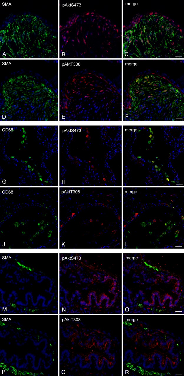
Figure 1 Akt phosphorylation co-localised within fibrotic foci, macrophages and bronchial epithelium in the idiopathic pulmonary fibrosis (IPF) lung. Immunofluorescence for α smooth muscle actin (αSMA, green, A and D), Akt phosphorylated at serine 473 (pAktS473, red, B) and threonine 308 (pAktT308, red, E) is shown co-localised (C and F) within fibrotic foci (A–F). Immunofluorescence for CD68 (green, G and J), pAktS473 (red, H), pAktT308 (red, K) is shown co-localised (I and L) within macrophages in airspaces (panels G–L). In panels M–R immunofluorescence for pAktS473 (red, N), pAktT308 (red, Q) is shown localised to unstained cells morphologically characteristic of bronchial epithelium (N, Q, O and R). Cells morphologically characteristic of airway smooth muscle are evident (green, M and P). DAPI-stained nuclei are highlighted in blue. 25 μm scale bars are shown.
Figure 1 Akt phosphorylation co-localised within fibrotic foci, macrophages and bronchial epithelium in the idiopathic pulmonary fibrosis (IPF) lung. Immunofluorescence for α smooth muscle actin (αSMA, green, A and D), Akt phosphorylated at serine 473 (pAktS473, red, B) and threonine 308 (pAktT308, red, E) is shown co-localised (C and F) within fibrotic foci (A–F). Immunofluorescence for CD68 (green, G and J), pAktS473 (red, H), pAktT308 (red, K) is shown co-localised (I and L) within macrophages in airspaces (panels G–L). In panels M–R immunofluorescence for pAktS473 (red, N), pAktT308 (red, Q) is shown localised to unstained cells morphologically characteristic of bronchial epithelium (N, Q, O and R). Cells morphologically characteristic of airway smooth muscle are evident (green, M and P). DAPI-stained nuclei are highlighted in blue. 25 μm scale bars are shown.CC BY · PubMed Central
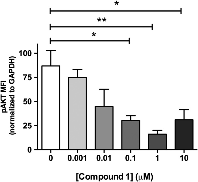
Figure 2 Pan-PI3 kinase (PI3K) dual mammalian target of rapamycin (mTOR) inhibition reduces Akt phosphorylation in human idiopathic pulmonary fibrosis (IPF) lung slices. Precision cut lung slices were treated with vehicle or increasing concentrations of PI3K inhibitor (Compound 1) for 2 h (0.1% DMSO vehicle was constant for all experimental conditions). Following harvest and homogenisation, pAkt levels were assessed by Milliplex Map transforming growth factor (TGF)-β signalling beads and normalised to glyceraldehyde 3-phosphate dehydrogenase (GAPDH). Data are shown for mean fluorescence intensity (MFI) normalised to GAPDH as ±SEM of n=4 technical replicate wells per condition; *p<0.05, **p<0.01 (one-way ANOVA with Tukey post-hoc testing).
Figure 2 Pan-PI3 kinase (PI3K) dual mammalian target of rapamycin (mTOR) inhibition reduces Akt phosphorylation in human idiopathic pulmonary fibrosis (IPF) lung slices. Precision cut lung slices were treated with vehicle or increasing concentrations of PI3K inhibitor (Compound 1) for 2 h (0.1% DMSO vehicle was constant for all experimental conditions). Following harvest and homogenisation, pAkt levels were assessed by Milliplex Map transforming growth factor (TGF)-β signalling beads and normalised to glyceraldehyde 3-phosphate dehydrogenase (GAPDH). Data are shown for mean fluorescence intensity (MFI) normalised to GAPDH as ±SEM of n=4 technical replicate wells per condition; *p<0.05, **p<0.01 (one-way ANOVA with Tukey post-hoc testing).CC BY · PubMed Central
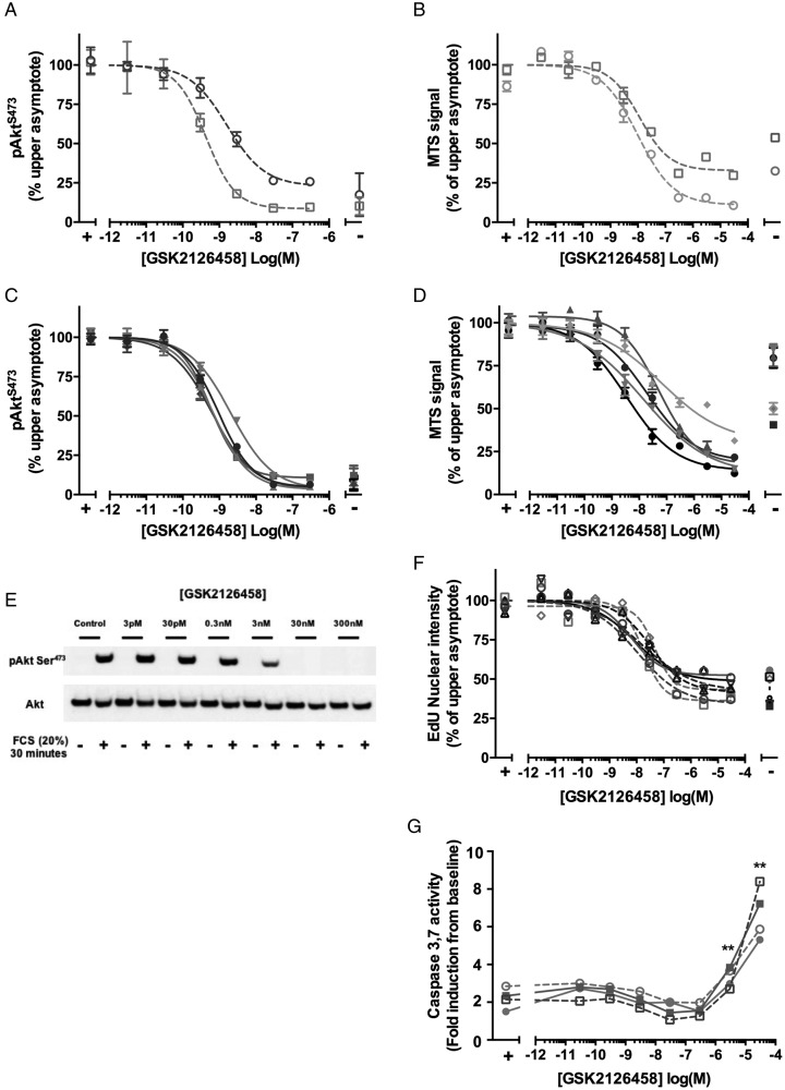
Figure 3 GSK2126458 inhibits Akt phosphorylation and proliferation in normal and idiopathic pulmonary fibrosis (IPF) primary human lung fibroblasts. Control (C-LF, n=2, A) or IPF lung fibroblasts (IPF-LF, n=5, C) were pre-incubated with increasing concentrations of GSK2126458. Following 30 min stimulation with 20% FCS, pAktS473 signal was normalised to total Akt and expressed as % maximum response (maximum asymptote) and is shown as mean±SEM of n=3 replicate wells per condition. A representative Western blot is shown visualising the effect of GSK2126458 on FCS-induced phosphorylation of AktS473 in a representative C-LF line (E). Subconfluent control (C-LF, n=2, B) or IPF lung fibroblasts (IPF-LF, n=5, D) were pre-incubated with increasing concentrations of GSK2126458. Following 72 h stimulation with 10% FCS, fibroblast proliferation was measured using an MTS assay at 490 nm. Data were expressed as % maximum response (maximum asymptote) and are shown as mean±n=6 replicate wells per condition. Incorporation of EdU (5-ethynyl-2′deoxyuridine) into the DNA of sub-confluent fibroblasts was also used as a measured of proliferation following 72 h of FCS stimulation (C-LF, n=5, dashed lines; IPF-LF, n=2, solid lines (F)). IC50 values (panels A–D and F) were calculated using four-parameter non-linear regression, and representative curves are shown for each fibroblast line. FCS stimulated and unstimulated DMSO vehicle control fibroblast data points are indicated as (+) and (−), respectively (panels A–F). DMSO was maintained at 0.1% for all conditions. Activation of fibroblast caspase 3 and 7 was assessed following incubation with increasing concentrations of GSK2126458 in the presence of 10% FCS by Caspase Glo (G). FCS-stimulated (+) vehicle control (0.1% DMSO) fibroblasts are indicated. Data are expressed as fold relative to baseline cells (no FCS, 0.1% DMSO) and shown as mean±SEM of n=3 replicate wells per condition. **p<0.0001 for differences in raw data values from FCS-stimulated controls (two-way ANOVA, Tukey's multiple comparisons test).
Figure 3 GSK2126458 inhibits Akt phosphorylation and proliferation in normal and idiopathic pulmonary fibrosis (IPF) primary human lung fibroblasts. Control (C-LF, n=2, A) or IPF lung fibroblasts (IPF-LF, n=5, C) were pre-incubated with increasing concentrations of GSK2126458. Following 30 min stimulation with 20% FCS, pAktS473 signal was normalised to total Akt and expressed as % maximum response (maximum asymptote) and is shown as mean±SEM of n=3 replicate wells per condition. A representative Western blot is shown visualising the effect of GSK2126458 on FCS-induced phosphorylation of AktS473 in a representative C-LF line (E). Subconfluent control (C-LF, n=2, B) or IPF lung fibroblasts (IPF-LF, n=5, D) were pre-incubated with increasing concentrations of GSK2126458. Following 72 h stimulation with 10% FCS, fibroblast proliferation was measured using an MTS assay at 490 nm. Data were expressed as % maximum response (maximum asymptote) and are shown as mean±n=6 replicate wells per condition. Incorporation of EdU (5-ethynyl-2′deoxyuridine) into the DNA of sub-confluent fibroblasts was also used as a measured of proliferation following 72 h of FCS stimulation (C-LF, n=5, dashed lines; IPF-LF, n=2, solid lines (F)). IC50 values (panels A–D and F) were calculated using four-parameter non-linear regression, and representative curves are shown for each fibroblast line. FCS stimulated and unstimulated DMSO vehicle control fibroblast data points are indicated as (+) and (−), respectively (panels A–F). DMSO was maintained at 0.1% for all conditions. Activation of fibroblast caspase 3 and 7 was assessed following incubation with increasing concentrations of GSK2126458 in the presence of 10% FCS by Caspase Glo (G). FCS-stimulated (+) vehicle control (0.1% DMSO) fibroblasts are indicated. Data are expressed as fold relative to baseline cells (no FCS, 0.1% DMSO) and shown as mean±SEM of n=3 replicate wells per condition. **p<0.0001 for differences in raw data values from FCS-stimulated controls (two-way ANOVA, Tukey's multiple comparisons test).CC BY · PubMed Central
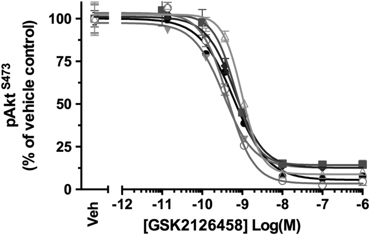
Figure 4 GSK2126458 inhibits Akt phosphorylation in bronchoalveolar lavage (BALF) cells from patients with idiopathic pulmonary fibrosis (IPF). IPF BALF cells (n=6) were incubated with 0.1% DMSO (vehicle control) or increasing concentrations of GSK2126458 for 30 min at 37°C (0.1% DMSO vehicle was constant for all experimental conditions). Cell lysates were assayed for total Akt and pAktS473 using Meso Scale Discovery (MSD) electro-chemiluminescence technology. pAktS473 signal was normalised to total Akt signal and expressed as % of a reference vehicle control well for each experiment (Veh). Data are shown as mean±SEM of triplicate wells per condition. IC50 values were calculated using four-parameter non-linear regression.
Figure 4 GSK2126458 inhibits Akt phosphorylation in bronchoalveolar lavage (BALF) cells from patients with idiopathic pulmonary fibrosis (IPF). IPF BALF cells (n=6) were incubated with 0.1% DMSO (vehicle control) or increasing concentrations of GSK2126458 for 30 min at 37°C (0.1% DMSO vehicle was constant for all experimental conditions). Cell lysates were assayed for total Akt and pAktS473 using Meso Scale Discovery (MSD) electro-chemiluminescence technology. pAktS473 signal was normalised to total Akt signal and expressed as % of a reference vehicle control well for each experiment (Veh). Data are shown as mean±SEM of triplicate wells per condition. IC50 values were calculated using four-parameter non-linear regression.CC BY · PubMed Central
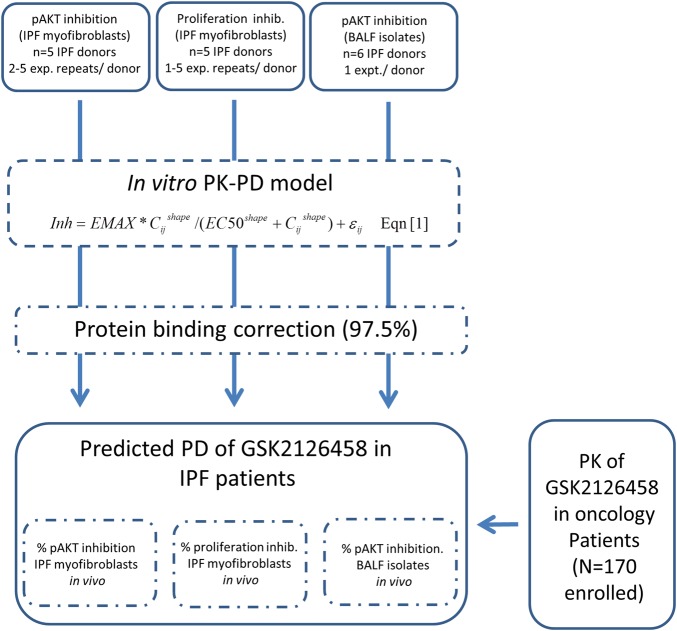
Figure 5 Construction of an integrated pharmacokinetic (PK)/pharmacodynamics (PD) model for GSK2126458 in patients with idiopathic pulmonary fibrosis (IPF). Schematic illustrating the strategy for integration of in vitro PK/PD data with human PK for clinical dose response estimation in IPF. Equation (1): Inh represents the % inhibition of pAkt (IPF myofibroblasts or bronchoalveolar lavage fluid (BALF) cell isolates) of fibroblast proliferation (IPF myofibroblasts), EMAX=maximum estimated inhibition, EC50=drug potency and shape=gradient of the response curve. ε refers to residual or unexplained variability, the subscript i indicates replicate cell line, j is individual drug concentration from each cell line.
Figure 5 Construction of an integrated pharmacokinetic (PK)/pharmacodynamics (PD) model for GSK2126458 in patients with idiopathic pulmonary fibrosis (IPF). Schematic illustrating the strategy for integration of in vitro PK/PD data with human PK for clinical dose response estimation in IPF. Equation (1): Inh represents the % inhibition of pAkt (IPF myofibroblasts or bronchoalveolar lavage fluid (BALF) cell isolates) of fibroblast proliferation (IPF myofibroblasts), EMAX=maximum estimated inhibition, EC50=drug potency and shape=gradient of the response curve. ε refers to residual or unexplained variability, the subscript i indicates replicate cell line, j is individual drug concentration from each cell line.CC BY · PubMed Central
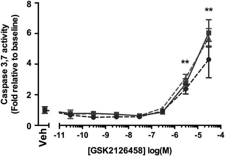
Figure 6 Effect of GSK2126458 on epithelial cell apoptosis. Activation of epithelial caspase 3 and 7 was assessed in primary bronchial epithelial cultures following incubation with increasing concentrations of GSK2126458 for 72 h (0.1% DMSO vehicle was constant for all experimental conditions). Human bronchial epithelial cell (HBEC) cultures were derived from n=2 control and n=1 idiopathic pulmonary fibrosis (IPF) donor lungs. Data are expressed fold relative to baseline (0.1% DMSO) controls (Veh) and shown as mean±SEM of n=6 replicate wells per condition; **p<0.0001 for differences in raw data values from no vehicle controls (two-way ANOVA, Tukey's multiple comparisons test).
Figure 6 Effect of GSK2126458 on epithelial cell apoptosis. Activation of epithelial caspase 3 and 7 was assessed in primary bronchial epithelial cultures following incubation with increasing concentrations of GSK2126458 for 72 h (0.1% DMSO vehicle was constant for all experimental conditions). Human bronchial epithelial cell (HBEC) cultures were derived from n=2 control and n=1 idiopathic pulmonary fibrosis (IPF) donor lungs. Data are expressed fold relative to baseline (0.1% DMSO) controls (Veh) and shown as mean±SEM of n=6 replicate wells per condition; **p<0.0001 for differences in raw data values from no vehicle controls (two-way ANOVA, Tukey's multiple comparisons test).CC BY · PubMed Central
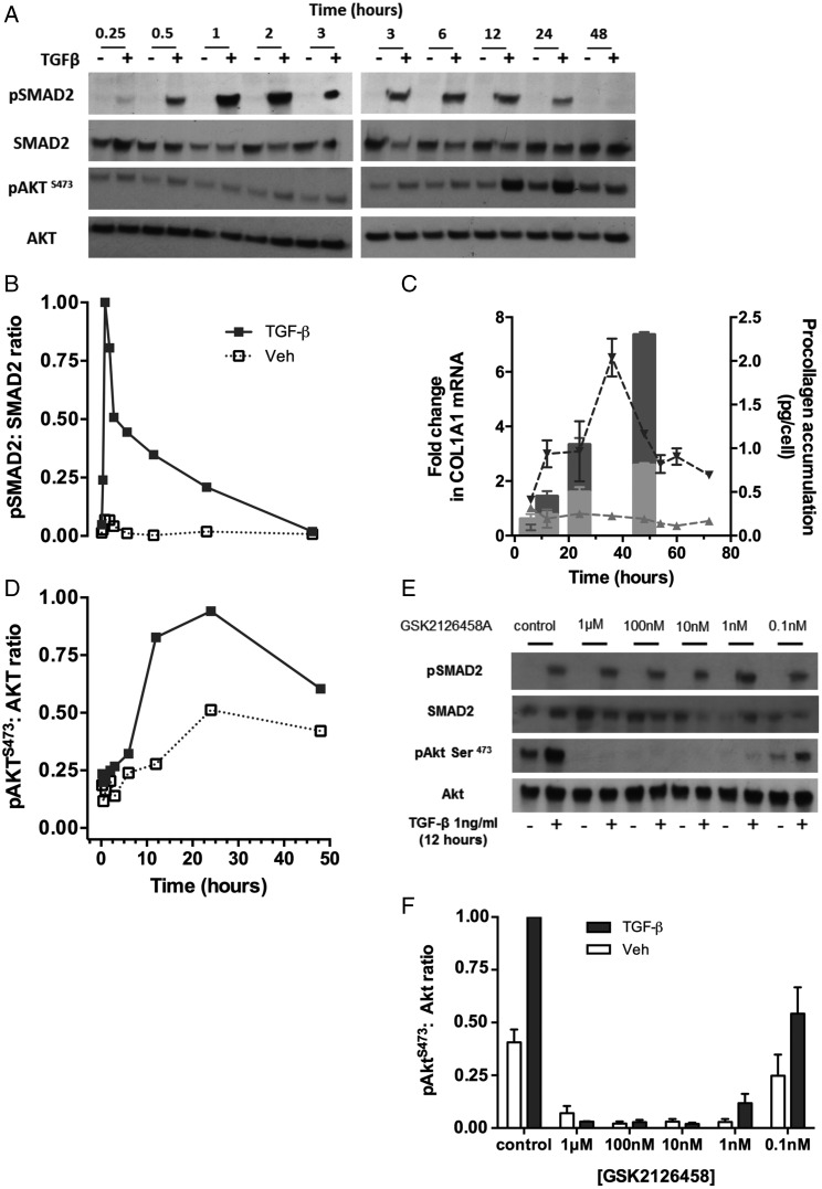
Figure 7 Time course of the primary human fibroblast response to transforming growth factor (TGF)-β1. Confluent lung fibroblasts (LFs) were stimulated with TGFβ1 (1 ng/mL) and lysates collected over the indicated timecourse (A, B and D). Phosphorylation of SMAD2 or AktS473 is shown by Western blot (A) and densitometry (B and D). Collagen gene expression in TGFβ1 (1 ng/mL) stimulated (C, dark triangles and dashed line) or unstimulated (C, light triangles and dashed line) was assessed in LFs over the indicated time course. Pro-collagen recovered from cell supernatants over the same time course in TGFβ1 stimulated (C, dark bar) or unstimulated (C, light bar) is also shown. Serum-free confluent primary human LFs (HLFs) were incubated with increasing concentrations of GSK2126458 or 0.1% DMSO and stimulated with TGFβ1 (1 ng/mL) for 12 h (0.1% DMSO vehicle was constant for all experimental conditions). Phosphorylation of SMAD2 and AktS473 is shown by representative Western blot (E). Densitometry illustrating the effects of GSK2126458 on pAktS473 in HLFs from n=3 donors is shown (F).
Figure 7 Time course of the primary human fibroblast response to transforming growth factor (TGF)-β1. Confluent lung fibroblasts (LFs) were stimulated with TGFβ1 (1 ng/mL) and lysates collected over the indicated timecourse (A, B and D). Phosphorylation of SMAD2 or AktS473 is shown by Western blot (A) and densitometry (B and D). Collagen gene expression in TGFβ1 (1 ng/mL) stimulated (C, dark triangles and dashed line) or unstimulated (C, light triangles and dashed line) was assessed in LFs over the indicated time course. Pro-collagen recovered from cell supernatants over the same time course in TGFβ1 stimulated (C, dark bar) or unstimulated (C, light bar) is also shown. Serum-free confluent primary human LFs (HLFs) were incubated with increasing concentrations of GSK2126458 or 0.1% DMSO and stimulated with TGFβ1 (1 ng/mL) for 12 h (0.1% DMSO vehicle was constant for all experimental conditions). Phosphorylation of SMAD2 and AktS473 is shown by representative Western blot (E). Densitometry illustrating the effects of GSK2126458 on pAktS473 in HLFs from n=3 donors is shown (F).CC BY · PubMed Central
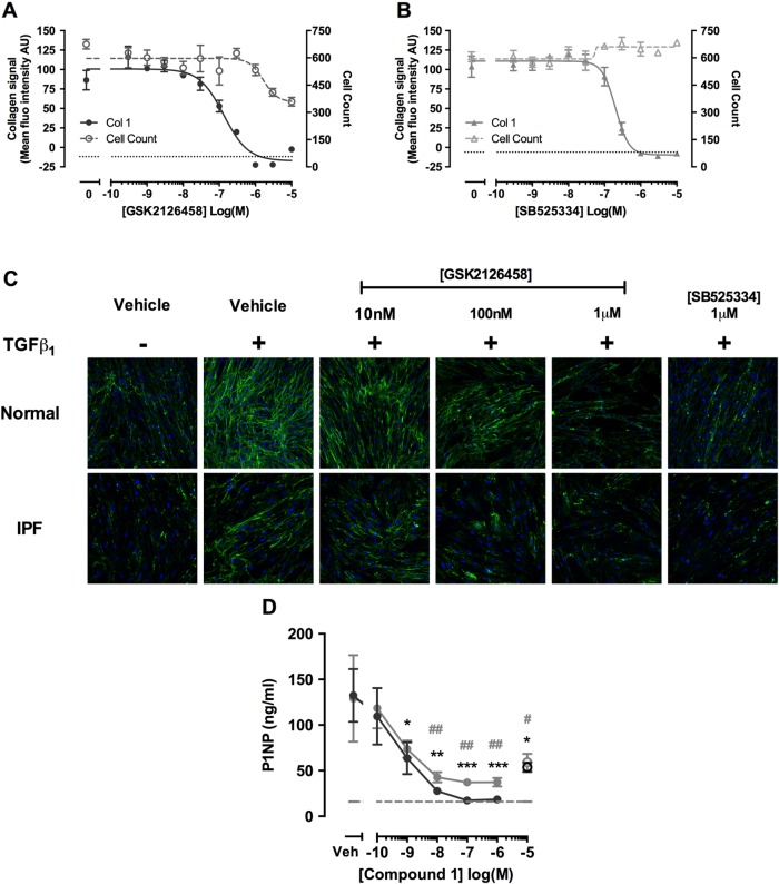
Figure 8 Pan-PI3 kinase (PI3K)/mammalian target of rapamycin (mTOR) inhibition attenuates transforming growth factor (TGF)-β1-induced collagen production in human lung fibroblasts (LFs), and collagen formation markers in idiopathic pulmonary fibrosis (IPF) lung tissue. Confluent LFs were incubated with increasing concentrations of GSK2126458 (A) or SB525334 (B) and stimulated for 48 h with TGFβ1 (1 ng/mL) and collagen biosynthesis assayed by molecular crowding assay (0.1% DMSO vehicle was constant for all experimental conditions). Data are expressed as mean fluorescent intensity (n=4 reads per well) and cell counts obtained from DAPI counterstaining (blue channel). Data are shown as mean±SEM of n=3 replicate wells per condition. IC50 values were calculated using four-parameter non-linear regression. Replicate experiments were carried out on human lung fibroblasts (HLFs) from a non-IPF donor and representative curves are shown. Images from each cell treatment are shown (C). 8 mm slices of IPF lung were cultured for 24 h in DMEM supplemented with 0.4% FCS and treated with vehicle, 10 μM SB525334 (open circles) or increasing concentrations of Compound 1 (inhibitor) for 0–3 days (filled grey circles) and further from 3 to 5 days (filled black circles) following media change. P1NP levels were measured in harvested supernatants (D). Data are shown as ±SEM of n=4 replicate wells per condition; *p<0.05, **p<0.01 (two-way ANOVA with Bonferroni post-hoc testing).
Figure 8 Pan-PI3 kinase (PI3K)/mammalian target of rapamycin (mTOR) inhibition attenuates transforming growth factor (TGF)-β1-induced collagen production in human lung fibroblasts (LFs), and collagen formation markers in idiopathic pulmonary fibrosis (IPF) lung tissue. Confluent LFs were incubated with increasing concentrations of GSK2126458 (A) or SB525334 (B) and stimulated for 48 h with TGFβ1 (1 ng/mL) and collagen biosynthesis assayed by molecular crowding assay (0.1% DMSO vehicle was constant for all experimental conditions). Data are expressed as mean fluorescent intensity (n=4 reads per well) and cell counts obtained from DAPI counterstaining (blue channel). Data are shown as mean±SEM of n=3 replicate wells per condition. IC50 values were calculated using four-parameter non-linear regression. Replicate experiments were carried out on human lung fibroblasts (HLFs) from a non-IPF donor and representative curves are shown. Images from each cell treatment are shown (C). 8 mm slices of IPF lung were cultured for 24 h in DMEM supplemented with 0.4% FCS and treated with vehicle, 10 μM SB525334 (open circles) or increasing concentrations of Compound 1 (inhibitor) for 0–3 days (filled grey circles) and further from 3 to 5 days (filled black circles) following media change. P1NP levels were measured in harvested supernatants (D). Data are shown as ±SEM of n=4 replicate wells per condition; *p<0.05, **p<0.01 (two-way ANOVA with Bonferroni post-hoc testing).CC BY · PubMed Central

Verification

Claims the paper makes

The claims and statistics of this paper haven’t been checked yet.

Run the check — free, no account needed. It reads the open-access text and checks the sources it cites.

A few free checks a day · papers already verified open instantly · results are public

Claims other papers make about this one

Each is checked against this paper’s own text: does it say what the citing paper says it does?

Self-check

Percentages, sums and reported differences re-computed from the paper’s own numbers.

The self-check needs the paper’s full text.

Citations

Cites

Show all 39 references

Cited by

Sorted by citations

See all 206

Metadata

Authors
Mercer, P. F., Woodcock, H. V., Eley, J. D., Platé, M., Sulikowski, M. G., Durrenberger, P. F., Franklin, L., Nanthakumar, C. B., Man, Y., Genovese, F., McAnulty, R. J., Yang, S., Maher, T. M., Nicholson, A. G., Blanchard, A. D., Marshall, R. P., Lukey, P. T., & Chambers, R. C.
Affiliations
  • University College London (GB)
  • GlaxoSmithKline (United Kingdom) (GB)
  • Nordic Bioscience (Denmark) (DK)
  • Royal Brompton Hospital (GB)
Corresponding author
Chambers, Rachel C
Venue
Thorax
Publisher
BMJ
Published
2016-04-21
Volume
71
Issue
8
Pages
701-711
ISSN
  • 0040-6376 (ISSN-L)
  • 1468-3296
OpenAlex
W2336895811
Cited by
206
References
39
Citations by year
2026: 13 · 2025: 16 · 2024: 15 · 2023: 19 · 2022: 18 · 2021: 24 · 2020: 32 · 2019: 25 · 2018: 21 · 2017: 20 · 2016: 3
FWCI
20.56
Type
journal article
Language
en
Open access
Licence
cc-by
Locations
  • Thorax (publishedVersion, cc-by)
  • Europe PMC (PubMed Central) (publishedVersion, cc-by)
  • PubMed Central (publishedVersion, cc-by)
  • PubMed (publishedVersion)
  • UCL Discovery (University College London) (submittedVersion)
  • HighWire Press Open Archive (submittedVersion)
  • Spiral (Imperial College London) (submittedVersion, other-oa)
Funders
  • GlaxoSmithKline
  • Medical Research Council (MR/K024078/1)
  • Biotechnology and Biological Sciences Research Council (BB/L502133/1)
Topics
  • Health Sciences › Medicine › Pulmonary and Respiratory Medicine › Interstitial Lung Diseases and Idiopathic Pulmonary Fibrosis
  • Health Sciences › Medicine › Pulmonary and Respiratory Medicine › Occupational and environmental lung diseases
  • Health Sciences › Medicine › Pulmonary and Respiratory Medicine › Pulmonary Hypertension Research and Treatments
Keywords
idiopathic pulmonary fibrosis, GSK2126458, PI3K/mTOR inhibition, antifibrotic therapy, PI3K signaling, Akt phosphorylation, lung fibroblasts, bronchoalveolar lavage cells, pharmacodynamic biomarkers
MeSH terms
Benzenesulfonamides, Clinical Trials as Topic, Fibroblasts, Humans, Pyridazines, Quinolines, Sulfonamides, Signal Transduction, Treatment Outcome, Phosphatidylinositol 3-Kinases, Protein Kinase Inhibitors, Cell Proliferation, Idiopathic Pulmonary Fibrosis, TOR Serine-Threonine Kinases

About these checks

Registry, retraction, Cites and statistics results are computed, not judged, with one exception: a printed reference that no index lists is read once by a model before it counts as a citation error. Claims are extracted by a model and kept only when their quote appears verbatim in the text; each cited source is then checked, with a model’s help, for whether it says what the claim relies on. Those verdicts can be wrong — read the claim’s quote and the cited source before relying on one.

Statistics and arithmetic are recomputed, not judged.

Confidence bands are shown only for fields calibrated on our annotated test set. None is calibrated yet, so no band is shown.

What counts as a claim ›How we measure confidence ›How we check this ›

Are you an author, or do you see a problem with this page? Request a review