

Motor Control and Neural Plasticity through Interhemispheric Interactions
Open accessThe DOI matches its Crossref record; 135 of the 135 references it cites were found in a registry or an index.
Abstract
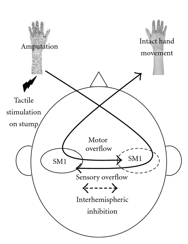
The corpus callosum, which is the largest white matter structure in the human brain, connects the 2 cerebral hemispheres. It plays a crucial role in maintaining the independent processing of the hemispheres and in integrating information between both hemispheres. The functional integrity of interhemispheric interactions can be tested electrophysiologically in humans by using transcranial magnetic stimulation, electroencephalography, and functional magnetic resonance imaging. As a brain structural imaging, diffusion tensor imaging has revealed the microstructural connectivity underlying interhemispheric interactions. Sex, age, and motor training in addition to the size of the corpus callosum influence interhemispheric interactions. Several neurological disorders change hemispheric asymmetry directly by impairing the corpus callosum. Moreover, stroke lesions and unilateral peripheral impairments such as amputation alter interhemispheric interactions indirectly. Noninvasive brain stimulation changes the interhemispheric interactions between both motor cortices. Recently, these brain stimulation techniques were applied in the clinical rehabilitation of patients with stroke by ameliorating the deteriorated modulation of interhemispheric interactions. Here, we review the interhemispheric interactions and mechanisms underlying the pathogenesis of these interactions and propose rehabilitative approaches for appropriate cortical reorganization.
Overview
The claims and statistics of this paper haven’t been checked yet.
Free: it reads the open-access text and checks the first 5 claims that cite a source we can check. Create a free account to check every claim.
Integrity signals
Registry
Cites
Statistics
Figures

Verification
Claims the paper makes
The claims and statistics of this paper haven’t been checked yet.
Free: it reads the open-access text and checks the first 5 claims that cite a source we can check. Create a free account to check every claim.
Claims other papers make about this one
Each is checked against this paper’s own text: does it say what the citing paper says it does?
The source backs part of the claim, not all of it — or backs it for a different population, condition or comparison.
In the citing paper
“This suggests that while the initiation of the reflex is slower in the elderly, the overall reflex duration stays comparable. Elderly individuals may rely on compensatory mechanisms, such as greater reliance on experience or adaptations to physiological changes.”
From Assessing neuromuscular system via patellar tendon reflex analysis using EMG in healthy individuals (2025) · cites this paper as [23]
Experimental — accuracy not yet measuredThe cited source says what the claim attributes to it.
In the citing paper
“Similarly, studies by Takeuchi et al. and Goble et al. highlighted how overactivation of specific brain regions, such as the prefrontal cortex and supplementary motor areas, may compensate for declines in motor control and cognitive tasks in older adults (23, 24).”
From Assessing neuromuscular system via patellar tendon reflex analysis using EMG in healthy individuals (2025) · cites this paper as [23]
Self-check
Percentages, sums and reported differences re-computed from the paper’s own numbers.
The self-check needs the paper’s full text.
Citations
Cites
Aging Gracefully: Compensatory Brain Activity in High-Performing Older Adults
Cabeza, R., Anderson, N. D., Locantore, J. K., & McIntosh, A. R. (2002) · NeuroImage · doi:10.1006/nimg.2002.1280 · Cited by 1,964 · cited 1×
“The results of previous studies supported the idea that overrecruitment of bilateral prefrontal activation compensates cognitive tasks in older adults [56, 57].”
ResultMemory and Executive Function in Aging and AD
Buckner, R. L. (2004) · Neuron · doi:10.1016/j.neuron.2004.09.006 · Cited by 1,508 · cited 2×
“As mentioned previously, it is thought that a reduction of hemispheric asymmetry may partly result from the failed inhibition of the contralateral hemisphere in older adults because of an age-related dysfunction of the corpus callosum [54, 55].”
BackgroundInterhemispheric inhibition of the human motor cortex.
Ferbert, A., Priori, A., Rothwell, J. C., Day, B. L., Colebatch, J. G., & Marsden, C. D. (1992) · The Journal of Physiology · doi:10.1113/jphysiol.1992.sp019243 · Cited by 1,486 · cited 2×
“Moreover, its effect is strongly dependent on the timing of the delivery of the stimulus pairs (8 ms), corresponding to the interval time between the double-pulse TMS that induces the interhemispheric inhibition [4].”
BackgroundInfluence of interhemispheric interactions on motor function in chronic stroke
Murase, N., Duque, J., Mazzocchio, R., & Cohen, L. G. (2004) · Annals of Neurology · doi:10.1002/ana.10848 · Cited by 1,472 · cited 7×
“TMS studies have showed that interhemispheric inhibition persisted from the unaffected to the affected hemisphere around the onset of the movement of the paretic hand in stroke patients, whereas the interhemispheric interaction in healthy controls changed from inhibitory to excitatory influence on the active motor cor…”
BackgroundResultInfluentialNeuroplasticity Subserving Motor Skill Learning
Dayan, E., & Cohen, L. G. (2011) · Neuron · doi:10.1016/j.neuron.2011.10.008 · Cited by 1,408 · cited 1×
“Therefore, it may be more efficient to use one hemisphere and inhibit the other hemisphere during simple tasks (e.g., physical identity and face-matching tasks); this promotes intrahemispheric processing and brain lateralization [2, 18, 19].”
BackgroundAge differences in the frontal lateralization of verbal and spatial working memory revealed by PET
Reuter-Lorenz, P. A., Jonides, J., Smith, E. E., Hartley, A., Miller, A., Marshuetz, C., & Koeppe, R. A. (2000) · Journal of Cognitive Neuroscience · doi:10.1162/089892900561814 · Cited by 998 · cited 1×
“The results of previous studies supported the idea that overrecruitment of bilateral prefrontal activation compensates cognitive tasks in older adults [56, 57].”
ResultNon-invasive brain stimulation: a new strategy to improve neurorehabilitation after stroke?
Hummel, F. C., & Cohen, L. G. (2006) · The Lancet Neurology · doi:10.1016/s1474-4422(06)70525-7 · Cited by 838 · cited 2×
“In fact, it has been reported that inhibitory NIBS over the unaffected hemisphere in patients with stroke decreases the interhemispheric inhibition from the unaffected hemisphere to the affected hemisphere and increases the excitability of the affected hemisphere, resulting in facilitated motor learning and motor reco…”
BackgroundEarly consolidation in human primary motor cortex
Muellbacher, W., Ziemann, U., Wissel, J., Dang, N., Kofler, M., Facchini, S., Boroojerdi, B., Poewe, W., & Hallett, M. (2002) · Nature · doi:10.1038/nature712 · Cited by 803 · cited 1×
“In addition, the increased motor cortical excitability induced by inhibitory NIBS enhances the effects of motor training on the ipsilateral side [120, 121], as the increase in excitability in the motor cortex appears to be a necessity for motor learning [122, 123].”
BackgroundGrefkes, C., & Fink, G. R. (2011) · Brain · doi:10.1093/brain/awr033 · Cited by 632 · cited 3×
“Therefore, longitudinal neuroimaging and electrophysiological studies must be performed to demonstrate the dynamic change in interhemispheric interaction between both hemispheres during the process of functional recovery [14, 103].”
BackgroundDynamic functional reorganization of the motor execution network after stroke
Wang, L., Yu, C., Chen, H., Qin, W., He, Y., Fan, F., Zhang, Y., Wang, M., Li, K., Zang, Y., et al. (2010) · Brain · doi:10.1093/brain/awq043 · Cited by 629 · cited 2×
“Moreover, functional magnetic resonance imaging (fMRI) studies have revealed interhemispheric interactions using resting-state functional and activity-dependent effective connectivity analyses [13, 14].”
BackgroundResult
Research works printed but not linked: 8
- 2008, Reviews in the Neurosciences, 19, 451
- 1995, Experimental Brain Research, 104, 527
- 1999, Electroencephalography and Clinical Neurophysiology. Supplement, 51, 243
- 2003, American Journal of Neuroradiology, 24, 410
- 2003, American Journal of Neuroradiology, 24, 1049
- 2013, Neuropharmacology, 64, 579
- 2007, Restorative Neurology and Neuroscience, 25, 461
- 2010, Clinical Neurophysiology, 121, s316
Cited by
Items 101–186 of 186
Dhamoon, M. S., Cheung, Y.-K. K., Bagci, A. M., Alperin, N. J., Sacco, R. L., Elkind, M. S. V., & Wright, C. B. (2017) · Frontiers in Aging Neuroscience · doi:10.3389/fnagi.2017.00305 · Cited by 10
“…reliably found, either in gray or white matter volume, or sulcal or gyral sizes; the functional basis of the asymmetry may reside in lateralized synaptic networks or microstructural white matter tract differences (Büchel et al., 2004; de Schotten et al., 2011; Thiebaut de Schotten et al., 2011).”
BackgroundLiu, C., Liao, W., Su, S.-Y., Chen, W.-L., & Hsieh, C. (2021) · Evidence-based Complementary and Alternative Medicine · doi:10.1155/2021/5564494 · Cited by 9
“In addition, stroke lesions can indirectly break off interhemispheric inhibition, contributing to poststroke recovery of motor function [32].”
BackgroundNeurofeedback-gestütztes Bewegungsvorstellungstraining zur Rehabilitation nach einem Schlaganfall
Dettmers, C., Braun, N., Büsching, I., Hassa, T., Debener, S., & Liepert, J. (2016) · Der Nervenarzt · doi:10.1007/s00115-016-0185-y · Cited by 9
Romkema, S., Bongers, R. M., & van der Sluis, C. K. (2015) · PLoS ONE · doi:10.1371/journal.pone.0128747 · Cited by 10
“These changes not only take place in the cortex but occur also in other parts of the brain, like the corpus callosum [47].”
BackgroundNeuroadaptation in neurodegenerative diseases: compensatory mechanisms and therapeutic approaches
Kopalli, S. R., Behl, T., Baldaniya, L. H., Ballal, S., Joshi, K. K., Arya, R., Chaturvedi, B., Chauhan, A. S., Verma, R. K., Patel, M., et al. (2025) · Progress in Neuro-Psychopharmacology and Biological Psychiatry · doi:10.1016/j.pnpbp.2025.111375 · Cited by 8
Baduni, K., Khan, O. A., Modlesky, C. M., & Maitre, N. L. (2025) · Developmental Medicine & Child Neurology · doi:10.1111/dmcn.70041 · Cited by 9
“35 Although many infants born preterm do not develop CP, a subset can be identified in infancy as high-risk for CP based on a combination of CP-specific early markers (e.g. atypical general movements, Hammersmith Infant Neurological Examination findings, or CP-predictive brain lesions).”
Kwon, B. M., Lee, Y., Lee, H. H., Ko, N., Kim, H.-T., Kim, B.-R., Moon, W., & Lee, J. (2022) · Brain & Neurorehabilitation · doi:10.12786/bn.2022.15.e7 · Cited by 8
“For future studies, we suggest that interhemispheric inhibition also needs to be evaluated in order to understand the precise roles of the CC in the ipsilesional hand function of stroke patients because handedness might be related to inhibitory interhemispheric interactions [33].”
BackgroundGirondini, M., Montanaro, M., Lega, C., & Gallace, A. (2024) · European Journal of Neuroscience · doi:10.1111/ejn.16481 · Cited by 8
“…the ipsilateral somatosensory area during the motor task, temporally bound with the movement due to the virtual interaction, might have led to functional (plasticity) changes between the somatosensory and the motor area of the two hemispheres via the subcortical connections (Takeuchi et al., 2012).”
Stroke rehabilitation and research: consideration of the role of the cortico-reticulospinal system
Silva, A., Vaughan‐Graham, J., Silva, C., Sousa, A. S. P., Cunha, C., Ferreira, R., & Barbosa, P. M. (2018) · Somatosensory & Motor Research · doi:10.1080/08990220.2018.1500363 · Cited by 9
Zhong, Y., Fan, J., Wang, H., & He, R. (2021) · Restorative Neurology and Neuroscience · doi:10.3233/rnn-211172 · Cited by 8
“The asymmetry or imbalance as a result of unilateral brain injury is not only the result of local lesions, but also because of the interaction of the con-tralesionalsideviathecorpuscallosum(Muraseetal., 2004;Takeuchietal.,2012).”
Han, M., Kim, Y. D., Park, H., Hwang, I. G., Choi, J., Ha, J., Heo, J. H., & Nam, H. S. (2019) · PLoS ONE · doi:10.1371/journal.pone.0208918 · Cited by 7
The impact of aging on interhemispheric transfer time and respective sex differences
Riedel, D., Fellerhoff, T., Mierau, A., Strüder, H. K., Wolf, D. G. F., Fischer, F. U., Fellgiebel, A., Tüscher, O., Kollmann, B., & Knaepen, K. (2022) · Aging Brain · doi:10.1016/j.nbas.2022.100040 · Cited by 7
“Female hormones may have beneficial effects with regards to interhemispheric communication [67] and, consequently,”
BackgroundDevelopment of a Non-invasive Brain Computer Interface for Neurorehabilitation
Pinto, R. D., & Ferreira, H. A. (2015) · Workshop on ICTs for improving Patients Rehabilitation Research Techniques (REHAB) · doi:10.1145/2838944.2838975 · Cited by 8
“In neurological disorders, this balance shifts in favour of plastic changes, resulting in the reorganization of network connectivity [49], [80], [83].”
BackgroundConessa, A., Léger, D., & Boutin, A. (2026) · Communications Biology · doi:10.1038/s42003-025-09425-6 · Cited by 6
“Given that upper-limb immobilization disrupts the balance of inter-hemispheric inhibition 40 , a process closely tied to motor learning and generalization 43 , it can be hypothesized that transient upper-limb immobilization would impact the consolidation and generalization of memory traces.”
Albishi, A. M. (2024) · Annals of Medicine and Surgery · doi:10.1097/ms9.0000000000002287 · Cited by 6
“The mechanism of controlling motor over fl ow, is interrupted in patients with stroke, resulting in a sequence of events referred to as interhemispheric compensatory communication [19 – 21] .”
BackgroundCallosal apraxia: a 34-year follow-up study
Falchook, A. D., Watson, R. T., & Heilman, K. M. (2016) · Neurocase · doi:10.1080/13554794.2016.1148743 · Cited by 6
Flores, A., Ribó, M., Rubiera, M., Cuevas, M. G., Pagola, J., Rodríguez‐Luna, D., Muchada, M., KALLAS, J. A., Meler, P., Sanjuán, E., et al. (2014) · Journal of Neuroimaging · doi:10.1111/jon.12113 · Cited by 6
Kim, D., Kwon, G. H., Lee, S., & Kim, L. (2025) · Brain and Behavior · doi:10.1002/brb3.70492 · Cited by 5
“On the other hand, the ipsilateral MA of HC showed a lack of local changes due to its inhibitory properties during motor task performance in the non-damaged brain (Takeuchi et al. 2012).”
Monaghan, A. S., Ofori, E., Fling, B. W., & Peterson, D. S. (2024) · Brain Imaging and Behavior · doi:10.1007/s11682-024-00867-w · Cited by 5
“However, the corpus callosum plays a significant role in mediating interhemispheric inhibition (Takeuchi et al., 2012), and intracortical inhibition has been related to motor learning (Stagg et al., 2011).”
Effects of rTMS to primary motor cortex and cerebellum on balance control in healthy adults
Parikh, V., Medley, A., & Goh, H. (2024) · European Journal of Neuroscience · doi:10.1111/ejn.16386 · Cited by 5
“Therefore, interventions such as non-invasive brain stimulation (NIBS) that can modulate the activity of M1 and the cerebellum could improve balance performance (Takeuchi et al., 2012).”
Aune, M. A., Lorås, H. W., Nynes, A., & Aune, T. K. (2021) · Frontiers in Psychology · doi:10.3389/fpsyg.2021.680268 · Cited by 5
Wang, B. J., Hui, S., Pan, X., Ma, W., Dong, L., Wang, Q., & Meng, P. (2023) · Frontiers in Human Neuroscience · doi:10.3389/fnhum.2023.1100320 · Cited by 6
Modulation of Alpha Band Brain Connectivity during Motor Imagery via Bimanual Motor Training
Phunruangsakao, C., Budsuren, J., & Hayashibe, M. (2024) · International Winter Conference on Brain-Computer Interface (BCI) · doi:10.1109/bci60775.2024.10480525 · Cited by 5
“The two hemispheres of the brain communicate and exchange information to facilitate these movements, allowing for precise spatial and temporal coordination between both hands [1].”
BackgroundBirgani, P. M., Ashtiyani, M., Jameie, S. B., Shahrokhi, A., Rahimian, E., Deevband, M. R., & Mirbagheri, M. M. (2025) · Basic and Clinical Neuroscience Journal · doi:10.32598/bcn.2023.4679.1 · Cited by 4
“The interhemispheric exchange role of CC could be inhibitory, excitatory, or a combination of both, which may alter through neurological diseases such as stroke and CP (Takeuchi et al., 2012).”
Experience‐driven competition in neural reorganization after stroke
Jones, T. A., Nemchek, V., & Fracassi, M. (2024) · The Journal of Physiology · doi:10.1113/jp285565 · Cited by 5
“There is support for the general capacity of behavioural experiences to influence interhemispheric interactions (Takeuchi et al., 2012), e.g. training the paretic arm can reduce interhemispheric inhibition (Harris-Love et al., 2011).”
Modulation of motor cortices on manual asymmetries
Fernandes, L. A., Romano‐Silva, M. A., Figueiredo, L. S., Parma, J. O., Apolinário‐Souza, T., & Lage, G. M. (2022) · Kinesiology · doi:10.26582/k.54.1.2 · Cited by 4
Esht, V., Alshehri, M. M., Balasubramanian, K., Sanjeevi, R. R., Shaphe, M. A., Alhowimel, A., Alenazi, A. M., Alqahtani, B. A., & Alhwoaimel, N. A. (2024) · Neurophysiologie Clinique · doi:10.1016/j.neucli.2024.102976 · Cited by 5
Cognition in adults with cerebral palsy: A systematic review
Baduni, K., Perry, B., Warschausky, S. A., & Maitre, N. L. (2026) · Developmental Medicine & Child Neurology · doi:10.1111/dmcn.70280 · Cited by 3
“15–17 These interactions are particularly evident in adulthood because cognitive and motor demands intensify, exacerbating existing impairments.”
Kotov, S. V., Романова, М. В., Kondur, A. A., Biryukova, Е. V., Frolov, A., Григорьевна, Т. Л., Валентиновна, И. Е., & Zaitseva, E. V. (2020) · Neuroscience and Behavioral Physiology · doi:10.1007/s11055-020-01017-7 · Cited by 3
“…using fMRI and EEG methods are parallel continue to appear; the fi rst of these methods provides objective evidence mainly of the spatial characteristics of the process, while the latter provides mainly the time characteristics [Takeuchi et al., 2012; Chen et al., 2017; Pichiorri et al., 2018].”
MethodWei, J., Liu, Z., Su, H., She, Y., Wang, J., Liu, Y., Zhong, Y., Mo, L., Tan, C., Liu, X., et al. (2025) · Neurological Sciences · doi:10.1007/s10072-024-07951-5 · Cited by 4
“Furthermore, interhemispheric communication between motor-related cortices including premotor cortices is crucial in the initiation and planning of voluntary movements [25].”
Hung, D. V., Toan, P. Q., Ngọc, N. T. B., Thang, L. V., Thuan, D. D., Quan, L. V., Tai, P. T., Nhat, D. V., Lang, H. N., Xuan, N. T., et al. (2025) · International Urology and Nephrology · doi:10.1007/s11255-025-04598-9 · Cited by 3
“It was reported that functional coupling between different brain areas, mediated by interhemispheric and intrahemispheric connections, influences excitatory and inhibitory processes [44, 45].”
Jin, H., & Gu, G. (2015) · Journal of Child and Adolescent Behaviour · doi:10.4172/2375-4494.1000260 · Cited by 4
“The test has been translated and revised in Chinese version (China-Wechlser Yonger Children Scale of Intelligence, C-WYCSI) which has appeared to have great unwavering quality in Chinese kids [16-20].”
MethodLench, D. H., Hutchinson, S., Woodbury, M. L., & Hanlon, C. A. (2020) · Archives of Rehabilitation Research and Clinical Translation · doi:10.1016/j.arrct.2020.100075 · Cited by 5
“The corpus callosum (CC) in particular plays a central role in the effective coordination of bimanual movements(8) by mediating interhemispheric communication.(9,10) Interhemispheric coordination is observed in non-human primates in which neural synchrony of homologous motor cortex (M1) is greatest during bimanual sym…”
BackgroundA Neuro-Integrative Assessment of Perceptual-Motor Performance and Wellness in ROTC Cadets
Wilkerson, G. B., Colston, M. A., Grillo, A. N., Rogers, A., Perry, T. S., & Acocello, S. N. (2022) · Perceptual and Motor Skills · doi:10.1177/00315125211067359 · Cited by 3
“…and stimulus evaluation (Serrien et al., 2006; Wilkerson et al., 2018, 2020, 2021b), and (b) disrupted structural integrity of the corpus callosum white matter tracts that convey both excitatory and inhibitory signals between the brain hemispheres (Locke et al., 2020; Takeuchi et al., 2012).”
BackgroundJiang, Y., Chen, S., Wu, D., He, W., Ma, X., Zhang, L., & Zhang, Q. (2024) · Frontiers in Neurology · doi:10.3389/fneur.2024.1330975 · Cited by 3
“. is to maintain independent processing and integration of information between the two hemispheres (3, 4).”
BackgroundStructural Models for the Design of PKMzeta Inhibitors with Neurobiological Indications
Purkayastha, P., Alokam, R., Malapati, A., Sriram, D., & Yogeeswari, P. (2015) · Molecular Informatics · doi:10.1002/minf.201500003 · Cited by 3
Lavrador, J. P., Rajwani, K., Patel, S., Kalaitzoglou, D., Soumpasis, C., GULLAN, R. W., Ashkan, K., Bhangoo, R. S., Dell’Acqua, F., & Vergani, F. (2024) · Cerebral Cortex · doi:10.1093/cercor/bhae251 · Cited by 2
Ravestyn, C. V., Riga, A., Bihin, B., Dricot, L., Dessain, Q., Delinte, N., Devis, L., Herman, B., Coene, B. D., Deltombe, T., et al. (2026) · Neuroscience · doi:10.1016/j.neuroscience.2026.04.018 · Cited by 2
Comparing neuromotor functions in 45- and 65-year-old adults with 18-year-old adolescents
Kakebeeke, T. H., Chaouch, A., Caflisch, J., Eichelberger, D. A., Wehrle, F. M., & Jenni, O. G. (2023) · Frontiers in Human Neuroscience · doi:10.3389/fnhum.2023.1286393 · Cited by 2
“The excitatory role consists mainly of the exchange and integration of information, and the inhibitory role allows functional specialization and independent processing of both hemispheres (Takeuchi et al., 2012; Gooijers and Swinnen, 2014).”
Speed as a Complex Conditional Ability
Radak, Z. O. (2018) · Elsevier eBooks · doi:10.1016/b978-0-12-815137-2.00006-1 · Cited by 2
Xiloyannis, M., Dhinh, K. B., Cappello, L., Antuvan, C. W., & Masia, L. (2018) · Elsevier eBooks · doi:10.1016/b978-0-12-811810-8.00010-5 · Cited by 2
Ghazwani, Y. Q., Patay, Z. J., Sadighi, Z. S., Sparrow, J., Upadhyaya, S. A., Boop, F. A., Gajjar, A. J., & Qaddoumi, I. A. (2020) · Journal of Pediatric Rehabilitation Medicine · doi:10.3233/prm-190637 · Cited by 2
“The corpus callosum functions by controlling interhemispheric interactions through inhibitory and excitatory interactions [7].”
Vidal, A. C., Banca, P., Pascoal, A. G., Cordeiro, G., Sargento‐Freitas, J., Gouveia, A., & Castelo-Branco, M. S. (2018) · International Journal of Stroke · doi:10.1177/1747493018767164 · Cited by 2
RetractedBruce, S. L., & Wilkerson, G. B. (2021) · Frontiers in Sports and Active Living · doi:10.3389/fspor.2021.733567 · Cited by 1
“One’s ability to adjust and accommodate to ever changing environments is often the difference between success and failure (Serrien et al., 2006; Takeuchi et al., 2012; Garrett et al., 2013; Grady and Garrett, 2018; Wilkerson et al., 2020).”
BackgroundTheory of Repetitive Facilitative Exercise
Kawahira, K., Shimodozono, M., & Noma, T. (2022) · Springer eBooks · doi:10.1007/978-981-19-0789-0_1 · Cited by 1
Neuromodulation of Impaired Spoken Discourse
Bakhtiar, M., Carthery‐Goulart, M. T., & Kong, A. P. (2023) · Springer eBooks · doi:10.1007/978-3-031-45190-4_20 · Cited by 1
Música, lateralidade e aprendizagem: uma revisão integrativa
Estevan, F. A., & Loureiro, C. M. V. (2020) · Percepta - Revista de Cognição Musical · doi:10.34018/2318-891x.8(1)11-33 · Cited by 1
Changes in hemispheric dominance following targeted muscle reinnervation: a case study
AboElnour, T. M., Fraser, K., Yang, K., Borrell, J. A., & Zuñiga, J. M. (2025) · Frontiers in Human Neuroscience · doi:10.3389/fnhum.2025.1665931 · Cited by 1
“Interhemispheric functional connectivity supports coordination of movement and sensory integration across hemispheres and is disrupted in stroke and amputation (Takeuchi et al., 2012).”
Investigating Brain Connectivity Patterns in Varying Degrees of Bimanual Task Asymmetry
Tantawanich, P., Phunruangsakao, C., & Hayashibe, M. (2025) · Annual International Conference of the IEEE Engineering in Medicine and Biology Society (EMBC) · doi:10.1109/embc58623.2025.11254153 · Cited by 1
“Symmetric bimanual tasks, where both hands perform identical or mirrored movements, rely on increased neural synchronization between hemispheres [1]– [4], facilitated by the corpus callosum [5], [6].”
InfluentialBrain Stimulation and Behavioral Change
Lucchiari, C., Kelley, N., Vanutelli, M., & Ferrucci, R. (2019) · Frontiers research topics · doi:10.3389/978-2-88945-838-7 · Cited by 1
Effects of bimanual vs unimanual motor actions on consumer behavior intention and attitude
Ramchandani, M., Bhattacharya, S., & Coste-Manière, I. (2024) · Journal of Consumer Marketing · doi:10.1108/jcm-04-2023-5980 · Cited by 1
Guo, X., Wu, W., & Tong, S. (2017) · Annual International Conference of the IEEE Engineering in Medicine and Biology Society (EMBC) · doi:10.1109/embc.2017.8037822 · Cited by 1
“, interhemispheric inhibition and integration, to complete simple or complicated tasks [3-5].”
BackgroundHe, R., Zhong, Y., Zhang, J., & Fan, J. (2020) · Research Square · doi:10.21203/rs.3.rs-106005/v1 · Cited by 1
Encoding social preference by interhemispheric neurons in the InsulaPreprint
Glangetas, C., Ladevèze, E., Guillaumin, A., Gauthier, M., Doudnikoff, É., Bézard, E., Taupignon, A., Baufreton, J., & Georges, F. E. (2022) · bioRxiv (Cold Spring Harbor Laboratory) · doi:10.1101/2022.12.15.520538 · Cited by 1
Assessing neuromuscular system via patellar tendon reflex analysis using EMG in healthy individuals
Khatun, Z., Kristinsdóttir, S., Thóra Thórisdóttir, A., Björk Halldórsdóttir, L., Tortorella, F., Gargiulo, P., & Helgason, T. (2025) · Frontiers in Neurology · doi:10.3389/fneur.2024.1522121
“Similarly, studies by Takeuchi et al. and Goble et al. highlighted how overactivation of specific brain regions, such as the prefrontal cortex and supplementary motor areas, may compensate for declines in motor control and cognitive tasks in older adults (23, 24).”
Atypical motor behavior in a poststroke subject with agenesis of the corpus callosum: A case report
Pandian, S. S. R., & Arya, K. N. (2014) · Annals of Physical and Rehabilitation Medicine · doi:10.1016/j.rehab.2014.02.003
Spizzirri, G. (2016) · Digital Library of Theses and Dissertations (Universidade de São Paulo) · doi:10.11606/t.5.2016.tde-18112016-123020 · Cited by 1
Unraveling psychiatric sub-phenotypes: The price of the reduction of myelin basic protein
Poggi, G. (2016) · eDiss (Georg-August-Universität Göttingen) · doi:10.53846/goediss-5777
EFFECT OF WEIGHTED HAND MOVEMENTS ON HAND FUNCTION IN CHRONIC STROKE PATIENTS
Zakaria, H. M., Ragab, W. M., & Ali, O. I. (2017) · International Journal of Physiotherapy and Research · doi:10.16965/ijpr.2016.214
Brain Imaging Correlates of Developmental Coordination Disorder and Associated Impairments
Bonthrone, A. F. (2018) · UCL Discovery (University College London) · Cited by 1
Bimanual coordination and motor learning in children with unilateral motor disorders
Rudisch, J. (2016) · Radar (Oxford Brookes University) · doi:10.24384/6tzh-5337
Análise da atividade motora em hemiplégicos submetidos à terapia espelho
Lima, E. D. O., de Andrade, T. M., de Melo, G. A., Clementino, A. C. C. R., Lemos, M. T. M., & Silva, C. A. G. (2015) · Revista Neurociências · doi:10.34024/rnc.2015.v23.8015
von Mechow, J. (2016) · Universitätsbibliothek der FU Berlin Hochschulschriftenstelle u. Dokumentenserver · doi:10.17169/refubium-7276
Clael, S., Wells, C., Olegário, R. L., Brandão, E. C. D. S., de Paiva, T. A. F., Rodrigues, J. C., Caland, L. M. Q., Mota, M. R., & Bezerra, L. M. A. (2019) · doi:10.20873/10.20873/abef.2595-0096.v2n1p62.2019 · Cited by 1
Padova: la rete stradale extraurbana e gli accesi urbani
Madugno, I. (1997) · Quaderni di archeologia del Veneto · Cited by 1
Advances in Electroencephalography and Brain Connectome
Poon, T. L. (2023) · IntechOpen eBooks · doi:10.5772/intechopen.104301
Transcranial Magnetic Stimulation, Connectome and Its Clinical Applications
Yuen, M.-H. (2023) · IntechOpen eBooks · doi:10.5772/intechopen.109963
Cross-education in people with Parkinson’s disease, a short-term randomized controlled trial
Clael, S., David, F. J., Brandão, E. C. D. S., & Bezerra, L. M. A. (2023) · Journal of Bodywork and Movement Therapies · doi:10.1016/j.jbmt.2023.04.026
Directional Neural Connectivity during Robot Mirror Therapy in Patients with Stroke
Kanaizuka, Y., Manabe, T., Huang, J., Hung, J.-W., & Ono, Y. (2023) · Asia-Pacific Signal and Information Processing Association Annual Summit and Conference (APSIPA ASC) · doi:10.1109/apsipaasc58517.2023.10317207
“In the case of dominant hand movement, both inhibitory and excitatory interhemispheric control mechanisms [8, 10] may contribute to the summed inward connectivity.”
BackgroundAphasie – kognitive Steuerungssysteme des Gehirns
Sidiropoulos, K. (2025) · Springer eBooks · doi:10.1007/978-3-662-70454-7_5
Phunruangsakao, C., Hosoda, C., & Hayashibe, M. (2025) · Cognitive Neurodynamics · doi:10.1007/s11571-025-10336-9
“Despite these bilateral contributions, bimanual motor behavior is predominantly governed by the left hemisphere, which may transfer control to the right hemisphere via inter-manual transfer (Mutha et al. 2011; Haaland et al. 2004; Takeuchi et al. 2012).”
Ong, J. X., Premchand, B., Lim, R. Y., Chew, E., Jiang, M., Tang, N., & Ang, K. K. (2025) · Annual International Conference of the IEEE Engineering in Medicine and Biology Society (EMBC) · doi:10.1109/embc58623.2025.11254631
Facial motor system: voluntary and emotional control
Ginatempo, F. (2019) · UnissResearch (Università degli Studi di Sassari)
Wu, J., Xu, Y., Yin, T., Zhang, N., Ge, J., Fan, D., & Ye, S. (2026) · Brain Imaging and Behavior · doi:10.1007/s11682-026-01089-y
“On the other hand, these connections may be associated with functional compensation to maintain the integrity and stability of impaired functions as much as possible(Agosta et al., 2011; Menke et al., 2018; Takeuchi et al., 2012).”
Sensorimotor predictors of upper-limb recovery after stroke : analysis of a reach-to-grasp movement
Metrot, J. (2013) · theses.fr (ABES) · doi:10.70675/fb3d0787z98ddz4944zbc63z5e238e0080fd
Kotov, S. V., Валентиновна, И. Е., & Ponomareva, E. S. (2026) · S S Korsakov Journal of Neurology and Psychiatry · doi:10.17116/jnevro202612603262
Case report: Forced walking for treating lower limb paralysis after corpus callosum injury
Xu, X., Zhang, S., Gu, C., Feng, W., Wang, X., & Bi, W. (2026) · Medicine · doi:10.1097/md.0000000000048767
“[17] Research has found that corpus callosum synapses exhibit significant plasticity.”
Стабілізація емоційного стану шляхом підсилення міжпівкульної взаємодії
Олексіївна, П. А. (2026) · eKNUTSHIR
Robert, T., Shaeri, M., & Shoaran, M. (2025) · International IEEE/EMBS Conference on Neural Engineering · doi:10.1109/ner61569.2025.11589063
“Nevertheless, neural plasticity has been shown to alter these representations, promoting greater symmetry through bimanual training, as observed in musicians [34].”
Altawalbeh, G., Ursu, E., Colella, F. E., Goldberg, M., Liang, R., Schröder, A., Ille, S., Krieg, S. M., Zavaglia, M., Ganguly, A., et al. (2026) · Journal of NeuroEngineering and Rehabilitation · doi:10.1186/s12984-026-02120-5
“Unilateral limb loss disrupts this balance through permanent deafferentation and asymmetric cortical reorganization, resulting in altered interhemispheric inhibition (IHI) and excitability [13–15].”
de Riedmatten, I., Spencer, A. P. C., Martuzzi, R., Rochas, V., Pérot, J., Szczepankiewicz, F., & Jelescu, I. (2026) · Human Brain Mapping · doi:10.1002/hbm.70629
Albluwi, A. A. A. A. A. (2019) · Journal of Advances and Scholarly Researches in Allied Education
Berger, A., Pixa, N. H., Steinberg, F., & Doppelmayr, M. (2018) · doi:10.3389/fnbeh.2018.00067.s001
Kakebeeke, T. H., Chaouch, A., Caflisch, J., Eichelberger, D. A., Wehrle, F. M., & Jenni, O. G. (2023) · doi:10.3389/fnhum.2023.1286393.s001
Metadata
- Authors
- Takeuchi, N., Oouchida, Y., & Izumi, S.-I.
- Affiliations
- Tohoku University (JP)
- Corresponding author
- Takeuchi, Naoyuki
- Venue
- Neural Plasticity
- Publisher
- Wiley
- Published
- 2012
- Volume
- 2012
- Pages
- 1-13
- ISSN
- 1687-5443 (ISSN-L)
- 2090-5904
- PMC
- PMC3541646
- OpenAlex
- W1964353232
- Cited by
- 171
- References
- 134
- Citations by year
- 2026: 7 · 2025: 16 · 2024: 14 · 2023: 12 · 2022: 12 · 2021: 16 · 2020: 17 · 2019: 13 · 2018: 13 · 2017: 11 · 2016: 12 · 2015: 14 · 2014: 9 · 2013: 4
- FWCI
- 0.80
- Type
- journal article
- Language
- en
- Open access
- gold
- doi.org
- Locations
- Neural Plasticity (publishedVersion, cc-by)
- Europe PMC (PubMed Central) (publishedVersion, cc-by)
- PubMed Central (publishedVersion, cc-by)
- PubMed (publishedVersion)
- DOAJ (DOAJ: Directory of Open Access Journals) (submittedVersion, cc-by-sa)
- Funders
- Japan Society for the Promotion of Science (23650314)
- Topics
- Life Sciences › Neuroscience › Neurology › Transcranial Magnetic Stimulation Studies
- Life Sciences › Neuroscience › Cognitive Neuroscience › Hemispheric Asymmetry in Neuroscience
- Health Sciences › Medicine › Radiology, Nuclear Medicine and Imaging › Advanced Neuroimaging Techniques and Applications
- Keywords
- interhemispheric interaction, corpus callosum, motor control, hemispheric lateralization, motor cortex, stroke rehabilitation, diffusion tensor imaging, non-invasive brain stimulation, cortical reorganization, amputation, sex differences, motor training
- MeSH terms
- Aging, Brain Diseases, Corpus Callosum, Humans, Functional Laterality, Learning, Magnetic Resonance Imaging, Motor Skills, Neuronal Plasticity, Sex Factors, Stroke
About these checks
Registry, retraction, Cites and statistics results are computed, not judged, with one exception: a printed reference that no index lists is read once by a model before it counts as a citation error. Claims are extracted by a model and kept only when their quote appears verbatim in the text; each cited source is then checked, with a model’s help, for whether it says what the claim relies on. Those verdicts can be wrong — read the claim’s quote and the cited source before relying on one.
Statistics and arithmetic are recomputed, not judged.
Confidence bands are shown only for fields calibrated on our annotated test set. None is calibrated yet, so no band is shown.
What counts as a claim ›How we measure confidence ›How we check this ›
Are you an author, or do you see a problem with this page? Request a review
Data sources
Bibliographic data: Semantic Scholar, OpenAlex, Crossref.
Contains information from the Semantic Scholar Academic Graph Datasets, made available under the ODC Attribution License (ODC-BY 1.0).
Semantic Scholar’s data is described in: Kinney, R., et al. (2023). The Semantic Scholar Open Data Platform. arXiv:2301.10140.
