

The DOI matches its Crossref record; 164 of the 164 references it cites were found in a registry or an index.
Unraveling the Potential Role of Glutathione in Multiple Forms of Cell Death in Cancer Therapy
Open accessAbstract
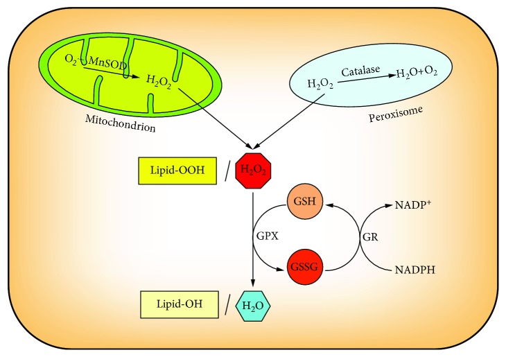
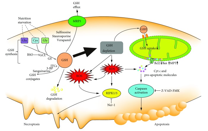
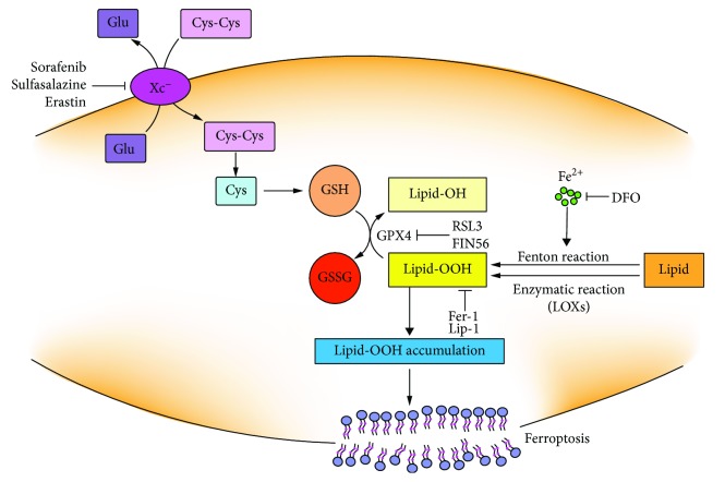
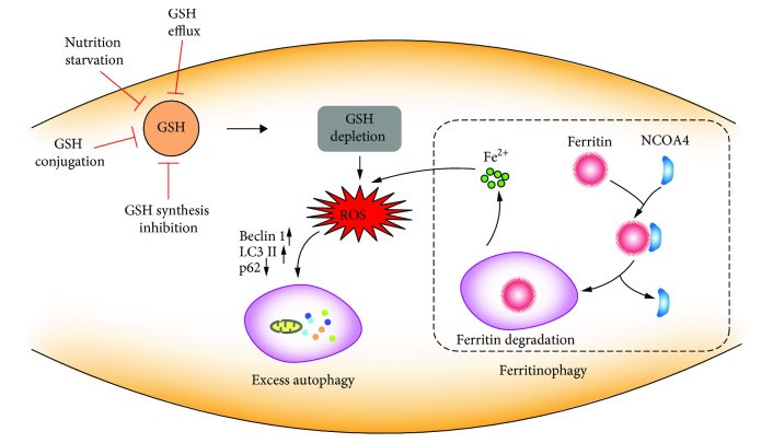
Glutathione is the principal intracellular antioxidant buffer against oxidative stress and mainly exists in the forms of reduced glutathione (GSH) and oxidized glutathione (GSSG). The processes of glutathione synthesis, transport, utilization, and metabolism are tightly controlled to maintain intracellular glutathione homeostasis and redox balance. As for cancer cells, they exhibit a greater ROS level than normal cells in order to meet the enhanced metabolism and vicious proliferation; meanwhile, they also have to develop an increased antioxidant defense system to cope with the higher oxidant state. Growing numbers of studies have implicated that altering the glutathione antioxidant system is associated with multiple forms of programmed cell death in cancer cells. In this review, we firstly focus on glutathione homeostasis from the perspectives of glutathione synthesis, distribution, transportation, and metabolism. Then, we discuss the function of glutathione in the antioxidant process. Afterwards, we also summarize the recent advance in the understanding of the mechanism by which glutathione plays a key role in multiple forms of programmed cell death, including apoptosis, necroptosis, ferroptosis, and autophagy. Finally, we highlight the glutathione-targeting therapeutic approaches toward cancers. A comprehensive review on the glutathione homeostasis and the role of glutathione depletion in programmed cell death provide insight into the redox-based research concerning cancer therapeutics.
Overview
The claims and statistics of this paper haven’t been checked yet.
Run the check — free, no account needed. It reads the open-access text and checks the sources it cites.
Integrity signals
Registry
Cites
Statistics
Figures
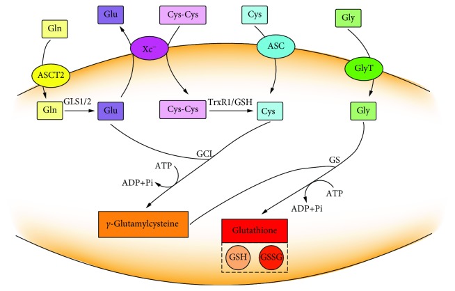
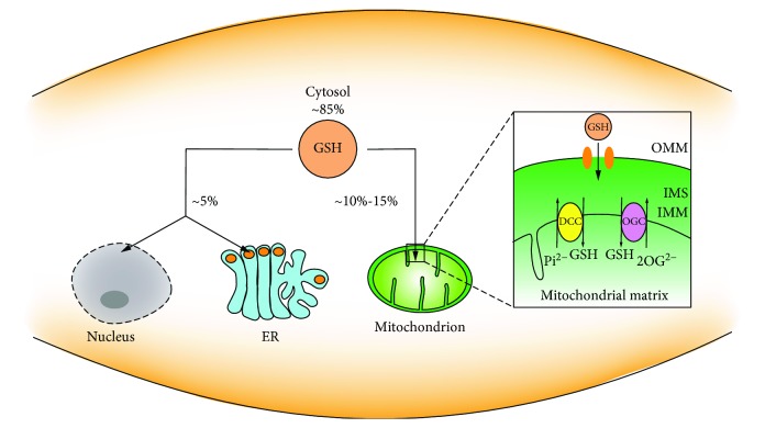
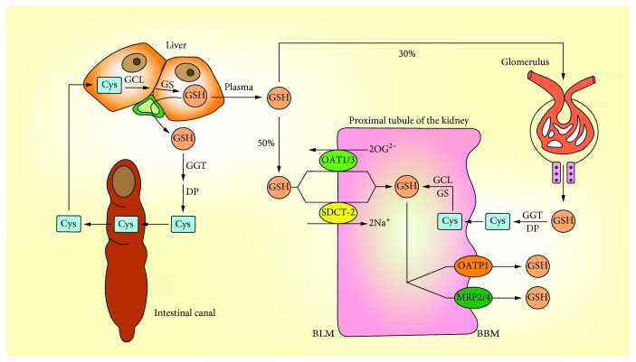
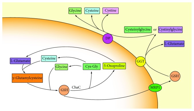
Verification
Claims the paper makes
The claims and statistics of this paper haven’t been checked yet.
Run the check — free, no account needed. It reads the open-access text and checks the sources it cites.
Claims other papers make about this one
Each is checked against this paper’s own text: does it say what the citing paper says it does?
In the citing paper
“Being the main intracellular antioxidant buffer, both the levels of GSH and GSH/GSSH ratio are tightly controlled through a fine regulation of their synthesis, metabolism, transport, and degradation [176,177].”
From The Role of Antioxidants in the Interplay between Oxidative Stress and Senescence (2022) · cites this paper as [177]
In the citing paper
“Glutathione can exist in two forms: reduced glutathione (GSH) and oxidized glutathione (GSSG), which are converted into each other by the enzymatic activity of GPx (that links two GSH in one GSSG through the formation of a disulphide bond) and glutathione reductase (that catalyses the reduction of one GSSG into two GSH to the expenses of NADPH) [177].”
From The Role of Antioxidants in the Interplay between Oxidative Stress and Senescence (2022) · cites this paper as [177]
Self-check
Percentages, sums and reported differences re-computed from the paper’s own numbers.
The self-check needs the paper’s full text.
Citations
Cites
Ferroptosis: An Iron-Dependent Form of Nonapoptotic Cell Death
Dixon, S. J., Lemberg, K. M., Lamprecht, M. R., Skouta, R., Zaitsev, E. M., Gleason, C. E., Patel, D. N., Bauer, A. J., Cantley, A. M., Yang, W. S., et al. (2012) · Cell · doi:10.1016/j.cell.2012.03.042 · Cited by 18,691
Free radicals and antioxidants in normal physiological functions and human disease
Valko, M., Leibfritz, D., Moncol, J., Cronin, M. T. D., Mazur, M., & Telser, J. (2007) · The International Journal of Biochemistry & Cell Biology · doi:10.1016/j.biocel.2006.07.001 · Cited by 14,269
Apoptosis: A Review of Programmed Cell Death
Elmore, S. (2007) · Toxicologic Pathology · doi:10.1080/01926230701320337 · Cited by 13,853
How mitochondria produce reactive oxygen species
Murphy, M. P. (2009) · Biochemical Journal · doi:10.1042/bj20081386 · Cited by 8,207
Regulation of Ferroptotic Cancer Cell Death by GPX4
Yang, W. S., SriRamaratnam, R., Welsch, M. E., Shimada, K., Skouta, R., Viswanathan, V. S., Cheah, J. H., Clemons, P. A., Shamji, A. F., Clish, C. B., et al. (2014) · Cell · doi:10.1016/j.cell.2013.12.010 · Cited by 7,713
Meister, A., & Anderson, M. E. (1983) · Annual Review of Biochemistry · doi:10.1146/annurev.bi.52.070183.003431 · Cited by 5,694
Glutathione Metabolism and Its Implications for Health
Wu, G., Lupton, J. R., Turner, N. D., Fang, Y.-Z., & Yang, S. (2004) · The Journal of Nutrition · doi:10.1093/jn/134.3.489 · Cited by 3,842
Inactivation of the ferroptosis regulator Gpx4 triggers acute renal failure in mice
Friedmann Angeli, J. P., Schneider, M., Proneth, B., Tyurina, Y. Y., Tyurin, V. A., Hammond, V. J., Herbach, N., Aichler, M., Walch, A., Eggenhofer, E., et al. (2014) · Nature Cell Biology · doi:10.1038/ncb3064 · Cited by 3,732
Modulation of oxidative stress as an anticancer strategy
Gorrini, C., Harris, I. S., & Mak, T. W. (2013) · Nature Reviews Drug Discovery · doi:10.1038/nrd4002 · Cited by 3,579
Reactive oxygen species, cellular redox systems, and apoptosis
Circu, M. L., & Aw, T. Y. (2010) · Free Radical Biology and Medicine · doi:10.1016/j.freeradbiomed.2009.12.022 · Cited by 3,340
Research works printed but not linked: 3
- 2003, Journal of Biological Chemistry, 281, 6372
- 2012, Toxicologic Pathology, 29, 997
- 2015, Anticancer Research, 35, 677
Cited by
Sorted by citations
Items 301–320 of 320
Effects of Heyndrickxia coagulans on Waterborne Copper Toxicity
Huang, C.-T., Chen, C., & Liang, Y. (2025) · Life · doi:10.3390/life15020300
Lin, X., Yin, X., Li, B., Liu, Y., & Li, M. (2025) · Medicine · doi:10.1097/md.0000000000043350
Banerjee, S., Banerjee, A., Singhvi, G., Taliyan, R., & Dhar, P. (2025) · Beni-Suef University Journal of Basic and Applied Sciences · doi:10.1186/s43088-025-00706-x
Chen, J., Wang, Z., Fu, J., Cai, Y., Cheng, H., Cui, X., Sun, M., Liu, M., & Zhang, X. (2024) · Figshare · doi:10.6084/m9.figshare.26711942
Li, J., Tang, Y., Lv, B., Wang, J., Wang, Z., & Song, Q. (2024) · Figshare · doi:10.6084/m9.figshare.26182251
Harnessing the ROS for Cancer Treatment
Osama, A., Zhang, J., & Fang, J. (2025) · Springer eBooks · doi:10.1007/978-3-032-00771-1_255
Malard, E. (2023) · theses.fr (ABES) · doi:10.70675/74964e84zb117z490dzb121zf85fa183d480
Characterization of an auranofin-resistant ovarian cancer cell line A2780
Limaj, X. (2024) · theses.fr (ABES) · doi:10.70675/5f21b24bzd436z4168z8555zdc660e2b2aca
Feng, B., Wang, J., Shang, W., 马会浩, Fu, Y., Lv, H., Xu, Y., & Dong, B. (2026) · International Journal of Genomics · doi:10.1155/ijog/7575181
Self-citationThe glutathione metabolism in cancer
Grusac, E., Sardari, V., Cebotari, D., Railean, N., Garbuz, O., Tagadiuc, O., & Gulea, A. (2026) · Advanced Materials in Biopharmaceuticals and Technics · doi:10.59295/mabt2026.23
Yıldırım, T., Ayar, A., Şenkuytu, E., Mesci̇, S., Fidan, M., ÇAKAL, H., Davarcı, D., & Yazgan, B. (2026) · Journal of Molecular Structure · doi:10.1016/j.molstruc.2026.147062
Çetin, S. (2026) · Etlik Veteriner Mikrobiyoloji Dergisi · doi:10.35864/evmd.1953299
Loges, L., Gabel, M., Knauss, A., Atreya, R. N., Gessner, A., Bartels, N., Neurath, M. F., & Weigmann, B. (2026) · The Journal of Immunology · doi:10.1093/jimmun/vkag175
Zhang, H., Cao, F., Wang, J., Yun, C., Qian, A., & Lin, X. (2026) · Antioxidants · doi:10.3390/antiox15080941
Zhao, H., Chen, Y., Liu, L., Peng, F., Guan, B., Li, J., Wang, Q., Liao, L., Chi, B., & Zhang, R. (2026) · Small · doi:10.1002/smll.75188
Ouyang, Y., Hu, Y., Chen, C., Zhao, S., Wu, M., Pan, H., Zhang, W., & Liu, H. (2026) · ACS Materials Letters · doi:10.1021/acsmaterialslett.6c00853
BindingDB (2025) · UC San Diego · doi:10.7270/q21z492q
Metadata
- Authors
- Lv, H., Zhen, C., Liu, J., Yang, P., Hu, L., & Shang, P.
- Affiliations
- Northwestern Polytechnical University (CN)
- Zhejiang Heye Health Technology Co. Ltd., Anji, Zhejiang 313300, China
- Corresponding author
- Shang, Peng
- Venue
- Oxidative Medicine and Cellular Longevity
- Publisher
- Wiley
- Published
- 2019-06-10
- Volume
- 2019
- Pages
- 1-16
- ISSN
- 1942-0994 (ISSN-L)
- 1942-0900
- PMC
- PMC6590529
- OpenAlex
- W2949432489
- Cited by
- 320
- References
- 164
- Citations by year
- 2026: 15 · 2025: 34 · 2024: 78 · 2023: 64 · 2022: 43 · 2021: 46 · 2020: 35 · 2019: 4
- FWCI
- 3.50
- Type
- journal article
- Language
- en
- Open access
- hybrid
- doi.org
- Locations
- Oxidative Medicine and Cellular Longevity (publishedVersion, cc-by)
- PubMed Central (publishedVersion, cc-by)
- PubMed (publishedVersion)
- DOAJ (DOAJ: Directory of Open Access Journals) (submittedVersion)
- Funders
- Northwestern Polytechnical University Foundation for Fundamental Research (3102018JGC012, 3102017OQD111, 51777171, 11872316, 81803032)
- Fundamental Research Funds for the Central Universities (3102018JGC012, 3102017OQD111, 51777171, 11872316, 81803032)
- National Natural Science Foundation of China (3102018JGC012, 3102017OQD111, 51777171, 11872316, 81803032)
- Topics
- Life Sciences › Biochemistry, Genetics and Molecular Biology › Biochemistry › Sulfur Compounds in Biology
- Life Sciences › Biochemistry, Genetics and Molecular Biology › Molecular Biology › Redox biology and oxidative stress
- Life Sciences › Biochemistry, Genetics and Molecular Biology › Molecular Biology › Cell death mechanisms and regulation
- Keywords
- glutathione, glutathione homeostasis, redox homeostasis, oxidative stress, cancer cells, programmed cell death, apoptosis, necroptosis, ferroptosis, autophagy, glutathione depletion, glutathione metabolism
- MeSH terms
- Glutathione, Humans, Neoplasms, Cell Death
About these checks
Registry, retraction, Cites and statistics results are computed, not judged, with one exception: a printed reference that no index lists is read once by a model before it counts as a citation error. Claims are extracted by a model and kept only when their quote appears verbatim in the text; each cited source is then checked, with a model’s help, for whether it says what the claim relies on. Those verdicts can be wrong — read the claim’s quote and the cited source before relying on one.
Statistics and arithmetic are recomputed, not judged.
Confidence bands are shown only for fields calibrated on our annotated test set. None is calibrated yet, so no band is shown.
What counts as a claim ›How we measure confidence ›How we check this ›
Are you an author, or do you see a problem with this page? Request a review
