

The DOI matches its Crossref record; 32 of the 32 references it cites were found in a registry or an index.
Extensive Proliferation of a Subset of Differentiated, yet Plastic, Medial Vascular Smooth Muscle Cells Contributes to Neointimal Formation in Mouse Injury and Atherosclerosis Models
Open accessAbstract
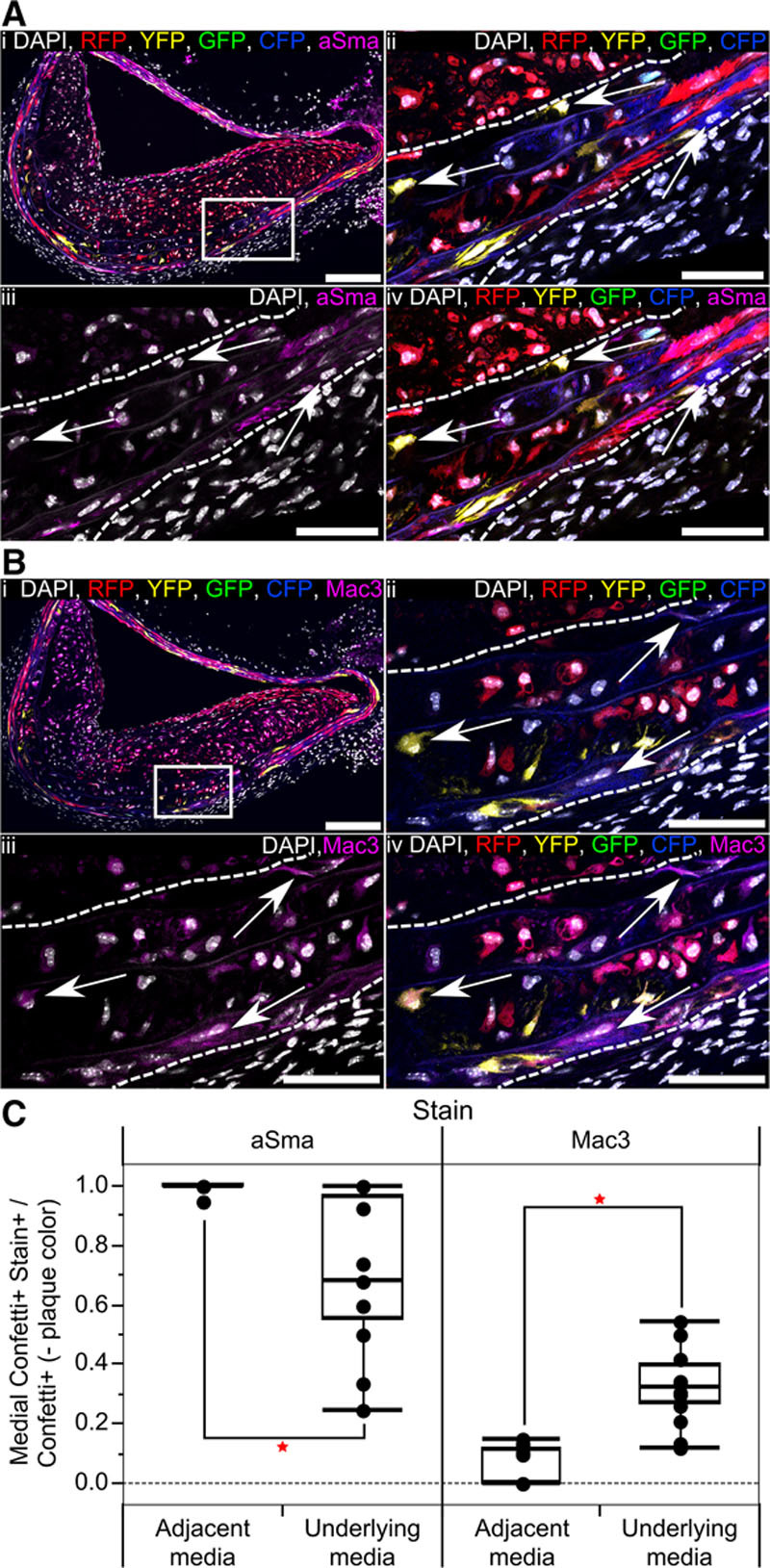
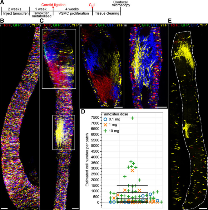
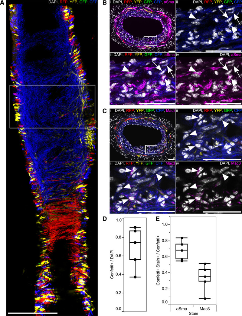
Rationale: Vascular smooth muscle cell (VSMC) accumulation is a hallmark of atherosclerosis and vascular injury. However, fundamental aspects of proliferation and the phenotypic changes within individual VSMCs, which underlie vascular disease, remain unresolved. In particular, it is not known whether all VSMCs proliferate and display plasticity or whether individual cells can switch to multiple phenotypes. Objective: To assess whether proliferation and plasticity in disease is a general characteristic of VSMCs or a feature of a subset of cells. Methods and Results: Using multicolor lineage labeling, we demonstrate that VSMCs in injury-induced neointimal lesions and in atherosclerotic plaques are oligoclonal, derived from few expanding cells. Lineage tracing also revealed that the progeny of individual VSMCs contributes to both alpha smooth muscle actin (aSma)–positive fibrous cap and Mac3-expressing macrophage-like plaque core cells. Costaining for phenotypic markers further identified a double-positive aSma+ Mac3+ cell population, which is specific to VSMC-derived plaque cells. In contrast, VSMC-derived cells generating the neointima after vascular injury generally retained the expression of VSMC markers and the upregulation of Mac3 was less pronounced. Monochromatic regions in atherosclerotic plaques and injury-induced neointima did not contain VSMC-derived cells expressing a different fluorescent reporter protein, suggesting that proliferation-independent VSMC migration does not make a major contribution to VSMC accumulation in vascular disease. Conclusions: We demonstrate that extensive proliferation of a low proportion of highly plastic VSMCs results in the observed VSMC accumulation after injury and in atherosclerotic plaques. Therapeutic targeting of these hyperproliferating VSMCs might effectively reduce vascular disease without affecting vascular integrity.
Overview
The claims and statistics of this paper haven’t been checked yet.
Run the check — free, no account needed. It reads the open-access text and checks the sources it cites.
Integrity signals
Registry
Cites
Statistics
Figures
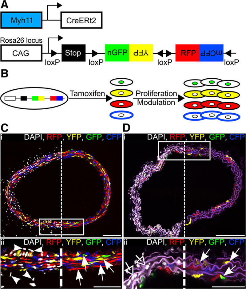
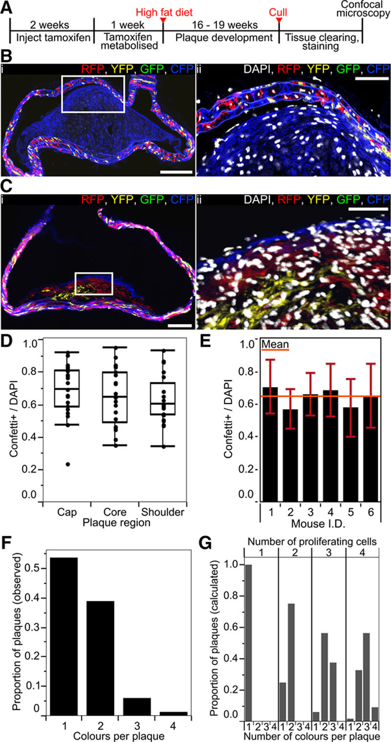
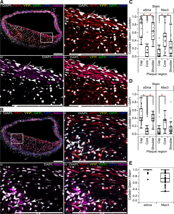
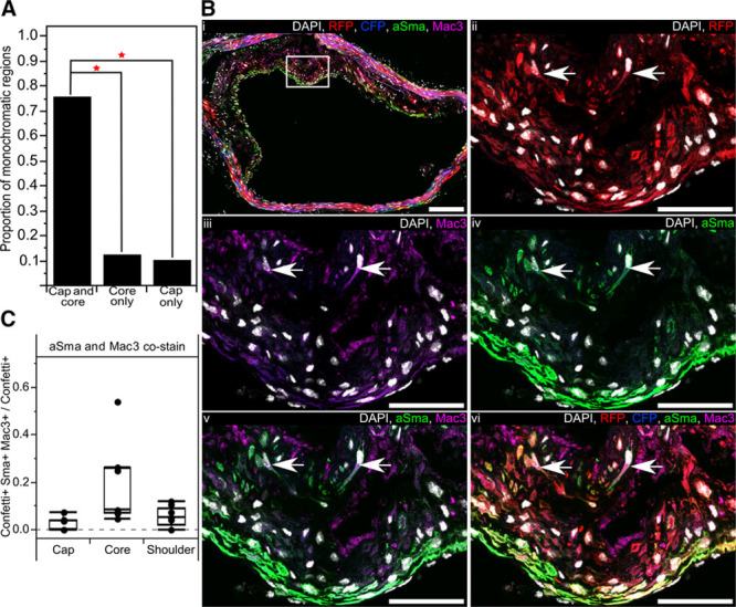
Verification
Claims the paper makes
The claims and statistics of this paper haven’t been checked yet.
Run the check — free, no account needed. It reads the open-access text and checks the sources it cites.
Claims other papers make about this one
Each is checked against this paper’s own text: does it say what the citing paper says it does?
In the citing paper
“Lineage tracing studies have unambiguously demonstrated that VSMCs contribute substantially to plaque formation in murine models of atherosclerosis, generating 30-70% of all plaque cells 11,18-20,22,23 .”
From Vascular smooth muscle cells in atherosclerosis (2019) · cites this paper as [20]
In the citing paper
“Within plaques a large proportion of reporter-expressing VSMC-derived cells do not have detectable levels of the contractile smooth muscle cell marker α SMA 11,20 .”
From Vascular smooth muscle cells in atherosclerosis (2019) · cites this paper as [20]
In the citing paper
“Instead, some plaque reporter-expressing cells were positive for Mac-3 20 , Lgals3 11 and CD68 17 - markers that have been previously used to study macrophages in atherosclerosis.”
From Vascular smooth muscle cells in atherosclerosis (2019) · cites this paper as [20]
Self-check
Percentages, sums and reported differences re-computed from the paper’s own numbers.
The self-check needs the paper’s full text.
Citations
Cites
Vascular smooth muscle cells in atherosclerosis
Bennett, M. R., Sinha, S., & Owens, G. K. (2016) · Circulation Research · doi:10.1161/circresaha.115.306361 · Cited by 2,179
Self-citationSnippert, H. J., van der Flier, L. G., Sato, T., van Es, J. H., van den Born, M., Kroon-Veenboer, C., Barker, N., Klein, A. M., van Rheenen, J., Simons, B. D., et al. (2010) · Cell · doi:10.1016/j.cell.2010.09.016 · Cited by 1,951
Self-citationClowes, A. W., Reidy, M. A., & Mm, C. (1983) · PubMed · Cited by 1,439
Shankman, L. S., Gomez, D., Cherepanova, O. A., Salmon, M., Alencar, G. F., Haskins, R. M., Swiatlowska, P., Newman, A. A. C., Greene, E. S., Straub, A. C., et al. (2015) · Nature Medicine · doi:10.1038/nm.3866 · Cited by 1,218
Piedrahita, J. A., Zhang, S. H., Hagaman, J. R., Oliver, P. M., & Maeda, N. (1992) · Proceedings of the National Academy of Sciences · doi:10.1073/pnas.89.10.4471 · Cited by 944
Regulation and characteristics of vascular smooth muscle cell phenotypic diversity
Rensen, S. S., Doevendans, P. A., & van Eys, G. J. J. M. (2007) · Netherlands Heart Journal · doi:10.1007/bf03085963 · Cited by 917
Evidence for a Monoclonal Origin of Human Atherosclerotic Plaques
Benditt, E. P., & Benditt, J. M. (1973) · Proceedings of the National Academy of Sciences · doi:10.1073/pnas.70.6.1753 · Cited by 913
Alexander, M. R., & Owens, G. K. (2011) · Annual Review of Physiology · doi:10.1146/annurev-physiol-012110-142315 · Cited by 796
Shankman, L. S., Gomez, D. A., Cherepanova, O. A., Salmon, M., Alencar, G. F., Haskins, R. M., Swiatlowska, P., Newman, A. A. C., Greene, E. S., Straub, A. C., et al. (2016) · Nature Medicine · doi:10.1038/nm0216-217a · Cited by 775
G12-G13–LARG–mediated signaling in vascular smooth muscle is required for salt-induced hypertension
Wirth, A., Benyó, Z., Lukasova, M., Leutgeb, B., Wettschureck, N., Gorbey, S., Örsy, P., Horváth, B., Maser‐Gluth, C., Greiner, E. F., et al. (2007) · Nature Medicine · doi:10.1038/nm1666 · Cited by 692
Clarke, M. C. H., Figg, N. L., Maguire, J. J., Davenport, A. P., Goddard, M. J., Littlewood, T. D., & Bennett, M. R. (2006) · Nature Medicine · doi:10.1038/nm1459 · Cited by 645
Self-citationMatthews, C., Gorenne, I., Scott, S., Figg, N., Kirkpatrick, P., Ritchie, A., Goddard, M., & Bennett, M. (2006) · Circulation Research · doi:10.1161/01.res.0000233315.38086.bc · Cited by 615 · cited 1×
“replicative exhaustion, telomere shortening, and increased senescence observed in human plaques.(22,23) Consistent with”
Self-citationTransdifferentiation of Vascular Smooth Muscle Cells to Macrophage-Like Cells During Atherogenesis
Feil, S., Fehrenbacher, B., Lukowski, R., Essmann, F., Schulze-Osthoff, K., Schaller, M., & Feil, R. (2014) · Circulation Research · doi:10.1161/circresaha.115.304634 · Cited by 578
Significance of quiescent smooth muscle migration in the injured rat carotid artery.
CLOWES, A. W., & Schwartz, S. M. (1985) · Circulation Research · doi:10.1161/01.res.56.1.139 · Cited by 575
Remodeling With Neointima Formation in the Mouse Carotid Artery After Cessation of Blood Flow
Kumar, A. F., & Lindner, V. (1997) · Arteriosclerosis Thrombosis and Vascular Biology · doi:10.1161/01.atv.17.10.2238 · Cited by 532
Significance of quiescent smooth muscle migration in the injured rat carotid artery.
WClowes, A., & MSchwartz, S. (1985) · Circulation Research · Cited by 337
Gardner, S. E., Humphry, M., Bennett, M. R., & Clarke, M. C. H. (2015) · Arteriosclerosis, Thrombosis, and Vascular Biology · doi:10.1161/atvbaha.115.305896 · Cited by 285
Self-citationDetection of histone modifications at specific gene loci in single cells in histological sections
Gomez, D., Shankman, L. S., Nguyen, A. T., & Owens, G. K. (2013) · Nature Methods · doi:10.1038/nmeth.2332 · Cited by 253
Multiple repressor pathways contribute to phenotypic switching of vascular smooth muscle cells
Kawai‐Kowase, K., & Owens, G. K. (2006) · American Journal of Physiology-Cell Physiology · doi:10.1152/ajpcell.00394.2006 · Cited by 245
Smooth muscle cell progenitors are primed to muscularize in pulmonary hypertension
Sheikh, A. Q., Misra, A., Rosas, I. O., Adams, R. H., & Greif, D. M. (2015) · Science Translational Medicine · doi:10.1126/scitranslmed.aaa9712 · Cited by 174
Herring, B. P., Hoggatt, A. M., Burlak, C., & Offermanns, S. (2014) · Vascular Cell · doi:10.1186/2045-824x-6-21 · Cited by 140
Nemenoff, R. A., Horita, H. N., Ostriker, A. C., Furgeson, S. B., Simpson, P. A., VanPutten, V., Crossno, J. T., Offermanns, S., & Weiser‐Evans, M. C. M. (2011) · Arteriosclerosis Thrombosis and Vascular Biology · doi:10.1161/atvbaha.111.223701 · Cited by 133
Smooth Muscle Phenotypic Modulation—A Personal Experience
Campbell, J. H., & CAMPBELL, G. R. (2012) · Arteriosclerosis Thrombosis and Vascular Biology · doi:10.1161/atvbaha.111.243212 · Cited by 126
Lineage tracing of cells involved in atherosclerosis
Albarrán-Juárez, J., Kaur, H., Grimm, M., Offermanns, S., & Wettschureck, N. (2016) · Atherosclerosis · doi:10.1016/j.atherosclerosis.2016.06.012 · Cited by 90
Yu, H., Clarke, M. C. H., Figg, N. L., Littlewood, T. D., & Bennett, M. R. (2011) · Arteriosclerosis Thrombosis and Vascular Biology · doi:10.1161/atvbaha.111.235622 · Cited by 76
Self-citationMechanical properties and structure of carotid arteries in mice lacking desmin
Lacolley, P. J., Challande, P., Boumaza, S., Cohuet, G., Laurent, S., Boutouyrie, P., Grimaud, J.-A., Paulin, D., Lamazière, J. D., & Li, Z. (2001) · Cardiovascular Research · doi:10.1016/s0008-6363(01)00278-4 · Cited by 73
Yu, H., Stoneman, V. E. A., Clarke, M. C. H., Figg, N. L., Xin, H., Kotlikoff, M. I., Littlewood, T. D., & Bennett, M. R. (2011) · Arteriosclerosis Thrombosis and Vascular Biology · doi:10.1161/atvbaha.110.218578 · Cited by 69
Self-citationIntegrin β3 inhibition is a therapeutic strategy for supravalvular aortic stenosis
Misra, A., Sheikh, A. Q., Kumar, A., Luo, J., Zhang, J., Hinton, R. B., Smoot, L. B., Kaplan, P. B., Urbán, Z., Qyang, Y., et al. (2016) · The Journal of Experimental Medicine · doi:10.1084/jem.20150688 · Cited by 53
Alterations in population dynamics of arterial smooth muscle cells during atherogenesis
Thomas, W. A., Florentin, R. A., Reiner, J. M., Lee, W. M., & Lee, K. T. (1976) · Experimental and Molecular Pathology · doi:10.1016/0014-4800(76)90009-5 · Cited by 52
Characterization of Proliferating Lesion‐Resident Cells During All Stages of Atherosclerotic Growth
Lhoták, Š., Gyulay, G., Cutz, J., Al‐Hashimi, A., Trigatti, B. L., Richards, C. D., Igdoura, S. A., Steinberg, G. R., Bramson, J. L., Ask, K., et al. (2016) · Journal of the American Heart Association · doi:10.1161/jaha.116.003945 · Cited by 45
Yang, P., Hong, M. S., Fu, C., Schmit, B. M., Su, Y., Berceli, S. A., & Jiang, Z. (2015) · Surgery · doi:10.1016/j.surg.2015.08.015 · Cited by 24
Josephy, P. D., Bibeau, K. L., & Evans, D. H. (2000) · Environmental and Molecular Mutagenesis · doi:10.1002/1098-2280(2000)35:4 · Cited by 15
Research works printed but not linked: 1
- 10.1002/1098-2280 · DOI
Cited by
Sorted by citations
Borén, J., Chapman, M. J., Krauss, R. M., Packard, C. J., Bentzon, J. F., Binder, C. J., Daemen, M. J., Demer, L. L., Hegele, R. A., Nicholls, S. J., et al. (2020) · European Heart Journal · doi:10.1093/eurheartj/ehz962 · Cited by 1,601
Vascular smooth muscle cells in atherosclerosis
Basatemur, G. L., Jørgensen, H. F., Clarke, M. C. H., Bennett, M. R., & Mallat, Z. (2019) · Nature Reviews Cardiology · doi:10.1038/s41569-019-0227-9 · Cited by 1,099
Self-citationTargeting inflammation in atherosclerosis — from experimental insights to the clinic
Soehnlein, O., & Libby, P. (2021) · Nature Reviews Drug Discovery · doi:10.1038/s41573-021-00198-1 · Cited by 1,085
Wirka, R. C., Wagh, D. A., Paik, D. T., Pjanic, M., Nguyen, T., Miller, C. L., Kundu, R., Nagao, M., Coller, J., Koyano, T. K., et al. (2019) · Nature Medicine · doi:10.1038/s41591-019-0512-5 · Cited by 874
HDL and Reverse Cholesterol Transport
Ouimet, M. I., Barrett, T. J., & Fisher, E. A. A. (2019) · Circulation Research · doi:10.1161/circresaha.119.312617 · Cited by 816
Metadata
- Authors
- Chappell, J., Harman, J. L., Narasimhan, V. M., Yu, H., Foote, K., Simons, B. D., Bennett, M. R., & Jørgensen, H. F.
- Affiliations
- Wellcome/MRC Cambridge Stem Cell Institute (GB)
- From the Cardiovascular Medicine Division, Department of Medicine (J.C., J.L.H., H.Y., K.F., M.R.B., H.F.J.), Cavendish Laboratory, Department of Physics (B.D.S.), The Wellcome Trust/Cancer Research UK Gurdon Institute (B.D.S.), and Wellcome Trust-Medical Research Council Stem Cell Institute (B.D.S.), University of Cambridge, United Kingdom; and The Wellcome Trust Sanger Institute, Hinxton, Cambridge, United Kingdom (V.M.N.).
- Corresponding author
- Jørgensen, Helle F.
- ORCID
- Venue
- Circulation Research
- Published
- 2016-12-09
- Volume
- 119
- Issue
- 12
- Pages
- 1313-1323
- ISSN
- 0009-7330 (ISSN-L)
- 1524-4571
- PMC
- PMC5149073
- OpenAlex
- W2525459565
- Cited by
- 437
- References
- 32
- Citations by year
- 2026: 39 · 2025: 38 · 2024: 45 · 2023: 56 · 2022: 62 · 2021: 62 · 2020: 39 · 2019: 45 · 2018: 29 · 2017: 17 · 2016: 3
- FWCI
- 18.96
- Type
- journal article
- Language
- en
- Open access
- hybrid
- doi.org
- Licence
- cc-by
- Locations
- Circulation Research (publishedVersion, cc-by)
- Europe PMC (PubMed Central) (publishedVersion, cc-by)
- PubMed Central (publishedVersion, cc-by)
- PubMed (publishedVersion)
- Apollo (University of Cambridge) (submittedVersion, cc-by)
- Apollo (cc-by)
- Funders
- Wellcome Trust (098357/Z/12/Z, 100574/Z/12/Z)
- British Heart Foundation (PG/13/25/30014, PG/16/11/32021, PG/12/86/29930, RM/13/3/30159, RG/13/14/30314, FS/15/38/31516, RE/13/6/30180)
- Topics
- Life Sciences › Immunology and Microbiology › Immunology › Atherosclerosis and Cardiovascular Diseases
- Health Sciences › Medicine › Surgery › Cardiovascular, Neuropeptides, and Oxidative Stress Research
- Health Sciences › Medicine › Cardiology and Cardiovascular Medicine › Cerebrovascular and Carotid Artery Diseases
- Keywords
- vascular smooth muscle cells, vascular smooth muscle cell proliferation, phenotypic plasticity, intimal hyperplasia, atherosclerosis, atherosclerotic plaque, lineage tracing, alpha-smooth muscle actin, VSMC migration, vascular injury
- MeSH terms
- Animals, Disease Models, Animal, Mice, Inbred BALB C, Mice, Inbred C57BL, Mice, Transgenic, Muscle, Smooth, Vascular, Mice, Knockout, Myocytes, Smooth Muscle, Cell Proliferation, Atherosclerosis, Mice, Vascular System Injuries, Neointima
About these checks
Registry, retraction, Cites and statistics results are computed, not judged, with one exception: a printed reference that no index lists is read once by a model before it counts as a citation error. Claims are extracted by a model and kept only when their quote appears verbatim in the text; each cited source is then checked, with a model’s help, for whether it says what the claim relies on. Those verdicts can be wrong — read the claim’s quote and the cited source before relying on one.
Statistics and arithmetic are recomputed, not judged.
Confidence bands are shown only for fields calibrated on our annotated test set. None is calibrated yet, so no band is shown.
What counts as a claim ›How we measure confidence ›How we check this ›
Are you an author, or do you see a problem with this page? Request a review
