

The DOI matches its Crossref record; 32 of the 32 references it cites were found in a registry or an index.
Extensive Proliferation of a Subset of Differentiated, yet Plastic, Medial Vascular Smooth Muscle Cells Contributes to Neointimal Formation in Mouse Injury and Atherosclerosis Models
Open accessAbstract
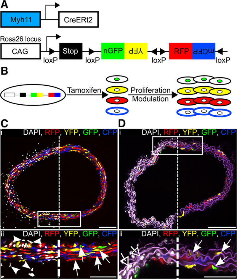
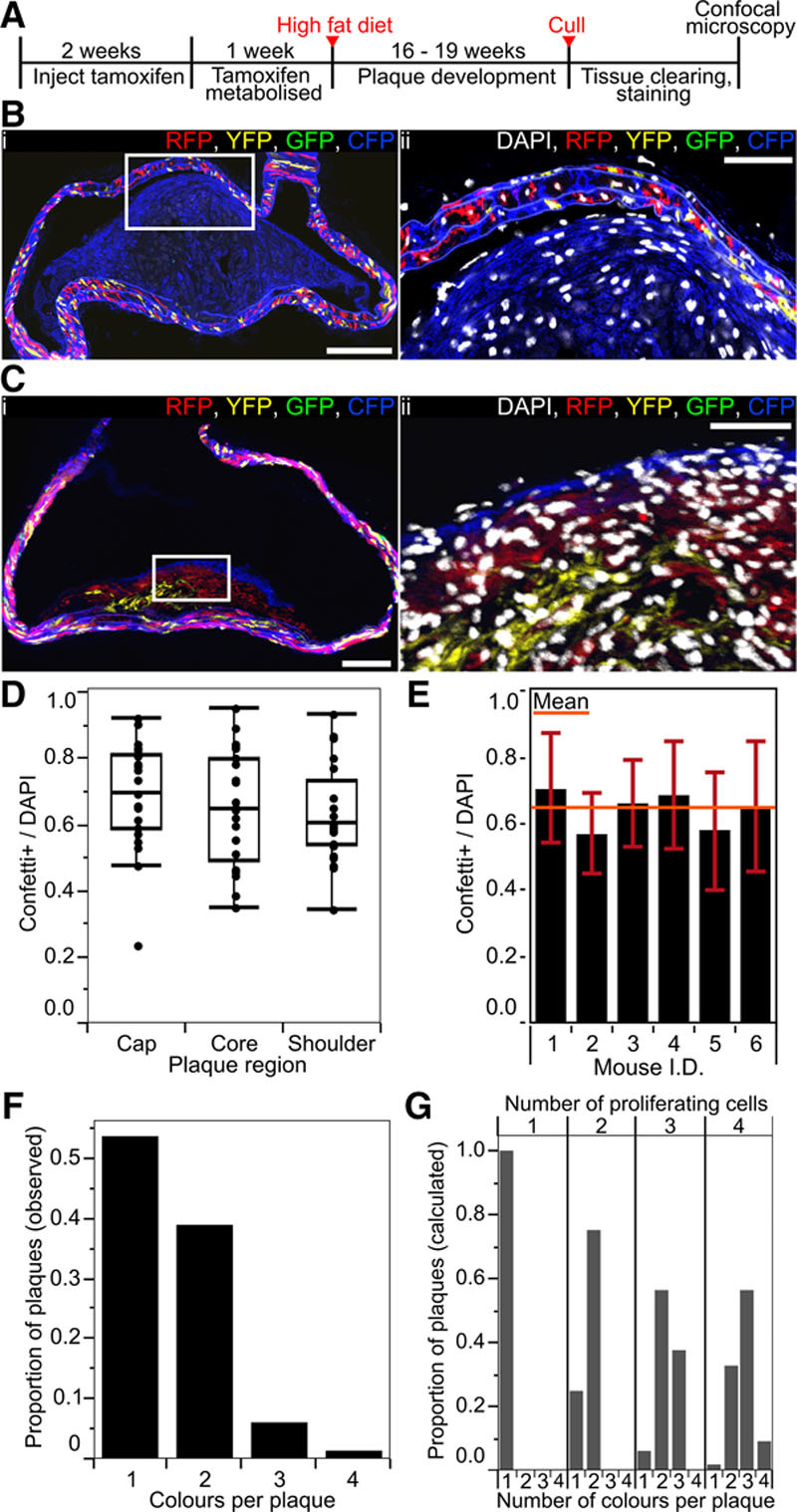
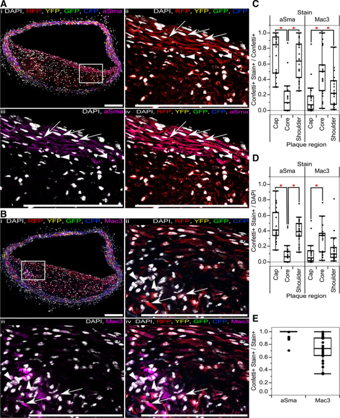
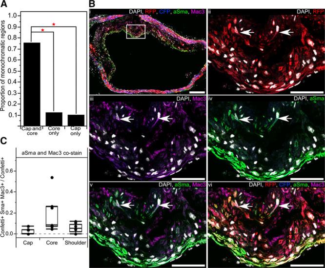
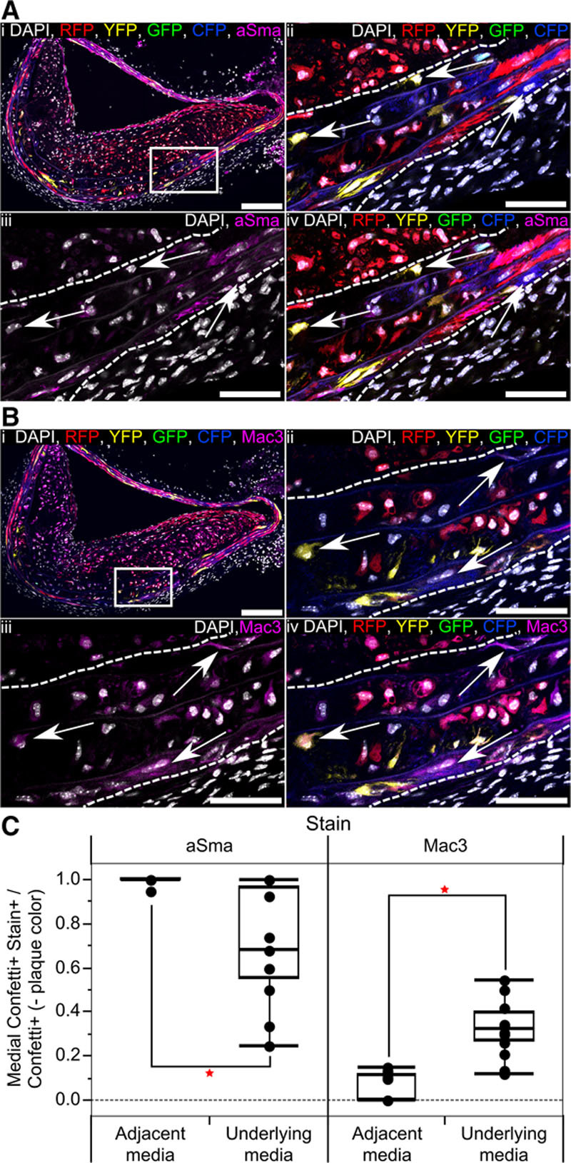
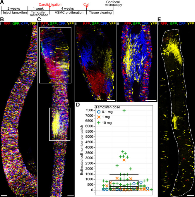
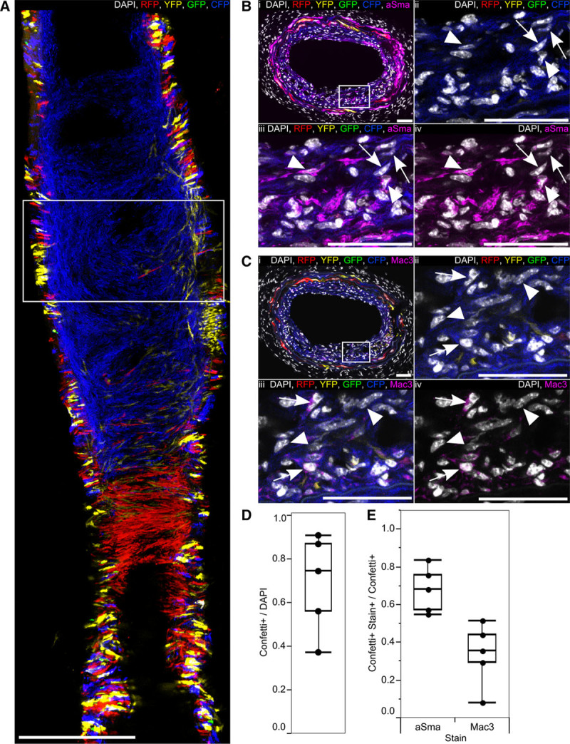
Rationale: Vascular smooth muscle cell (VSMC) accumulation is a hallmark of atherosclerosis and vascular injury. However, fundamental aspects of proliferation and the phenotypic changes within individual VSMCs, which underlie vascular disease, remain unresolved. In particular, it is not known whether all VSMCs proliferate and display plasticity or whether individual cells can switch to multiple phenotypes. Objective: To assess whether proliferation and plasticity in disease is a general characteristic of VSMCs or a feature of a subset of cells. Methods and Results: Using multicolor lineage labeling, we demonstrate that VSMCs in injury-induced neointimal lesions and in atherosclerotic plaques are oligoclonal, derived from few expanding cells. Lineage tracing also revealed that the progeny of individual VSMCs contributes to both alpha smooth muscle actin (aSma)–positive fibrous cap and Mac3-expressing macrophage-like plaque core cells. Costaining for phenotypic markers further identified a double-positive aSma+ Mac3+ cell population, which is specific to VSMC-derived plaque cells. In contrast, VSMC-derived cells generating the neointima after vascular injury generally retained the expression of VSMC markers and the upregulation of Mac3 was less pronounced. Monochromatic regions in atherosclerotic plaques and injury-induced neointima did not contain VSMC-derived cells expressing a different fluorescent reporter protein, suggesting that proliferation-independent VSMC migration does not make a major contribution to VSMC accumulation in vascular disease. Conclusions: We demonstrate that extensive proliferation of a low proportion of highly plastic VSMCs results in the observed VSMC accumulation after injury and in atherosclerotic plaques. Therapeutic targeting of these hyperproliferating VSMCs might effectively reduce vascular disease without affecting vascular integrity.
Overview
The claims and statistics of this paper haven’t been checked yet.
Run the check — free, no account needed. It reads the open-access text and checks the sources it cites.
Integrity signals
Registry
Cites
Statistics
Figures
Verification
Claims the paper makes
The claims and statistics of this paper haven’t been checked yet.
Run the check — free, no account needed. It reads the open-access text and checks the sources it cites.
Claims other papers make about this one
Each is checked against this paper’s own text: does it say what the citing paper says it does?
In the citing paper
“Lineage tracing studies have unambiguously demonstrated that VSMCs contribute substantially to plaque formation in murine models of atherosclerosis, generating 30-70% of all plaque cells 11,18-20,22,23 .”
From Vascular smooth muscle cells in atherosclerosis (2019) · cites this paper as [20]
In the citing paper
“Within plaques a large proportion of reporter-expressing VSMC-derived cells do not have detectable levels of the contractile smooth muscle cell marker α SMA 11,20 .”
From Vascular smooth muscle cells in atherosclerosis (2019) · cites this paper as [20]
In the citing paper
“Instead, some plaque reporter-expressing cells were positive for Mac-3 20 , Lgals3 11 and CD68 17 - markers that have been previously used to study macrophages in atherosclerosis.”
From Vascular smooth muscle cells in atherosclerosis (2019) · cites this paper as [20]
Self-check
Percentages, sums and reported differences re-computed from the paper’s own numbers.
The self-check needs the paper’s full text.
Citations
Cites
Vascular smooth muscle cells in atherosclerosis
Bennett, M. R., Sinha, S., & Owens, G. K. (2016) · Circulation Research · doi:10.1161/circresaha.115.306361 · Cited by 2,179
Self-citationSnippert, H. J., van der Flier, L. G., Sato, T., van Es, J. H., van den Born, M., Kroon-Veenboer, C., Barker, N., Klein, A. M., van Rheenen, J., Simons, B. D., et al. (2010) · Cell · doi:10.1016/j.cell.2010.09.016 · Cited by 1,951
Self-citationClowes, A. W., Reidy, M. A., & Mm, C. (1983) · PubMed · Cited by 1,439
Shankman, L. S., Gomez, D., Cherepanova, O. A., Salmon, M., Alencar, G. F., Haskins, R. M., Swiatlowska, P., Newman, A. A. C., Greene, E. S., Straub, A. C., et al. (2015) · Nature Medicine · doi:10.1038/nm.3866 · Cited by 1,218
Piedrahita, J. A., Zhang, S. H., Hagaman, J. R., Oliver, P. M., & Maeda, N. (1992) · Proceedings of the National Academy of Sciences · doi:10.1073/pnas.89.10.4471 · Cited by 944
Regulation and characteristics of vascular smooth muscle cell phenotypic diversity
Rensen, S. S., Doevendans, P. A., & van Eys, G. J. J. M. (2007) · Netherlands Heart Journal · doi:10.1007/bf03085963 · Cited by 917
Evidence for a Monoclonal Origin of Human Atherosclerotic Plaques
Benditt, E. P., & Benditt, J. M. (1973) · Proceedings of the National Academy of Sciences · doi:10.1073/pnas.70.6.1753 · Cited by 913
Alexander, M. R., & Owens, G. K. (2011) · Annual Review of Physiology · doi:10.1146/annurev-physiol-012110-142315 · Cited by 796
Shankman, L. S., Gomez, D. A., Cherepanova, O. A., Salmon, M., Alencar, G. F., Haskins, R. M., Swiatlowska, P., Newman, A. A. C., Greene, E. S., Straub, A. C., et al. (2016) · Nature Medicine · doi:10.1038/nm0216-217a · Cited by 775
G12-G13–LARG–mediated signaling in vascular smooth muscle is required for salt-induced hypertension
Wirth, A., Benyó, Z., Lukasova, M., Leutgeb, B., Wettschureck, N., Gorbey, S., Örsy, P., Horváth, B., Maser‐Gluth, C., Greiner, E. F., et al. (2007) · Nature Medicine · doi:10.1038/nm1666 · Cited by 692
Research works printed but not linked: 1
- 10.1002/1098-2280 · DOI
Cited by
Sorted by citations
Borén, J., Chapman, M. J., Krauss, R. M., Packard, C. J., Bentzon, J. F., Binder, C. J., Daemen, M. J., Demer, L. L., Hegele, R. A., Nicholls, S. J., et al. (2020) · European Heart Journal · doi:10.1093/eurheartj/ehz962 · Cited by 1,601
Vascular smooth muscle cells in atherosclerosis
Basatemur, G. L., Jørgensen, H. F., Clarke, M. C. H., Bennett, M. R., & Mallat, Z. (2019) · Nature Reviews Cardiology · doi:10.1038/s41569-019-0227-9 · Cited by 1,099
Self-citationTargeting inflammation in atherosclerosis — from experimental insights to the clinic
Soehnlein, O., & Libby, P. (2021) · Nature Reviews Drug Discovery · doi:10.1038/s41573-021-00198-1 · Cited by 1,085
Wirka, R. C., Wagh, D. A., Paik, D. T., Pjanic, M., Nguyen, T., Miller, C. L., Kundu, R., Nagao, M., Coller, J., Koyano, T. K., et al. (2019) · Nature Medicine · doi:10.1038/s41591-019-0512-5 · Cited by 874
HDL and Reverse Cholesterol Transport
Ouimet, M. I., Barrett, T. J., & Fisher, E. A. A. (2019) · Circulation Research · doi:10.1161/circresaha.119.312617 · Cited by 816
Pan, H., Xue, C., Auerbach, B. J., Fan, J., Bashore, A. C., Cui, J. G., Yang, D. Y., Trignano, S. B., Liu, W., Shi, J., et al. (2020) · Circulation · doi:10.1161/circulationaha.120.048378 · Cited by 619
Vascular smooth muscle cells in atherosclerosis: time for a re-assessment
Grootaert, M. O. J., & Bennett, M. R. (2021) · Cardiovascular Research · doi:10.1093/cvr/cvab046 · Cited by 531
Self-citationSmooth muscle cell fate and plasticity in atherosclerosis
Allahverdian, S., Chaâbane, C., Boukais, K., Francis, G. A., & Bochaton‐Piallat, M. (2018) · Cardiovascular Research · doi:10.1093/cvr/cvy022 · Cited by 513
Kim, K., Shim, D., Lee, J. S., Zaitsev, K., Williams, J. W., Kim, K.-W., Jang, M.-Y., Seok Jang, H., Yun, T. J., Lee, S. H., et al. (2018) · Circulation Research · doi:10.1161/circresaha.118.312804 · Cited by 434
How vascular smooth muscle cell phenotype switching contributes to vascular disease
Cao, G., Xuan, X., Hu, J. H., Zhang, R., Jin, H., & Dong, H. (2022) · Cell Communication and Signaling · doi:10.1186/s12964-022-00993-2 · Cited by 472
Smooth muscle cell-driven vascular diseases and molecular mechanisms of VSMC plasticity
Frismantiene, A., Philippova, M., Erné, P., & Resink, T. J. (2018) · Cellular Signalling · doi:10.1016/j.cellsig.2018.08.019 · Cited by 415
DNA hypermethylation in disease: mechanisms and clinical relevance
Ehrlich, M. (2019) · Epigenetics · doi:10.1080/15592294.2019.1638701 · Cited by 382
Fate and state of vascular smooth muscle cells in atherosclerosis
Miano, J. M., Fisher, E. A., & Majesky, M. W. (2021) · Circulation · doi:10.1161/circulationaha.120.049922 · Cited by 318
Dobnikar, L., Taylor, A. L., Chappell, J., Oldach, P., Harman, J. J., Oerton, E., Dzierzak, E. A., Bennett, M. R., Spivakov, M., & Jørgensen, H. F. (2018) · Nature Communications · doi:10.1038/s41467-018-06891-x · Cited by 298
Self-citationRole of Vascular Smooth Muscle Cell Plasticity and Interactions in Vessel Wall Inflammation
Sorokin, V., Vickneson, K., Kofidis, T., Woo, C. C., Lin, X. Y., Foo, R., & Shanahan, C. M. (2020) · Frontiers in Immunology · doi:10.3389/fimmu.2020.599415 · Cited by 292
Smooth Muscle Cell Phenotypic Diversity
Liu, M., & Gomez, D. A. (2019) · Arteriosclerosis Thrombosis and Vascular Biology · doi:10.1161/atvbaha.119.312131 · Cited by 281
Six Shades of Vascular Smooth Muscle Cells Illuminated by KLF4 (Krüppel-Like Factor 4)
Yap, C., Mieremet, A., de Vries, C. J. M., Micha, D., & de Waard, V. (2021) · Arteriosclerosis Thrombosis and Vascular Biology · doi:10.1161/atvbaha.121.316600 · Cited by 283
Resident and Monocyte-Derived Macrophages in Cardiovascular Disease
Honold, L., & Nahrendorf, M. (2018) · Circulation Research · doi:10.1161/circresaha.117.311071 · Cited by 283
Smooth muscle cell and arterial aging: basic and clinical aspects
Lacolley, P. J., Regnault, V., & Avolio, A. P. (2018) · Cardiovascular Research · doi:10.1093/cvr/cvy009 · Cited by 229
Integrin beta3 regulates clonality and fate of smooth muscle-derived atherosclerotic plaque cells
Misra, A., Feng, Z., Chandran, R. R., Kabir, I., Rotllan, N., Aryal, B., Sheikh, A. Q., Ding, L., Qin, L., Fernández-Hernando, C., et al. (2018) · Nature Communications · doi:10.1038/s41467-018-04447-7 · Cited by 207
Phenotypic Switching of Vascular Smooth Muscle Cells in Atherosclerosis
Chen, R., McVey, D. G., Shen, D., Huang, X., & Ye, S. (2023) · Journal of the American Heart Association · doi:10.1161/jaha.123.031121 · Cited by 210
Li, Z., Solomonidis, E. G., Meloni, M., Taylor, R. S., Duffin, R., Dobie, R., Pinto, M. M., Henderson, B. E. P., Louwe, P. A., D’Amico, G., et al. (2019) · European Heart Journal · doi:10.1093/eurheartj/ehz305 · Cited by 206
Self-citationTargeting smooth muscle cell phenotypic switching in vascular disease
Chakraborty, R., Chatterjee, P., Dave, J. M., Ostriker, A. C., Greif, D. M., Rzucidlo, E. M., & Martin, K. A. (2021) · JVS Vascular Science · doi:10.1016/j.jvssci.2021.04.001 · Cited by 182
Vascular Smooth Muscle Cell Plasticity and Autophagy in Dissecting Aortic Aneurysms
Clément, M., Chappell, J., Raffort, J., Lareyre, F., Vandestienne, M., Taylor, A. L., Finigan, A., Harrison, J., Bennett, M. R., Bruneval, P., et al. (2019) · Arteriosclerosis, Thrombosis, and Vascular Biology · doi:10.1161/atvbaha.118.311727 · Cited by 184
Self-citationJacobsen, K., Lund, M. B., Shim, J., Gunnersen, S., Füchtbauer, E.-M., Kjolby, M., Carramolino, L., & Bentzon, J. F. (2017) · JCI Insight · doi:10.1172/jci.insight.95890 · Cited by 172
Promoters to Study Vascular Smooth Muscle
Chakraborty, R., Saddouk, F. Z., Carrao, A. C. R., Krause, D. S., Greif, D. M., & Martin, K. A. (2019) · Arteriosclerosis Thrombosis and Vascular Biology · doi:10.1161/atvbaha.119.312449 · Cited by 171
Vascular Macrophages in Atherosclerosis
Xu, H., Jiang, J., Chen, W., Li, W., & Chen, Z. (2019) · Journal of Immunology Research · doi:10.1155/2019/4354786 · Cited by 176
Fasolo, F., Jin, H., Winski, G., Chernogubova, E., Pauli, J., Winter, H., Li, D. Y., Glukha, N., Bauer, S., Metschl, S., et al. (2021) · Circulation · doi:10.1161/circulationaha.120.052023 · Cited by 167
Zhang, F. S., Guo, X. Q., Xia, Y., & Mao, L. (2021) · Cellular and Molecular Life Sciences · doi:10.1007/s00018-021-04079-z · Cited by 177
Macrophages in vascular inflammation and atherosclerosis
Cochain, C., & Zernecke, A. (2017) · Pflügers Archiv - European Journal of Physiology · doi:10.1007/s00424-017-1941-y · Cited by 168
Atherosclerosis Is a Smooth Muscle Cell–Driven Tumor-Like Disease
Pan, H., Ho, S. E., Xue, C., Cui, J. G., Johanson, Q., Sachs, N., Ross, L. S., Li, F., Solomon, R. A., Connolly, E. S., et al. (2024) · Circulation · doi:10.1161/circulationaha.123.067587 · Cited by 157
Arterial Sca1+ Vascular Stem Cells Generate De Novo Smooth Muscle for Artery Repair and Regeneration
Tang, J., Wang, H., Huang, X., Li, F., Zhu, H., Li, Y., He, L., Zhang, H., Pu, W., Liu, K., et al. (2019) · Cell stem cell · doi:10.1016/j.stem.2019.11.010 · Cited by 151
Elmarasi, M., Elmakaty, I., Elsayed, B., Elsayed, A., Zein, J. A., Boudaka, A., & Eid, A. H. (2024) · Journal of Cellular Physiology · doi:10.1002/jcp.31200 · Cited by 159
The role of smooth muscle cells in plaque stability: Therapeutic targeting potential
Harman, J. J., & Jørgensen, H. F. (2019) · British Journal of Pharmacology · doi:10.1111/bph.14779 · Cited by 147
Self-citationAhmed, A. S. I., Dong, K., Liu, J., Wen, T., Yu, L., Xu, F., Kang, X., Osman, I., Hu, G., Bunting, K. M., et al. (2018) · Proceedings of the National Academy of Sciences · doi:10.1073/pnas.1803725115 · Cited by 145
Nox, Reactive Oxygen Species and Regulation of Vascular Cell Fate
Burtenshaw, D., Hakimjavadi, R., Redmond, E. M., & Cahill, P. A. (2017) · Antioxidants · doi:10.3390/antiox6040090 · Cited by 136
Vascular smooth muscle cells in intimal hyperplasia, an update
Déglise, S., Bechelli, C., & Allagnat, F. (2023) · Frontiers in Physiology · doi:10.3389/fphys.2022.1081881 · Cited by 133
Wang, Y., Nanda, V., Direnzo, D., Ye, J.-Q., Xiao, S., Kojima, Y., Howe, K., Jarr, K., Flores, A. M., Tsantilas, P., et al. (2020) · Proceedings of the National Academy of Sciences of the United States of America · doi:10.1073/pnas.2006348117 · Cited by 116
Role of Resident Stem Cells in Vessel Formation and Arteriosclerosis
Zhang, L., Bhaloo, S. I., Chen, T., Zhou, B., & Xu, Q. (2018) · Circulation Research · doi:10.1161/circresaha.118.313058 · Cited by 132
Aortic Aneurysms and Dissections Series
Shen, Y. H., LeMaire, S. A., Webb, N. R., Cassis, L. A., Daugherty, A., & Lu, H. S. (2020) · Arteriosclerosis Thrombosis and Vascular Biology · doi:10.1161/atvbaha.120.313991 · Cited by 130
TGFβ, smooth muscle cells and coronary artery disease: a review
Low, E. L., Baker, A. H., & Bradshaw, A. C. (2018) · Cellular Signalling · doi:10.1016/j.cellsig.2018.09.004 · Cited by 128
Reactive Oxygen Species (ROS), Intimal Thickening, and Subclinical Atherosclerotic Disease
Burtenshaw, D., Kitching, M., Redmond, E. M., Megson, I. L., & Cahill, P. A. (2019) · Frontiers in Cardiovascular Medicine · doi:10.3389/fcvm.2019.00089 · Cited by 125
Vulnerable Plaque, Characteristics, Detection, and Potential Therapies
Hafiane, A. (2019) · Journal of Cardiovascular Development and Disease · doi:10.3390/jcdd6030026 · Cited by 130
Cao, G., Xuan, X., Li, Y., Hu, J. H., Zhang, R., Jin, H., & Dong, H. (2023) · Cell Communication and Signaling · doi:10.1186/s12964-023-01120-5 · Cited by 116
Akoumianakis, I., Sanna, F., Margaritis, M., Badi, I., Akawi, N. A., Herdman, L., Coutinho, P., Fagan, H. A., Antonopoulos, A. S., Oikonomou, E. K., et al. (2019) · Science Translational Medicine · doi:10.1126/scitranslmed.aav5055 · Cited by 97
Translational opportunities of single-cell biology in atherosclerosis
de Winther, M. P. J., Bäck, M., Evans, P. C., Gomez, D. A., Gonçalves, I., Jørgensen, H. F., Koenen, R. R., Lutgens, E., Norata, G. D., Osto, E., et al. (2022) · European Heart Journal · doi:10.1093/eurheartj/ehac686 · Cited by 97
Self-citationMechanisms of Trained Innate Immunity in oxLDL Primed Human Coronary Smooth Muscle Cells
Schnack, L., Sohrabi, Y., Lagache, S. M. M., Kahles, F., Bruemmer, D., Waltenberger, J. L., & Findeisen, H. M. (2019) · Frontiers in Immunology · doi:10.3389/fimmu.2019.00013 · Cited by 97
Steffes, L. C., Froistad, A. A., Andruska, A., Boehm, M., McGlynn, M. N., Zhang, F., Zhang, W., Hou, D., Tian, X., Miquerol, L., et al. (2020) · Circulation · doi:10.1161/circulationaha.120.045750 · Cited by 93
CARMN Loss Regulates Smooth Muscle Cells and Accelerates Atherosclerosis in Mice
Vacante, F., Rodor, J., Lalwani, M. K., Mahmoud, A. D., Bennett, M., Pace, A. L. D., Miller, E., van Kuijk, K., de Bruijn, J., Gijbels, M. J., et al. (2021) · Circulation Research · doi:10.1161/circresaha.120.318688 · Cited by 88
Self-citationVascular smooth muscle cell phenotypic switching in atherosclerosis
Yu, Y., Cai, Y., Yang, F., Yang, Y., Cui, Z., Shi, D., & Bai, R. (2024) · Heliyon · doi:10.1016/j.heliyon.2024.e37727 · Cited by 99
Osman, I., He, X., Liu, J., Dong, K., Wen, T., Zhang, F., Yu, L., Hu, G., Xin, H., Zhang, W., et al. (2019) · Circulation Research · doi:10.1161/circresaha.118.314187 · Cited by 89
Ni, Z., Deng, J., Potter, C. M. F., Nowak, W. N., Gu, W., Zhang, Z., Chen, T., Chen, Q., Hu, Y., Zhou, B., et al. (2019) · Circulation Research · doi:10.1161/circresaha.119.314855 · Cited by 86
Mußbacher, M., Schossleitner, K., Kral-Pointner, J. B., Salzmann, M., Schrammel, A., & Schmid, J. A. (2022) · Current Atherosclerosis Reports · doi:10.1007/s11883-022-01023-9 · Cited by 86
Xu, X., Zhang, D., Kong, P., Gao, Y., Huang, X.-F., Song, Y., Zhang, W., Guo, R., Li, C., Chen, B., et al. (2023) · Cell Reports · doi:10.1016/j.celrep.2023.112869 · Cited by 86
Lee, G.-L., Yeh, C.-C., Wu, J.-Y., Lin, H., Wang, Y., Kuo, Y.-Y., Hsieh, Y.-T., Hsu, Y., & Kuo, C.-C. (2019) · Arteriosclerosis Thrombosis and Vascular Biology · doi:10.1161/atvbaha.118.311874 · Cited by 84
Efficacy and limitations of senolysis in atherosclerosis
Garrido, A. M., Kaistha, A., Uryga, A. K., Oc, S., Foote, K., Shah, A., Finigan, A., Figg, N., Dobnikar, L., Jørgensen, H., et al. (2022) · Cardiovascular Research · doi:10.1093/cvr/cvab208 · Cited by 83
“example in Myh11-Cre/Rosa26-Confetti mice; flow cytometric sorting of isolated confettiþ cells followed by single cell sequencing (scRNAseq) can then quantify mRNA expression specifically in VSMCs.(14,22) Normal healthy aortas of Myh11-Cre/Rosa26-Confetti mice showed cell clusters corresponding to VSMCs (Myh11þ), adve…”
BackgroundInfluentialSelf-citationTGFβ signaling pathways in human health and disease
Chen, P., Qin, L., & Simons, M. (2023) · Frontiers in Molecular Biosciences · doi:10.3389/fmolb.2023.1113061 · Cited by 89
Connexins: Key Players in the Control of Vascular Plasticity and Function
Pohl, U. (2020) · Physiological Reviews · doi:10.1152/physrev.00010.2019 · Cited by 78
Senescent cells suppress innate smooth muscle cell repair functions in atherosclerosis
Childs, B. G., Zhang, C., Shuja, F., Sturmlechner, I., Trewartha, S., Velasco, R. F., Baker, D. J., Li, H., & van Deursen, J. M. (2021) · Nature Aging · doi:10.1038/s43587-021-00089-5 · Cited by 76
Uryga, A. K., Grootaert, M. O. J., Garrido, A. M., Oc, S., Foote, K., Chappell, J., Finigan, A., Rossiello, F., d’Adda di Fagagna, F., Aravani, D., et al. (2021) · Communications Biology · doi:10.1038/s42003-021-02123-z · Cited by 73
Self-citationA role for collagen type IV in cardiovascular disease?
Steffensen, L. B., & Rasmussen, L. M. (2018) · American Journal of Physiology-Heart and Circulatory Physiology · doi:10.1152/ajpheart.00070.2018 · Cited by 75
Vendrov, A. E., Stevenson, M., Alahari, S., Pan, H., Wickline, S. A., Madamanchi, N. R., & Runge, M. S. (2017) · Journal of the American Heart Association · doi:10.1161/jaha.117.006775 · Cited by 73
Lineage tracking of origin and fate of smooth muscle cells in atherosclerosis
Bentzon, J. F., & Majesky, M. W. (2017) · Cardiovascular Research · doi:10.1093/cvr/cvx251 · Cited by 72
Li, B., Zang, G., Zhong, W. H., Chen, R., Zhang, Y., Yang, P., & Yan, J. (2019) · Biomedicine & Pharmacotherapy · doi:10.1016/j.biopha.2019.109593 · Cited by 72
Mechanisms of fibrous cap formation in atherosclerosis
Alonso-Herranz, L., Albarrán-Juárez, J., & Bentzon, J. F. (2023) · Frontiers in Cardiovascular Medicine · doi:10.3389/fcvm.2023.1254114 · Cited by 72
Novel Insights Regarding Nitric Oxide and Cardiovascular Diseases
Infante, T., Costa, D., & Claudio Napoli, A. (2021) · Angiology · doi:10.1177/0003319720979243 · Cited by 72
Redox control of vascular smooth muscle cell function and plasticity
Durgin, B. G., & Straub, A. C. (2018) · Laboratory Investigation · doi:10.1038/s41374-018-0032-9 · Cited by 72
cGMP Signaling and Vascular Smooth Muscle Cell Plasticity
Lehners, M., Dobrowinski, H., Feil, S. C., & Feil, R. (2018) · Journal of Cardiovascular Development and Disease · doi:10.3390/jcdd5020020 · Cited by 72
Härdtner, C., Kornemann, J., Krebs, K., Ehlert, C. A., Jander, A., Zou, J., Starz, C., Rauterberg, S., Sharipova, D., Dufner, B., et al. (2020) · Basic Research in Cardiology · doi:10.1007/s00395-020-00838-4 · Cited by 69
Xue, Y., Luo, M., Hu, X., Li, X., Shen, J., Zhu, W., Huang, L., Hu, Y., Guo, Y., Liu, L., et al. (2022) · Communications Biology · doi:10.1038/s42003-022-04255-2 · Cited by 69
Vendrov, A. E., Sumida, A., Canugovi, C., Lozhkin, A. P., Hayami, T., Madamanchi, N. R., & Runge, M. S. (2018) · Redox Biology · doi:10.1016/j.redox.2018.11.021 · Cited by 63
Lim, W., Corden, B., Ng, B., Vanezis, K., D’Agostino, G. A., Widjaja, A. A., Song, W., Xie, C., Su, L., Kwek, X.-Y., et al. (2020) · Scientific Reports · doi:10.1038/s41598-020-74944-7 · Cited by 60
Santini, M. P., Malide, D. A., Hoffman, G. E., Pandey, G. S., d’Escamard, V., Nomura-Kitabayashi, A., Rovira, I. I., Kataoka, H., Ochando, J., Harvey, R. P., et al. (2020) · Cell Reports · doi:10.1016/j.celrep.2019.12.045 · Cited by 58
Wu, W., Zhang, W., Choi, M., Zhao, J., Gao, P. T., Xue, M., Singer, H. A., Jourd’heuil, D., & Long, X. (2019) · Redox Biology · doi:10.1016/j.redox.2019.101137 · Cited by 62
Harman, J. J., Dobnikar, L., Chappell, J., Stokell, B. G., Dalby, A., Foote, K., Finigan, A. J., Freire-Pritchett, P., Taylor, A. L., Worssam, M. D., et al. (2019) · Arteriosclerosis Thrombosis and Vascular Biology · doi:10.1161/atvbaha.119.312765 · Cited by 66
Self-citationNi, H., Haemmig, S., Deng, Y., Chen, J., Simion, V., Yang, D., Sukhova, G. K., Shvartz, E., Wara, A. K., Cheng, H. S., et al. (2021) · Arteriosclerosis Thrombosis and Vascular Biology · doi:10.1161/atvbaha.120.315911 · Cited by 60
Cellular mechanisms of oligoclonal vascular smooth muscle cell expansion in cardiovascular disease
Worssam, M. D., Lambert, J., Oc, S., Taylor, J. C. K., Taylor, A. L., Dobnikar, L., Chappell, J., Harman, J. J., Figg, N. L., Finigan, A. J., et al. (2022) · Cardiovascular Research · doi:10.1093/cvr/cvac138 · Cited by 61
Self-citationCheng, P. P., Wirka, R. C., Kim, J. B., Kim, H.-J., Nguyen, T., Kundu, R. K., Zhao, Q., Sharma, D., Pedroza, A. J., Nagao, M., et al. (2022) · Nature Cardiovascular Research · doi:10.1038/s44161-022-00042-8 · Cited by 59
Immune Mechanisms of Plaque Instability
Gerhardt, T., Haghikia, A., Stapmanns, P., & Leistner, D. M. (2022) · Frontiers in Cardiovascular Medicine · doi:10.3389/fcvm.2021.797046 · Cited by 66
Cai, Y., Wen, J., Ma, S., Mai, Z., Zhan, Q., Wang, Y., Zhang, Y., Chen, H., Li, H., Wu, W., et al. (2021) · Frontiers in Physiology · doi:10.3389/fphys.2021.666449 · Cited by 56
Cho, D. I., Ahn, M. J., Cho, H. H., Cho, M., Jun, J. H., Kang, B. G., Lim, S. Y., Yoo, S. J., Kim, M. R., Kim, H.-S., et al. (2023) · Experimental & Molecular Medicine · doi:10.1038/s12276-023-00937-x · Cited by 55
Lipid accumulation and novel insight into vascular smooth muscle cells in atherosclerosis
Liu, Y., Yuan, P.-Z., Wu, J., & Hu, B. (2021) · Journal of Molecular Medicine · doi:10.1007/s00109-021-02109-8 · Cited by 56
Burtenshaw, D., Regan, B., Owen, K., Collins, D. A., MCENEANEY, D. J., Megson, I. L., Redmond, E. M., & Cahill, P. A. (2022) · Frontiers in Cell and Developmental Biology · doi:10.3389/fcell.2022.853451 · Cited by 56
Perivascular cell-specific knockout of the stem cell pluripotency gene Oct4 inhibits angiogenesis
Hess, D. L., Kelly‐Goss, M. R., Cherepanova, O. A., Nguyen, A. T., Baylis, R. A., Tkachenko, S., Annex, B. H., Peirce, S. M., & Owens, G. K. (2019) · Nature Communications · doi:10.1038/s41467-019-08811-z · Cited by 52
van Kuijk, K., Kuppe, C., Betsholtz, C., Vanlandewijck, M., Kramann, R., & Sluimer, J. C. (2019) · Cardiovascular Research · doi:10.1093/cvr/cvz185 · Cited by 52
Silencing Myeloid Netrin-1 Induces Inflammation Resolution and Plaque Regression
Schlegel, M., Sharma, M., Brown, E. J., Newman, A. A. C., Cyr, Y., Afonso, M. S., Corr, E. M., Koelwyn, G. J., Solingen, C. V., Guzman, J., et al. (2021) · Circulation Research · doi:10.1161/circresaha.121.319313 · Cited by 51
Deaton, R. A., Bulut, G. B., Serbulea, V., Salamon, A., Shankman, L. S., Nguyen, A. T., & Owens, G. K. (2022) · Arteriosclerosis Thrombosis and Vascular Biology · doi:10.1161/atvbaha.122.318160 · Cited by 51
Knockout rat models mimicking human atherosclerosis created by Cpf1-mediated gene targeting
Lee, J. G., Ha, C. H., Yoon, B., Cheong, S.-A., Kim, G., Lee, D. J., Woo, D., Kim, Y., Nam, S.-Y., Lee, S.-W., et al. (2019) · Scientific Reports · doi:10.1038/s41598-019-38732-2 · Cited by 50
Fibrocytes: A Critical Review and Practical Guide
Reinhardt, J. W., & Breuer, C. K. (2021) · Frontiers in Immunology · doi:10.3389/fimmu.2021.784401 · Cited by 43
Erythrocyte Efferocytosis by the Arterial Wall Promotes Oxidation in Early-Stage Atheroma in Humans
Delbosc, S., Bayles, R., Khallou‐Laschet, J., Ollivier, V., Ho‐Tin‐Noé, B., Touat, Z., Deschildre, C., Morvan, M., Louedec, L., Gouya, L., et al. (2017) · Frontiers in Cardiovascular Medicine · doi:10.3389/fcvm.2017.00043 · Cited by 50
Worssam, M. D., & Jørgensen, H. F. (2021) · Biochemical Society Transactions · doi:10.1042/bst20210138 · Cited by 46
Self-citationDiRenzo, D. M., Owens, G. K., & Leeper, N. J. (2017) · Circulation Research · doi:10.1161/circresaha.116.310091 · Cited by 44
Cao, K., Zhang, T., Li, Z., Song, M., Li, A., Yan, J., Guo, S., Wang, L., Huang, S., Li, Z., et al. (2023) · The Journal of Pathology · doi:10.1002/path.6052 · Cited by 45
Gong, X., Liu, Y., Liu, H., Cao, N., Zeng, L., Tian, M., Zeng, C., Hu, Y., Zhang, R., Chen, Y., et al. (2024) · Theranostics · doi:10.7150/thno.87201 · Cited by 43
Smooth Muscle Heterogeneity and Plasticity in Health and Aortic Aneurysmal Disease
Hu, Y., Cai, Z., & He, B. (2023) · International Journal of Molecular Sciences · doi:10.3390/ijms241411701 · Cited by 42
An update on ox-LDL-inducing vascular smooth muscle cell-derived foam cells in atherosclerosis
Guo, J., & Du, L. (2024) · Frontiers in Cell and Developmental Biology · doi:10.3389/fcell.2024.1481505 · Cited by 44
Smooth muscle cells in atherosclerosis: essential but overlooked translational perspectives
Azar, P., Jarr, K.-U., Gomez, D. A., Jørgensen, H. F., Leeper, N. J., & Bochaton‐Piallat, M. (2025) · European Heart Journal · doi:10.1093/eurheartj/ehaf337 · Cited by 45
Self-citationAdult Stem Cells in Vascular Remodeling
Wang, D., Li, L. K., Dai, T., Wang, A., & Li, S. (2017) · Theranostics · doi:10.7150/thno.19577 · Cited by 43
Yao, D., Xu, L., Xu, O., Li, R., Chen, M., Shen, H., Zhu, H., Zhang, F., Yao, D., Chen, Y.-F., et al. (2018) · Arteriosclerosis Thrombosis and Vascular Biology · doi:10.1161/atvbaha.117.310468 · Cited by 41
Lacey, M. R., Baribault, C., Ehrlich, K. C., & Ehrlich, M. (2018) · Atherosclerosis · doi:10.1016/j.atherosclerosis.2018.11.031 · Cited by 45
Metadata
- Authors
- Chappell, J., Harman, J. L., Narasimhan, V. M., Yu, H., Foote, K., Simons, B. D., Bennett, M. R., & Jørgensen, H. F.
- Affiliations
- Wellcome/MRC Cambridge Stem Cell Institute (GB)
- From the Cardiovascular Medicine Division, Department of Medicine (J.C., J.L.H., H.Y., K.F., M.R.B., H.F.J.), Cavendish Laboratory, Department of Physics (B.D.S.), The Wellcome Trust/Cancer Research UK Gurdon Institute (B.D.S.), and Wellcome Trust-Medical Research Council Stem Cell Institute (B.D.S.), University of Cambridge, United Kingdom; and The Wellcome Trust Sanger Institute, Hinxton, Cambridge, United Kingdom (V.M.N.).
- Corresponding author
- Jørgensen, Helle F.
- ORCID
- Venue
- Circulation Research
- Published
- 2016-12-09
- Volume
- 119
- Issue
- 12
- Pages
- 1313-1323
- ISSN
- 0009-7330 (ISSN-L)
- 1524-4571
- PMC
- PMC5149073
- OpenAlex
- W2525459565
- Cited by
- 437
- References
- 32
- Citations by year
- 2026: 39 · 2025: 38 · 2024: 45 · 2023: 56 · 2022: 62 · 2021: 62 · 2020: 39 · 2019: 45 · 2018: 29 · 2017: 17 · 2016: 3
- FWCI
- 18.96
- Type
- journal article
- Language
- en
- Open access
- hybrid
- doi.org
- Licence
- cc-by
- Locations
- Circulation Research (publishedVersion, cc-by)
- Europe PMC (PubMed Central) (publishedVersion, cc-by)
- PubMed Central (publishedVersion, cc-by)
- PubMed (publishedVersion)
- Apollo (University of Cambridge) (submittedVersion, cc-by)
- Apollo (cc-by)
- Funders
- Wellcome Trust (098357/Z/12/Z, 100574/Z/12/Z)
- British Heart Foundation (PG/13/25/30014, PG/16/11/32021, PG/12/86/29930, RM/13/3/30159, RG/13/14/30314, FS/15/38/31516, RE/13/6/30180)
- Topics
- Life Sciences › Immunology and Microbiology › Immunology › Atherosclerosis and Cardiovascular Diseases
- Health Sciences › Medicine › Surgery › Cardiovascular, Neuropeptides, and Oxidative Stress Research
- Health Sciences › Medicine › Cardiology and Cardiovascular Medicine › Cerebrovascular and Carotid Artery Diseases
- Keywords
- vascular smooth muscle cells, vascular smooth muscle cell proliferation, phenotypic plasticity, intimal hyperplasia, atherosclerosis, atherosclerotic plaque, lineage tracing, alpha-smooth muscle actin, VSMC migration, vascular injury
- MeSH terms
- Animals, Disease Models, Animal, Mice, Inbred BALB C, Mice, Inbred C57BL, Mice, Transgenic, Muscle, Smooth, Vascular, Mice, Knockout, Myocytes, Smooth Muscle, Cell Proliferation, Atherosclerosis, Mice, Vascular System Injuries, Neointima
About these checks
Registry, retraction, Cites and statistics results are computed, not judged, with one exception: a printed reference that no index lists is read once by a model before it counts as a citation error. Claims are extracted by a model and kept only when their quote appears verbatim in the text; each cited source is then checked, with a model’s help, for whether it says what the claim relies on. Those verdicts can be wrong — read the claim’s quote and the cited source before relying on one.
Statistics and arithmetic are recomputed, not judged.
Confidence bands are shown only for fields calibrated on our annotated test set. None is calibrated yet, so no band is shown.
What counts as a claim ›How we measure confidence ›How we check this ›
Are you an author, or do you see a problem with this page? Request a review
