Student or educator? Sign up with your school email and get 6 months of Pro free.
Cited by 35

The DOI matches its Crossref record; 53 of the 53 references it cites were found in a registry or an index.

Neural Stem Cell Transplantation Is Associated with Inhibition of Apoptosis, Bcl-xL Upregulation, and Recovery of Neurological Function in a Rat Model of Traumatic Brain Injury

Pang, Ai-Lan, Xiong, Liu-Lin, Xia, Qing-Jie, Liu, Fen, Wang, You-Cui, Liu, Fei, Zhang, Piao, Meng, Bu-Liang, Tan, Sheng, Wang, Ting-Hua·2017·Cell Transplantation

Abstract

Traumatic brain injury (TBI) is a common disease that usually causes severe neurological damage, and current treatment is far from satisfactory. The neuroprotective effects of neural stem cell (NSC) transplantation in the injured nervous system have largely been known, but the underlying mechanisms remain unclear, and their limited sources impede their clinical application. Here, we established a rat model of TBI by dropping a weight onto the cortical motor area of the brain and explored the effect of engrafted NSCs (passage 3, derived from the hippocampus of embryonic 12- to 14-d green fluorescent protein transgenic mice) on TBI rats. Moreover, RT-PCR and Western blotting were employed to investigate the possible mechanism associated with NSC grafts. We found rats with TBI exhibited a severe motor and equilibrium dysfunction, while NSC transplantation could partly improve the motor function and significantly reduce cell apoptosis and increase B-cell lymphoma–extra large (Bcl-xL) expression at 7 d postoperation. However, other genes including Bax, B-cell lymphoma 2, Fas ligand, and caspase3 did not exhibit significant differences in expression. Moreover, to test whether Bcl-xL could be used as a therapeutic target, herpes simplex virus (HSV) 1 carrying Bcl-xL recombinant was constructed and injected into the pericontusional cortices. Bcl-xL overexpression not only resulted in a significant improvement in neurological function but also inhibits cell apoptosis, as compared with the TBI rats, and exhibits the same effects as the administration of NSC. The present study therefore indicated that NSC transplantation could promote the recovery of TBI rats in a manner similar to that of Bcl-xL overexpression. Therefore, Bcl-xL overexpression, to some extent, could be considered as a useful strategy to replace NSC grafting in the treatment of TBI in future clinical practices.

Overview

The claims and statistics of this paper haven’t been checked yet.

Run the check — free, no account needed. It reads the open-access text and checks the sources it cites.

A few free checks a day · papers already verified open instantly · results are public

Integrity signals

Registry

Crossref · 10.1177/0963689717715168

Cites

53 of 53 references verified0 citation errors · 0 not checkable

Statistics

Figures

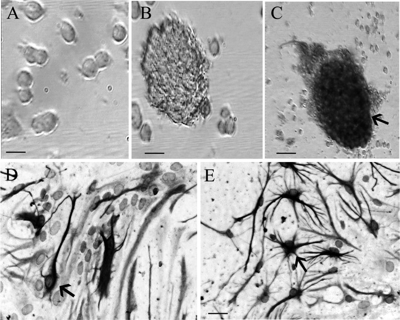
Figure 1. Morphology and identification of neural stem cells (NSCs). (a) The morphology of cells at 12 h of primary culture. (b) The neurospheres composed of hundreds of cells at 3 d after culture under microscope in bright field. (c, d, and e) Three days after culture, respectively. Nestin (marker of NSCs), NeuN (marker of neurons), and glial fibrillary acidic protein (GFAP; marker of astrocytes) immunopositive cells were observed under the light microscope, respectively. Scale bar = 25 μm in a, b, d, e. Scale bar = 50 μm in c.
Figure 1. Morphology and identification of neural stem cells (NSCs). (a) The morphology of cells at 12 h of primary culture. (b) The neurospheres composed of hundreds of cells at 3 d after culture under microscope in bright field. (c, d, and e) Three days after culture, respectively. Nestin (marker of NSCs), NeuN (marker of neurons), and glial fibrillary acidic protein (GFAP; marker of astrocytes) immunopositive cells were observed under the light microscope, respectively. Scale bar = 25 μm in a, b, d, e. Scale bar = 50 μm in c.CC BY-NC · PubMed Central
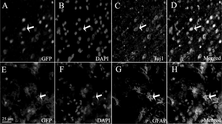
Figure 2. Survival and differentiation of transplanted neural stem cells (NSCs) in host brain. (a) Green fluorescent protein (GFP)–positive cells were observed in host brain under fluorescence microscope. (b) Cell nucleus stained by 4,6-diamidino-2-phenyl indole (DAPI). (c) The number of Tuj1-positive cells. (d) The merged picture. (e) Green fluorescent protein (GFP)–positive (green) cells observed in host brain under fluorescence microscope. (f) The cell nucleus stained by DAPI. (g) The differentiated GFAP-like cells. (h) The merged picture. Scale bar = 25 μm in a to h.
Figure 2. Survival and differentiation of transplanted neural stem cells (NSCs) in host brain. (a) Green fluorescent protein (GFP)–positive cells were observed in host brain under fluorescence microscope. (b) Cell nucleus stained by 4,6-diamidino-2-phenyl indole (DAPI). (c) The number of Tuj1-positive cells. (d) The merged picture. (e) Green fluorescent protein (GFP)–positive (green) cells observed in host brain under fluorescence microscope. (f) The cell nucleus stained by DAPI. (g) The differentiated GFAP-like cells. (h) The merged picture. Scale bar = 25 μm in a to h.CC BY-NC · PubMed Central
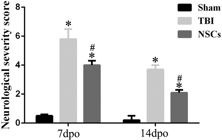
Figure 3. Effect of neural stem cell (NSC) transplantation on neurological functional repair. The figure shows neurological severity score (NSS) test 7 and 14 d after traumatic brain injury (TBI) surgery. The findings showed that TBI induced a significant increase in NSS, while the value of NSS was significantly decreased by NSC implantation. Data are presented as means ± standard deviation (SD). #P < 0.05 versus TBI; *P < 0.05 versus sham.
Figure 3. Effect of neural stem cell (NSC) transplantation on neurological functional repair. The figure shows neurological severity score (NSS) test 7 and 14 d after traumatic brain injury (TBI) surgery. The findings showed that TBI induced a significant increase in NSS, while the value of NSS was significantly decreased by NSC implantation. Data are presented as means ± standard deviation (SD). #P < 0.05 versus TBI; *P < 0.05 versus sham.CC BY-NC · PubMed Central
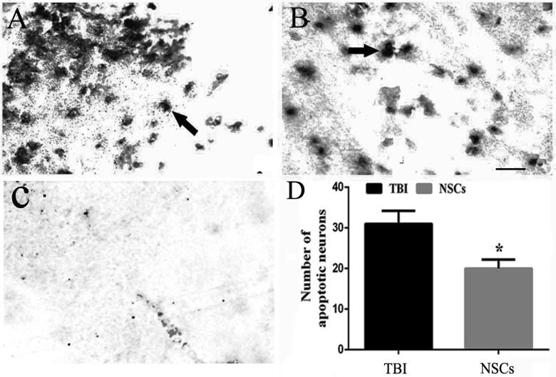
Figure 4. Cell apoptosis in host brain. (a and b) Terminal deoxynucleotidyl transferase 2′-deoxyuridine 5′-triphosphate nick end labeling (TUNEL) staining of the cerebral cortex in the traumatic brain injury (TBI) group and the NSC group, respectively. (c) The staining in negative control. (d) The representative bar graph of the TUNEL-positive cells. Data are presented as means ± standard deviation (SD). *P < 0.05 versus TBI. Arrow showed the apoptotic cells. Scale bars = 25 μm.
Figure 4. Cell apoptosis in host brain. (a and b) Terminal deoxynucleotidyl transferase 2′-deoxyuridine 5′-triphosphate nick end labeling (TUNEL) staining of the cerebral cortex in the traumatic brain injury (TBI) group and the NSC group, respectively. (c) The staining in negative control. (d) The representative bar graph of the TUNEL-positive cells. Data are presented as means ± standard deviation (SD). *P < 0.05 versus TBI. Arrow showed the apoptotic cells. Scale bars = 25 μm.CC BY-NC · PubMed Central
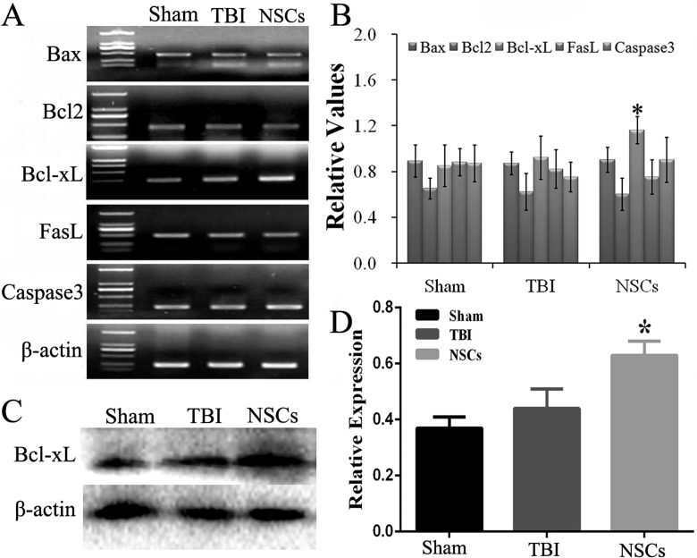
Figure 5. Change of apoptosis genes in host brain. (a) The changes on mRNA of B-cell lymphoma-extra large (Bcl-xL), BCL2-associated X protein (Bax), B-cell lymphoma 2 (Bcl2), Fas ligand (FasL), and caspase3 in host cerebral cortex, which were examined by reverse transcription-polymerase chain reaction (RT-PCR), and quantitative analysis was shown in (b). (c and d) The evaluation of Bcl-xL protein by Western blot and quantitative analysis, respectively. Data are presented as means ± standard deviation (SD). #P < 0.05 versus TBI; *P < 0.05 versus sham.
Figure 5. Change of apoptosis genes in host brain. (a) The changes on mRNA of B-cell lymphoma-extra large (Bcl-xL), BCL2-associated X protein (Bax), B-cell lymphoma 2 (Bcl2), Fas ligand (FasL), and caspase3 in host cerebral cortex, which were examined by reverse transcription-polymerase chain reaction (RT-PCR), and quantitative analysis was shown in (b). (c and d) The evaluation of Bcl-xL protein by Western blot and quantitative analysis, respectively. Data are presented as means ± standard deviation (SD). #P < 0.05 versus TBI; *P < 0.05 versus sham.CC BY-NC · PubMed Central
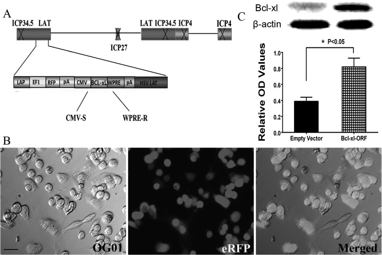
Figure 6. Construction and identification of the herpes simplex virus (HSV) B-cell lymphoma-extra large (Bcl-xL) overexpression vector. (a) Clone representation of the HSV-1-based Bcl-xL recombinant vector. The schematic of the defective HSV-based vector was produced by deleting the infectious cell protein 4 (ICP4), ICP27, and ICP34.5 genes in the wild-type HSV-1. The vector also contained the elements of human cytomegalovirus (CMV) promoter, woodchuck hepatitis posttranscriptional regulatory element (WPRE), and a polyA (pA) region. (b) Vero cells were transfected by HSV-Bcl-xL-open reading frame (ORF). Scale bar = 25 μm. (c) The protein expression of Bcl-xL after HSV-empty vector and HSV-Bcl-xL-ORF was transfected into the cerebral cortex, indicated by Western blot, and the protein expression of Bcl-xL in the Bcl-xL-ORF group is significantly higher than that in the empty vector group. *P < 0.05.
Figure 6. Construction and identification of the herpes simplex virus (HSV) B-cell lymphoma-extra large (Bcl-xL) overexpression vector. (a) Clone representation of the HSV-1-based Bcl-xL recombinant vector. The schematic of the defective HSV-based vector was produced by deleting the infectious cell protein 4 (ICP4), ICP27, and ICP34.5 genes in the wild-type HSV-1. The vector also contained the elements of human cytomegalovirus (CMV) promoter, woodchuck hepatitis posttranscriptional regulatory element (WPRE), and a polyA (pA) region. (b) Vero cells were transfected by HSV-Bcl-xL-open reading frame (ORF). Scale bar = 25 μm. (c) The protein expression of Bcl-xL after HSV-empty vector and HSV-Bcl-xL-ORF was transfected into the cerebral cortex, indicated by Western blot, and the protein expression of Bcl-xL in the Bcl-xL-ORF group is significantly higher than that in the empty vector group. *P < 0.05.CC BY-NC · PubMed Central
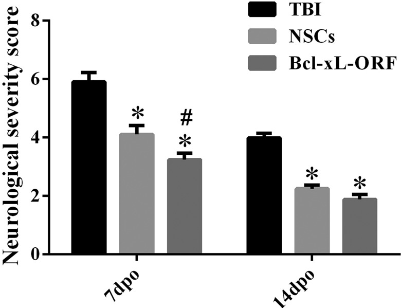
Figure 7. Effect of B-cell lymphoma-extra large (Bcl-xL)-open reading frame (ORF) on neurological functional repair as compared with neural stem cell (NSC) transplantation. The representative bar graph shows neurological severity score (NSS) test after 7 and 14 d of traumatic brain injury (TBI) surgery. The results exhibited that TBI induced a significant increase in NSS, while the value of NSS was significantly decreased by NSC implantation or Bcl-xL-ORF injection. Moreover, the NSS scores in the Bcl-xL-ORF group were lower than that in the NSC group at 7 d postoperation (dpo). Data are presented as means ± standard deviation (SD). #P < 0.05 versus TBI; *P < 0.05 versus sham.
Figure 7. Effect of B-cell lymphoma-extra large (Bcl-xL)-open reading frame (ORF) on neurological functional repair as compared with neural stem cell (NSC) transplantation. The representative bar graph shows neurological severity score (NSS) test after 7 and 14 d of traumatic brain injury (TBI) surgery. The results exhibited that TBI induced a significant increase in NSS, while the value of NSS was significantly decreased by NSC implantation or Bcl-xL-ORF injection. Moreover, the NSS scores in the Bcl-xL-ORF group were lower than that in the NSC group at 7 d postoperation (dpo). Data are presented as means ± standard deviation (SD). #P < 0.05 versus TBI; *P < 0.05 versus sham.CC BY-NC · PubMed Central
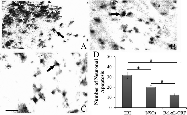
Figure 8. Effect of B-cell lymphoma-extra large (Bcl-xL)-open reading frame (ORF) on cell apoptosis. The changes of cells apoptosis by terminal deoxynucleotidyl transferase dUTP nick end labeling (TUNEL) staining in host brain are shown in TBI group (a) and neural stem cell (NSC) group (b) and Bcl-xL-ORF group (c). The arrows in (a-c) show the apoptotic cells. (d) The quantitative analysis of the cell apoptosis. Data are presented as means ± standard deviation (SD). #P < 0.01; *P < 0.05. Scale bars = 25 μm (a-c).
Figure 8. Effect of B-cell lymphoma-extra large (Bcl-xL)-open reading frame (ORF) on cell apoptosis. The changes of cells apoptosis by terminal deoxynucleotidyl transferase dUTP nick end labeling (TUNEL) staining in host brain are shown in TBI group (a) and neural stem cell (NSC) group (b) and Bcl-xL-ORF group (c). The arrows in (a-c) show the apoptotic cells. (d) The quantitative analysis of the cell apoptosis. Data are presented as means ± standard deviation (SD). #P < 0.01; *P < 0.05. Scale bars = 25 μm (a-c).CC BY-NC · PubMed Central
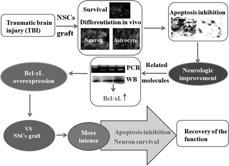
Figure 9. Model of the underlying mechanism of how neural stem cell (NSC) transplantation induced functional recovery in traumatic brain injury (TBI) rats as well as comparison with B-cell lymphoma-extra large (Bcl-xL) overexpression. At the first day of TBI, NSCs were transplanted into the host brain tissue and demonstrated to survive and differentiate into neurons and astrocytes in vivo. In addition, after NSC transplantation, the cell apoptosis was decreased and the neurologic function was improved. Real-time quantitative polymerase chain reaction (RT-PCR) and Western blot (WB) exhibited that Bcl-xL was closely related to the neuroprotective role of NSC transplantation after TBI. Thereafter, the comparison of Bcl-xL overexpressing and NSCs transplantation showed that Bcl-xL overexpression could inhibit cell apoptosis and promote neuronal survival more intensely. These indicate overexpressing Bcl-xL, as an alternative for stem cell therapy, may be a novel treatment of TBI patients.
Figure 9. Model of the underlying mechanism of how neural stem cell (NSC) transplantation induced functional recovery in traumatic brain injury (TBI) rats as well as comparison with B-cell lymphoma-extra large (Bcl-xL) overexpression. At the first day of TBI, NSCs were transplanted into the host brain tissue and demonstrated to survive and differentiate into neurons and astrocytes in vivo. In addition, after NSC transplantation, the cell apoptosis was decreased and the neurologic function was improved. Real-time quantitative polymerase chain reaction (RT-PCR) and Western blot (WB) exhibited that Bcl-xL was closely related to the neuroprotective role of NSC transplantation after TBI. Thereafter, the comparison of Bcl-xL overexpressing and NSCs transplantation showed that Bcl-xL overexpression could inhibit cell apoptosis and promote neuronal survival more intensely. These indicate overexpressing Bcl-xL, as an alternative for stem cell therapy, may be a novel treatment of TBI patients.CC BY-NC · PubMed Central

Verification

Claims the paper makes

The claims and statistics of this paper haven’t been checked yet.

Run the check — free, no account needed. It reads the open-access text and checks the sources it cites.

A few free checks a day · papers already verified open instantly · results are public

Claims other papers make about this one

Each is checked against this paper’s own text: does it say what the citing paper says it does?

Self-check

Percentages, sums and reported differences re-computed from the paper’s own numbers.

The self-check needs the paper’s full text.

Citations

Cites

Show all 53 references

Research works printed but not linked: 5

  1. Dewall J, 2010, JEMS, 35, 54
  2. Zhou FC, 2001, Restor Neurol Neurosci, 18, 95
  3. Xing RX, 2015, Ibrain, 1, 36
  4. Olanow CW, 1996, J Neural Transm Suppl, 48, 75
  5. Duan K, 2016, Nan Fang Yi Ke Da Xue Xue Bao, 36, 32

Cited by

See all 35

Metadata

Authors
Pang, A.-L., Xiong, L.-L., Xia, Q.-J., Liu, F., Wang, Y.-C., Liu, F., Zhang, P., Meng, B.-L., Tan, S., & Wang, T.-H.
Affiliations
  • Department of Neurology, Zhujiang Hospital Southern Medical University, Guangzhou, Guangdong, China
  • Institute of Neurological Disease, Department of Anesthesiology, Translational Neuroscience Center, West China Hospital, Sichuan University, Chengdu, China
  • Institute of Neuroscience, Kunming Medical University, Kunming, China
  • Department of Human Anatomy Histology and Embryology, Kunming Medical University, Kunming, China
Venue
Cell Transplantation
Published
2017-06-30
Volume
26
Issue
7
Pages
1262-1275
ISSN
  • 0963-6897
  • 1555-3892
Cited by
35
References
53
Language
en
Licence
journals.sagepub.com/page/policies/text-and-data-mining-license (tdm)

About these checks

Registry, retraction, Cites and statistics results are computed, not judged, with one exception: a printed reference that no index lists is read once by a model before it counts as a citation error. Claims are extracted by a model and kept only when their quote appears verbatim in the text; each cited source is then checked, with a model’s help, for whether it says what the claim relies on. Those verdicts can be wrong — read the claim’s quote and the cited source before relying on one.

Statistics and arithmetic are recomputed, not judged.

Confidence bands are shown only for fields calibrated on our annotated test set. None is calibrated yet, so no band is shown.

What counts as a claim ›How we measure confidence ›How we check this ›

Are you an author, or do you see a problem with this page? Request a review

How to cite

Cite CoChat for the checks on this page, and the paper for its own findings.

This page
CoChat. (2026). Citation check: Neural Stem Cell Transplantation Is Associated with Inhibition of Apoptosis, Bcl-xL Upregulation, and Recovery of Neurological Function in a Rat Model of Traumatic Brain Injury. https://app.cochat.ai/papers/10.1177/0963689717715168
The paper
Pang, A.-L., Xiong, L.-L., Xia, Q.-J., Liu, F., Wang, Y.-C., Liu, F., Zhang, P., Meng, B.-L., Tan, S., & Wang, T.-H. (2017). Neural Stem Cell Transplantation Is Associated with Inhibition of Apoptosis, Bcl-xL Upregulation, and Recovery of Neurological Function in a Rat Model of Traumatic Brain Injury. Cell Transplantation. https://doi.org/10.1177/0963689717715168

Data sources

Bibliographic data: Semantic Scholar, OpenAlex, Crossref.

Contains information from the Semantic Scholar Academic Graph Datasets, made available under the ODC Attribution License (ODC-BY 1.0).

Semantic Scholar’s data is described in: Kinney, R., et al. (2023). The Semantic Scholar Open Data Platform. arXiv:2301.10140.