

The DOI matches its Crossref record; 10 of the 10 references it cites were found in a registry or an index.
MRI with intrathecal MRI gadolinium contrast medium administration: a possible method to assess glymphatic function in human brain
Open accessAbstract
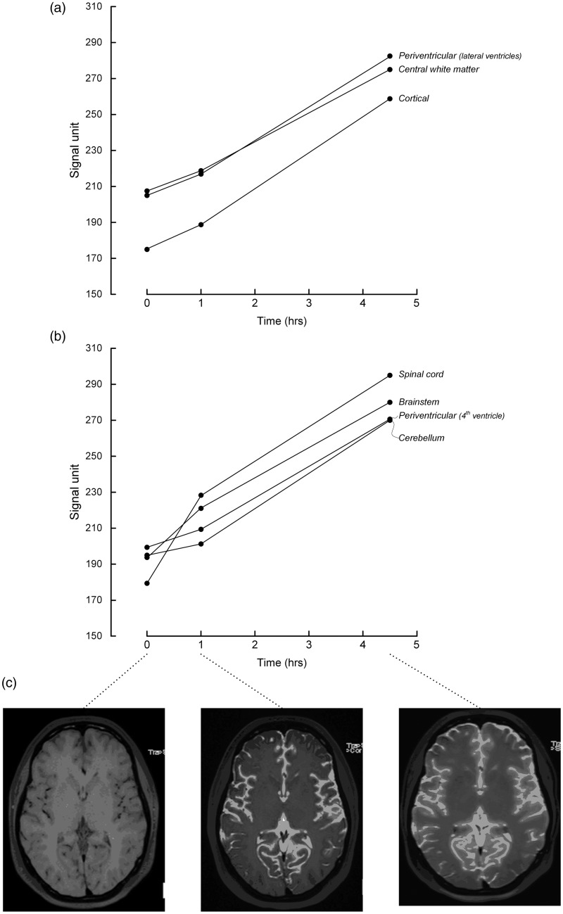
Recently, the “glymphatic system” of the brain has been discovered in rodents, which is a paravascular, transparenchymal route for clearance of excess brain metabolites and distribution of compounds in the cerebrospinal fluid. It has already been demonstrated that intrathecally administered gadolinium (Gd) contrast medium distributes along this route in rats, but so far not in humans. A 27-year-old woman underwent magnetic resonance imaging (MRI) with intrathecal administration of gadobutrol, which distributed throughout her entire brain after 1 and 4.5 h. MRI with intrathecal Gd may become a tool to study glymphatic function in the human brain.
Overview
The claims and statistics of this paper haven’t been checked yet.
Run the check — free, no account needed. It reads the open-access text and checks the sources it cites.
Integrity signals
Registry
Cites
Statistics
Figures
Verification
Claims the paper makes
The claims and statistics of this paper haven’t been checked yet.
Run the check — free, no account needed. It reads the open-access text and checks the sources it cites.
Claims other papers make about this one
Each is checked against this paper’s own text: does it say what the citing paper says it does?
The source backs part of the claim, not all of it — or backs it for a different population, condition or comparison.
In the citing paper
“However, literature reporting human in vivo CSF tracer studies is lacking, and observations made in animals have not been translated into humans.”
From Brain-wide glymphatic enhancement and clearance in humans assessed with MRI. (2018) · cites this paper as [12]
Experimental — accuracy not yet measuredThe cited source says what the claim attributes to it.
In the citing paper
“Two recent human studies demonstrated brain parenchyma enhancement subsequent to subarachnoid (intrathecal) administration of a MRI contrast agent, but observations were limited to selected ROIs (12, 13).”
From Brain-wide glymphatic enhancement and clearance in humans assessed with MRI. (2018) · cites this paper as [12]
Self-check
Percentages, sums and reported differences re-computed from the paper’s own numbers.
The self-check needs the paper’s full text.
Citations
Cites
Iliff, J. J., Wang, M., Liao, Y., Plogg, B. A., Peng, W., Gundersen, G. A., Benveniste, H., Vates, G. E., Deane, R., Goldman, S. A., et al. (2012) · Science Translational Medicine · doi:10.1126/scitranslmed.3003748 · Cited by 5,824
The glymphatic system: a beginner's guide
Jessen, N. A., Munk, A. S. F., Lundgaard, I., & Nedergaard, M. (2015) · Neurochemical Research · doi:10.1007/s11064-015-1581-6 · Cited by 2,075
Impairment of paravascular clearance pathways in the aging brain
Kress, B. T., Iliff, J. J., Xia, M., Wang, M., Wei, H. S., Zeppenfeld, D., Xie, L., Kang, H., Xu, Q., Liew, J. A., et al. (2014) · Annals of Neurology · doi:10.1002/ana.24271 · Cited by 1,521 · cited 1×
“Glymphatic function has also been shown to decrease dramatically with aging (2) and in the injured brain (3).”
BackgroundProduction of the Alzheimer Amyloid β Protein by Normal Proteolytic Processing
Shoji, M., Golde, T. E., Ghiso, J., Cheung, T. T., Estus, S., Shaffer, L. M., Cai, X.-D., McKay, D. M., Tintner, R., Frangione, B., et al. (1992) · Science · doi:10.1126/science.1439760 · Cited by 1,447
Impairment of Glymphatic Pathway Function Promotes Tau Pathology after Traumatic Brain Injury
Iliff, J. J., Chen, M. J., Plog, B. A., Zeppenfeld, D. M., Soltero, M., Yang, L., Singh, I., Deane, R., & Nedergaard, M. (2014) · The Journal of Neuroscience · doi:10.1523/jneurosci.3020-14.2014 · Cited by 1,159
Brain-wide pathway for waste clearance captured by contrast-enhanced MRI
Iliff, J. J., Lee, H., Yu, M., Feng, T., Logan, J., Nedergaard, M., & Benveniste, H. (2013) · Journal of Clinical Investigation · doi:10.1172/jci67677 · Cited by 1,148
Impaired Glymphatic Perfusion After Strokes Revealed by Contrast-Enhanced MRI
Gaberel, T., Gakuba, C., Goulay, R., de Lizarrondo, S. M., Hanouz, J., Emery, É., Touzé, E., Vivien, D., & Gauberti, M. (2014) · Stroke · doi:10.1161/strokeaha.114.006617 · Cited by 438
Akbar, J. J., Luetmer, P. H., Schwartz, K. M., et al. (2011) · American Journal of Neuroradiology · doi:10.3174/ajnr.a2815 · Cited by 119
LOCALIZATION OF CEREBROSPINAL FLUID LEAKS BY GADOLINIUM-ENHANCED MAGNETIC RESONANCE CISTERNOGRAPHY
Aydın, K., Terzibaşioğlu, E., Sencer, S., Sencer, A., Süoğlu, Y., Karasu, A., Kiris, T., & Turantan, M. I. (2008) · Neurosurgery · doi:10.1227/01.neu.0000317306.39203.24 · Cited by 68
Adam, G., Neuerburg, J. M., Spüntrup, E., et al. (1994) · Magnetic Resonance in Medicine · doi:10.1002/mrm.1910320511 · Cited by 45
Cited by
Sorted by citations
Verkhratsky, A., & Nedergaard, M. (2018) · Physiological Reviews · doi:10.1152/physrev.00042.2016 · Cited by 1,678
The glymphatic pathway in neurological disorders.
Rasmussen, M. K., Mestre, H., & Nedergaard, M. (2018) · The Lancet Neurology · doi:10.1016/s1474-4422(18)30318-1 · Cited by 1,560
Cerebral amyloid angiopathy and Alzheimer disease — one peptide, two pathways
Greenberg, S. M., Bacskai, B. J., Hernandez-Guillamon, M., Pruzin, J., Sperling, R., & van Veluw, S. J. (2020) · Nature Reviews Neurology · doi:10.1038/s41582-019-0281-2 · Cited by 947
Aquaporin-4-dependent glymphatic solute transport in the rodent brain
Mestre, H., Hablitz, L. M., Xavier, A. L., Feng, W., Zou, W., Pu, T., Monai, H., Murlidharan, G., Castellanos Rivera, R. M., Simon, M. J., et al. (2018) · eLife · doi:10.7554/elife.40070 · Cited by 738
Glymphatic MRI in idiopathic normal pressure hydrocephalus
Ringstad, G., Vatnehol, S. A. S., & Eide, P. K. (2017) · Brain · doi:10.1093/brain/awx191 · Cited by 722
Self-citation
Metadata
- Authors
- Eide, P. K., & Ringstad, G.
- Affiliations
- Oslo University Hospital (NO)
- Corresponding author
- Eide, Per Kristian
- Venue
- Acta Radiologica Open
- Publisher
- SAGE Publications
- Published
- 2015-11-01
- Volume
- 4
- Issue
- 11
- Pages
- 205846011560963
- ISSN
- 2047-9816 (ISSN-L)
- 2058-4601
- PMC
- PMC4652208
- OpenAlex
- W2252352100
- Cited by
- 218
- References
- 10
- Citations by year
- 2026: 17 · 2025: 33 · 2024: 22 · 2023: 22 · 2022: 15 · 2021: 22 · 2020: 26 · 2019: 17 · 2018: 22 · 2017: 15 · 2016: 7
- FWCI
- 6.84
- Type
- journal article
- Language
- en
- Open access
- gold
- doi.org
- Locations
- Acta Radiologica Open (publishedVersion, cc-by-nc)
- Europe PMC (PubMed Central) (publishedVersion, cc-by-nc)
- PubMed Central (publishedVersion, cc-by-nc)
- PubMed (publishedVersion)
- DOAJ (DOAJ: Directory of Open Access Journals) (submittedVersion, cc-by-sa)
- Topics
- Health Sciences › Medicine › Clinical Neurology › Cerebrospinal fluid and hydrocephalus
- Health Sciences › Medicine › Radiology, Nuclear Medicine and Imaging › Advanced MRI Techniques and Applications
- Life Sciences › Neuroscience › Neurology › Barrier Structure and Function Studies
- Keywords
- glymphatic system, gadobutrol, cerebrospinal fluid, human brain imaging
About these checks
Registry, retraction, Cites and statistics results are computed, not judged, with one exception: a printed reference that no index lists is read once by a model before it counts as a citation error. Claims are extracted by a model and kept only when their quote appears verbatim in the text; each cited source is then checked, with a model’s help, for whether it says what the claim relies on. Those verdicts can be wrong — read the claim’s quote and the cited source before relying on one.
Statistics and arithmetic are recomputed, not judged.
Confidence bands are shown only for fields calibrated on our annotated test set. None is calibrated yet, so no band is shown.
What counts as a claim ›How we measure confidence ›How we check this ›
Are you an author, or do you see a problem with this page? Request a review
