

The DOI matches its Crossref record; 10 of the 10 references it cites were found in a registry or an index.
MRI with intrathecal MRI gadolinium contrast medium administration: a possible method to assess glymphatic function in human brain
Open accessAbstract
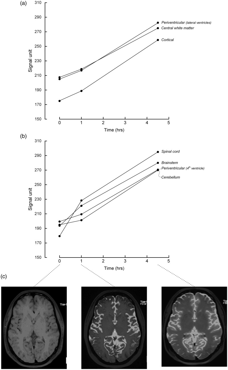
Recently, the “glymphatic system” of the brain has been discovered in rodents, which is a paravascular, transparenchymal route for clearance of excess brain metabolites and distribution of compounds in the cerebrospinal fluid. It has already been demonstrated that intrathecally administered gadolinium (Gd) contrast medium distributes along this route in rats, but so far not in humans. A 27-year-old woman underwent magnetic resonance imaging (MRI) with intrathecal administration of gadobutrol, which distributed throughout her entire brain after 1 and 4.5 h. MRI with intrathecal Gd may become a tool to study glymphatic function in the human brain.
Overview
The claims and statistics of this paper haven’t been checked yet.
Run the check — free, no account needed. It reads the open-access text and checks the sources it cites.
Integrity signals
Registry
Cites
Statistics
Figures
Verification
Claims the paper makes
The claims and statistics of this paper haven’t been checked yet.
Run the check — free, no account needed. It reads the open-access text and checks the sources it cites.
Claims other papers make about this one
Each is checked against this paper’s own text: does it say what the citing paper says it does?
The source backs part of the claim, not all of it — or backs it for a different population, condition or comparison.
In the citing paper
“However, literature reporting human in vivo CSF tracer studies is lacking, and observations made in animals have not been translated into humans.”
From Brain-wide glymphatic enhancement and clearance in humans assessed with MRI. (2018) · cites this paper as [12]
Experimental — accuracy not yet measuredThe cited source says what the claim attributes to it.
In the citing paper
“Two recent human studies demonstrated brain parenchyma enhancement subsequent to subarachnoid (intrathecal) administration of a MRI contrast agent, but observations were limited to selected ROIs (12, 13).”
From Brain-wide glymphatic enhancement and clearance in humans assessed with MRI. (2018) · cites this paper as [12]
Self-check
Percentages, sums and reported differences re-computed from the paper’s own numbers.
The self-check needs the paper’s full text.
Citations
Cites
Iliff, J. J., Wang, M., Liao, Y., Plogg, B. A., Peng, W., Gundersen, G. A., Benveniste, H., Vates, G. E., Deane, R., Goldman, S. A., et al. (2012) · Science Translational Medicine · doi:10.1126/scitranslmed.3003748 · Cited by 5,824
The glymphatic system: a beginner's guide
Jessen, N. A., Munk, A. S. F., Lundgaard, I., & Nedergaard, M. (2015) · Neurochemical Research · doi:10.1007/s11064-015-1581-6 · Cited by 2,075
Impairment of paravascular clearance pathways in the aging brain
Kress, B. T., Iliff, J. J., Xia, M., Wang, M., Wei, H. S., Zeppenfeld, D., Xie, L., Kang, H., Xu, Q., Liew, J. A., et al. (2014) · Annals of Neurology · doi:10.1002/ana.24271 · Cited by 1,521 · cited 1×
“Glymphatic function has also been shown to decrease dramatically with aging (2) and in the injured brain (3).”
BackgroundProduction of the Alzheimer Amyloid β Protein by Normal Proteolytic Processing
Shoji, M., Golde, T. E., Ghiso, J., Cheung, T. T., Estus, S., Shaffer, L. M., Cai, X.-D., McKay, D. M., Tintner, R., Frangione, B., et al. (1992) · Science · doi:10.1126/science.1439760 · Cited by 1,447
Impairment of Glymphatic Pathway Function Promotes Tau Pathology after Traumatic Brain Injury
Iliff, J. J., Chen, M. J., Plog, B. A., Zeppenfeld, D. M., Soltero, M., Yang, L., Singh, I., Deane, R., & Nedergaard, M. (2014) · The Journal of Neuroscience · doi:10.1523/jneurosci.3020-14.2014 · Cited by 1,159
Brain-wide pathway for waste clearance captured by contrast-enhanced MRI
Iliff, J. J., Lee, H., Yu, M., Feng, T., Logan, J., Nedergaard, M., & Benveniste, H. (2013) · Journal of Clinical Investigation · doi:10.1172/jci67677 · Cited by 1,148
Impaired Glymphatic Perfusion After Strokes Revealed by Contrast-Enhanced MRI
Gaberel, T., Gakuba, C., Goulay, R., de Lizarrondo, S. M., Hanouz, J., Emery, É., Touzé, E., Vivien, D., & Gauberti, M. (2014) · Stroke · doi:10.1161/strokeaha.114.006617 · Cited by 438
Akbar, J. J., Luetmer, P. H., Schwartz, K. M., et al. (2011) · American Journal of Neuroradiology · doi:10.3174/ajnr.a2815 · Cited by 119
LOCALIZATION OF CEREBROSPINAL FLUID LEAKS BY GADOLINIUM-ENHANCED MAGNETIC RESONANCE CISTERNOGRAPHY
Aydın, K., Terzibaşioğlu, E., Sencer, S., Sencer, A., Süoğlu, Y., Karasu, A., Kiris, T., & Turantan, M. I. (2008) · Neurosurgery · doi:10.1227/01.neu.0000317306.39203.24 · Cited by 68
Adam, G., Neuerburg, J. M., Spüntrup, E., et al. (1994) · Magnetic Resonance in Medicine · doi:10.1002/mrm.1910320511 · Cited by 45
Cited by
Sorted by citations
Verkhratsky, A., & Nedergaard, M. (2018) · Physiological Reviews · doi:10.1152/physrev.00042.2016 · Cited by 1,678
The glymphatic pathway in neurological disorders.
Rasmussen, M. K., Mestre, H., & Nedergaard, M. (2018) · The Lancet Neurology · doi:10.1016/s1474-4422(18)30318-1 · Cited by 1,560
Cerebral amyloid angiopathy and Alzheimer disease — one peptide, two pathways
Greenberg, S. M., Bacskai, B. J., Hernandez-Guillamon, M., Pruzin, J., Sperling, R., & van Veluw, S. J. (2020) · Nature Reviews Neurology · doi:10.1038/s41582-019-0281-2 · Cited by 947
Aquaporin-4-dependent glymphatic solute transport in the rodent brain
Mestre, H., Hablitz, L. M., Xavier, A. L., Feng, W., Zou, W., Pu, T., Monai, H., Murlidharan, G., Castellanos Rivera, R. M., Simon, M. J., et al. (2018) · eLife · doi:10.7554/elife.40070 · Cited by 738
Glymphatic MRI in idiopathic normal pressure hydrocephalus
Ringstad, G., Vatnehol, S. A. S., & Eide, P. K. (2017) · Brain · doi:10.1093/brain/awx191 · Cited by 722
Self-citationRasmussen, M. K., Mestre, H., & Nedergaard, M. (2021) · Physiological Reviews · doi:10.1152/physrev.00031.2020 · Cited by 642
Brain-wide glymphatic enhancement and clearance in humans assessed with MRI.
Ringstad, G., Valnes, L. M., Dale, A. M., Pripp, A. H., Vatnehol, S.-A. S., Emblem, K. E., Mardal, K.-A., & Eide, P. K. (2018) · JCI Insight · doi:10.1172/jci.insight.121537 · Cited by 533
“005 Basal ganglia [11, 12, 13, 50, 51, 52] –3 ± 8 –6 ± 14 1 ± 25 –4 ± 14 4 ± 18 32 –8 ± 18 0.”
Self-citationGlymphatic clearance function in patients with cerebral small vessel disease
Zhang, W., Zhou, Y., Wang, J., Gong, X., Chen, Z., Zhang, X., Cai, J., Chen, S., Fang, L., Sun, J., et al. (2021) · NeuroImage · doi:10.1016/j.neuroimage.2021.118257 · Cited by 492
Perivascular spaces, glymphatic dysfunction, and small vessel disease
Mestre, H., Kostrikov, S., Mehta, R. I., & Nedergaard, M. (2017) · Clinical Science · doi:10.1042/cs20160381 · Cited by 403
Taoka, T., Ito, R., Nakamichi, R., Nakane, T., Kawai, H., & Naganawa, S. (2024) · Magnetic Resonance in Medical Sciences · doi:10.2463/mrms.rev.2023-0175 · Cited by 423
Impairment of the Glymphatic Pathway and Putative Meningeal Lymphatic Vessels in the Aging Human
Zhou, Y., Cai, J., Zhang, W., Gong, X., Yan, S., Zhang, K., Luo, Z., Sun, J., Jiang, Q., & Lou, M. (2020) · Annals of Neurology · doi:10.1002/ana.25670 · Cited by 305
Impairment of the glymphatic system after diabetes
Jiang, Q., Zhang, L., Ding, G., Davoodi-Bojd, E., Li, Q., Li, L., Sadry, N., Nedergaard, M., Chopp, M., & Zhang, Z. (2017) · Journal of Cerebral Blood Flow & Metabolism · doi:10.1177/0271678x16654702 · Cited by 308
Emerging roles for dynamic aquaporin-4 subcellular relocalization in CNS water homeostasis
Salman, M. M., Kitchen, P., Halsey, A., Wang, M. X., Törnroth-Horsefield, S., Conner, A. C., Badaut, J., Iliff, J. J., & Bill, R. M. (2022) · Brain · doi:10.1093/brain/awab311 · Cited by 303
Glymphatic clearance controls state-dependent changes in brain lactate concentration
Lundgaard, I., Lu, M. L., Yang, E., Peng, W., Mestre, H., Hitomi, E., Deane, R., & Nedergaard, M. (2016) · Journal of Cerebral Blood Flow & Metabolism · doi:10.1177/0271678x16661202 · Cited by 309
Taoka, T., Ito, R., Nakamichi, R., Kamagata, K., Sakai, M., Kawai, H., Nakane, T., Abe, T., Ichikawa, K., Kikuta, J., et al. (2021) · Japanese Journal of Radiology · doi:10.1007/s11604-021-01187-5 · Cited by 296
Huang, S., Zhang, Y.-R., Guo, Y., Du, J., Ren, P., Wu, B., Feng, J., Initiative, A. D. N., Cheng, W., & Yu, J. (2024) · Alzheimer s & Dementia · doi:10.1002/alz.13789 · Cited by 305
Lymphatic drainage system of the brain: A novel target for intervention of neurological diseases
Sun, B., Wang, L.-H., Yang, T., Sun, J., Mao, L., Yang, M., Yuan, H., Colvin, R. A., & Yang, X. (2017) · Progress in Neurobiology · doi:10.1016/j.pneurobio.2017.08.007 · Cited by 264
The glymphatic hypothesis: the theory and the evidence
Hladky, S. B., & Barrand, M. A. (2022) · Fluids and Barriers of the CNS · doi:10.1186/s12987-021-00282-z · Cited by 283
Taoka, T., & Naganawa, S. (2019) · Journal of Magnetic Resonance Imaging · doi:10.1002/jmri.26892 · Cited by 244
The glymphatic system: implications for drugs for central nervous system diseases
Lohela, T. J., Lilius, T. O., & Nedergaard, M. (2022) · Nature Reviews Drug Discovery · doi:10.1038/s41573-022-00500-9 · Cited by 254
Xiong, Y., Mahmood, A., & Chopp, M. (2018) · Chinese Journal of Traumatology · doi:10.1016/j.cjtee.2018.02.003 · Cited by 253
Eide, P. K., & Ringstad, G. A. (2018) · Journal of Cerebral Blood Flow & Metabolism · doi:10.1177/0271678x18760974 · Cited by 239
Self-citationShen, T., Yue, Y., Ba, F., He, T., Tang, X., Hu, X., Pu, J., Huang, C., Lv, W., Zhang, B., et al. (2022) · npj Parkinson s Disease · doi:10.1038/s41531-022-00437-1 · Cited by 216
Gadolinium Deposition in the Brain: Current Updates
Choi, J. W., & Moon, W. (2018) · Korean Journal of Radiology · doi:10.3348/kjr.2018.0356 · Cited by 185
Glymphatic System Function in Relation to Anesthesia and Sleep States
Benveniste, H., Heerdt, P. M., Fontes, M. L., Rothman, D. L., & Volkow, N. D. (2019) · Anesthesia & Analgesia · doi:10.1213/ane.0000000000004069 · Cited by 182
Steward, C. E., Venkatraman, V. K., LUI, E. H., Malpas, C. B., Ellis, K. A., Cyarto, E., Vivash, L. E., O'Brien, T. J., Velakoulis, D., Ames, D. J., et al. (2021) · Journal of Neuroimaging · doi:10.1111/jon.12837 · Cited by 184
Interaction between blood-brain barrier and glymphatic system in solute clearance
Verheggen, I. C. M., van Boxtel, M. P. J., Verhey, F. R. J., Jansen, J. F. A., & Backes, W. H. (2018) · Neuroscience & Biobehavioral Reviews · doi:10.1016/j.neubiorev.2018.03.028 · Cited by 171
Boespflug, E. L., & Iliff, J. J. (2017) · Biological Psychiatry · doi:10.1016/j.biopsych.2017.11.031 · Cited by 179
Bacyinski, A., Xu, M., Wang, W., & Hu, J. (2017) · Frontiers in Neuroanatomy · doi:10.3389/fnana.2017.00101 · Cited by 170
Gouveia-Freitas, K., & de Bastos-Leite, A. J. (2021) · Neuroradiology · doi:10.1007/s00234-021-02718-7 · Cited by 166
Obstructive sleep apnea decreases central nervous system–derived proteins in the cerebrospinal fluid
Ju, Y. S., Finn, M. B., Sutphen, C. L., Herries, E. M., Jerome, G. M., Ladenson, J. H., Crimmins, D. L., Fagan, A. M., & Holtzman, D. M. (2016) · Annals of Neurology · doi:10.1002/ana.24672 · Cited by 168
Gadolinium deposition in the brain
Kanda, T., Nakai, Y., Oba, H., Toyoda, K., Kitajima, K., & Furui, S. (2016) · Magnetic Resonance Imaging · doi:10.1016/j.mri.2016.08.024 · Cited by 164
Sundaram, S., Hughes, R. L., Peterson, E., Müller-Oehring, E. M., Brontë-Stewart, H. M., Poston, K. L., Faerman, A., Bhowmick, C., & Schulte, T. (2019) · Neuroscience & Biobehavioral Reviews · doi:10.1016/j.neubiorev.2019.05.016 · Cited by 152
Goulay, R., Romo, L. M., Hol, E. M., & Dijkhuizen, R. M. (2019) · Translational Stroke Research · doi:10.1007/s12975-019-00755-2 · Cited by 144
Burfeind, K. G., Murchison, C. F., Westaway, S. K., Simon, M. J., Erten‐Lyons, D., Kaye, J. A., Quinn, J. F., & Iliff, J. J. (2017) · Alzheimer's & Dementia: Translational Research & Clinical Interventions · doi:10.1016/j.trci.2017.05.001 · Cited by 136
The homeostatic astroglia emerges from evolutionary specialization of neural cells
Verkhratsky, A., & Nedergaard, M. (2016) · Philosophical Transactions of the Royal Society B: Biological Sciences · doi:10.1098/rstb.2015.0428 · Cited by 133
Evaluation of Glymphatic System Using Diffusion MR Technique in T2DM Cases
Yang, G., Deng, N., Liu, Y., Gu, Y., & Yao, X. (2020) · Frontiers in Human Neuroscience · doi:10.3389/fnhum.2020.00300 · Cited by 130
Lundgaard, I., Wang, W., Eberhardt, A., Vinitsky, H. S., Reeves, B. C., Peng, S., Lou, N., Hussain, R., & Nedergaard, M. (2018) · Scientific Reports · doi:10.1038/s41598-018-20424-y · Cited by 136
Klostranec, J. M., Vucevic, D., Bhatia, K. D., Kortman, H. G. J., Krings, T., Murphy, K., terBrugge, K. G., & Mikulis, D. J. (2021) · Radiology · doi:10.1148/radiol.2021204088 · Cited by 121
Altered Brain Glymphatic Flow at Diffusion-Tensor MRI in Rapid Eye Movement Sleep Behavior Disorder
Bae, Y. J., Kim, J., Choi, B. S., Ryoo, N., Song, Y. S., Nam, Y., Yoon, I., Cho, S. J., & Kim, J. H. (2023) · Radiology · doi:10.1148/radiol.221848 · Cited by 121
Naganawa, S., & Taoka, T. (2020) · Magnetic Resonance in Medical Sciences · doi:10.2463/mrms.rev.2020-0122 · Cited by 122
Gadolinium Retention and Toxicity—An Update
Ramalho, M., Ramalho, J., Burke, L. M. B., & Semelka, R. C. (2017) · Advances in Chronic Kidney Disease · doi:10.1053/j.ackd.2017.03.004 · Cited by 115
The role of the immune system in Alzheimer’s disease
Wu, K., Zhang, Y.-R., Huang, Y., Dong, Q., Tan, L., & Yu, J. (2021) · Ageing Research Reviews · doi:10.1016/j.arr.2021.101409 · Cited by 119
Quantitative Gd‐DOTA uptake from cerebrospinal fluid into rat brain using 3D VFA‐SPGR at 9.4T
Lee, H., Mortensen, K., Sanggaard, S., Koch, P., Brunner, H., Quistorff, B., Nedergaard, M., & Benveniste, H. (2018) · Magnetic Resonance in Medicine · doi:10.1002/mrm.26779 · Cited by 110
Han, F., Brown, G. L., Zhu, Y., Belkin-Rosen, A., Lewis, M. M., Du, G., Gu, Y., Eslinger, P. J., Mailman, R. B., Huang, X., et al. (2021) · Movement Disorders · doi:10.1002/mds.28643 · Cited by 111
Decreased CSF clearance and increased brain amyloid in Alzheimer’s disease
Li, Y., Rusinek, H., Butler, T., Glodzik, L., Pirraglia, E., Babich, J. W., Mozley, P. D., Nehmeh, S. A., Pahlajani, S. S., Wang, X. H., et al. (2022) · Fluids and Barriers of the CNS · doi:10.1186/s12987-022-00318-y · Cited by 110
Glymphatic Cerebrospinal Fluid and Solute Transport Quantified by MRI and PET Imaging
Benveniste, H., Lee, H., Ozturk, B., Chen, X., Koundal, S., Vaska, P., Tannenbaum, A., & Volkow, N. D. (2020) · Neuroscience · doi:10.1016/j.neuroscience.2020.11.014 · Cited by 106
Impaired brain glymphatic flow in experimental hepatic encephalopathy
Hadjihambi, A., Harrison, I. F., Costas‐Rodríguez, M., Vanhaecke, F., Arias, N., Gallego‐Durán, R., Mastitskaya, S., Hosford, P. S., Damink, S. W. M. O., Davies, N. A., et al. (2018) · Journal of Hepatology · doi:10.1016/j.jhep.2018.08.021 · Cited by 105
Christensen, J., Yamakawa, G. R., Shultz, S. R., & Mychasiuk, R. (2020) · Progress in Neurobiology · doi:10.1016/j.pneurobio.2020.101917 · Cited by 110
The glymphatic system and meningeal lymphatics of the brain: new understanding of brain clearance
Yankova, G., Bogomyakova, O. B., & Тулупов, А. А. (2021) · Reviews in the Neurosciences · doi:10.1515/revneuro-2020-0106 · Cited by 110
Neurofluid Dynamics and the Glymphatic System: A Neuroimaging Perspective
Taoka, T., & Naganawa, S. (2020) · Korean Journal of Radiology · doi:10.3348/kjr.2020.0042 · Cited by 98
Measuring Glymphatic Flow in Man Using Quantitative Contrast-Enhanced MRI
Watts, R., Steinklein, J. M., Waldman, L. E., Zhou, X., & Filippi, C. G. (2019) · American Journal of Neuroradiology · doi:10.3174/ajnr.a5931 · Cited by 96
Patel, M., Atyani, A., Salameh, J., et al. (2020) · Radiology · doi:10.1148/radiol.2020191373 · Cited by 96
Glymphatic system: an emerging therapeutic approach for neurological disorders
Gao, Y., Liu, K., & Zhu, J. (2023) · Frontiers in Molecular Neuroscience · doi:10.3389/fnmol.2023.1138769 · Cited by 109
Harnessing cerebrospinal fluid circulation for drug delivery to brain tissues
Naseri Kouzehgarani, G., Feldsien, T., Engelhard, H. H., Mirakhur, K. K., Phipps, C., Nimmrich, V., Clausznitzer, D., & Lefebvre, D. R. (2021) · Advanced Drug Delivery Reviews · doi:10.1016/j.addr.2021.03.002 · Cited by 95
Taoka, T., & Naganawa, S. (2020) · Japanese Journal of Radiology · doi:10.1007/s11604-020-01017-0 · Cited by 88
Apolipoprotein E4 and meningeal lymphatics in Alzheimer disease: a conceptual framework
Mentis, A. A., Dardiotis, E., & Chrousos, G. P. (2020) · Molecular Psychiatry · doi:10.1038/s41380-020-0731-7 · Cited by 83
Plog, B. A., Lou, N., Pierre, C. A., Cove, A., Kenney, H. M., Hitomi, E., Kang, H., Iliff, J. J., Zeppenfeld, D. M., Nedergaard, M., et al. (2019) · Journal of neurosurgery · doi:10.3171/2019.2.jns182675 · Cited by 89
Altered Waste Disposal System in Aging and Alzheimer’s Disease: Focus on Astrocytic Aquaporin-4
Valenza, M., Facchinetti, R., Steardo, L., & Scuderi, C. (2020) · Frontiers in Pharmacology · doi:10.3389/fphar.2019.01656 · Cited by 90
Achieving brain clearance and preventing neurodegenerative diseases—A glymphatic perspective
Kylkilahti, T. M., Berends, E., Ramos, M., Shanbhag, N. C., Töger, J., Bloch, K. M., & Lundgaard, I. (2021) · Journal of Cerebral Blood Flow & Metabolism · doi:10.1177/0271678x20982388 · Cited by 91
Simon, M. J., Wang, M. X., Murchison, C. F., Roese, N. E., Boespflug, E. L., Woltjer, R. L., & Iliff, J. J. (2018) · Scientific Reports · doi:10.1038/s41598-018-30779-x · Cited by 71
In vivo assessment of cerebrospinal fluid efflux to nasal mucosa in humans
Melin, E., Eide, P. K., & Ringstad, G. A. (2020) · Scientific Reports · doi:10.1038/s41598-020-72031-5 · Cited by 78
Self-citationCarare, R. O., Aldea, R., Agarwal, N., Bacskai, B. J., Bechman, I., Boche, D., Bu, G., Bulters, D. O., Alt, C., Counts, S., et al. (2020) · Alzheimer s & Dementia Diagnosis Assessment & Disease Monitoring · doi:10.1002/dad2.12053 · Cited by 77
Self-citationCould dexmedetomidine be repurposed as a glymphatic enhancer?
Persson, N. D. Å., Uusalo, P., Nedergaard, M., Lohela, T. J., & Lilius, T. O. (2022) · Trends in Pharmacological Sciences · doi:10.1016/j.tips.2022.09.007 · Cited by 78
Intrathecal Contrast-Enhanced Magnetic Resonance Imaging–Related Brain Signal Changes
Öner, A. Y., Asfuroğlu, B. B., Aykol, Ş., & Talı, E. T. (2016) · Investigative Radiology · doi:10.1097/rli.0000000000000327 · Cited by 72
Chachaj, A., Gąsiorowski, K., Szuba, A., Sieradzki, A., & Leszek, J. (2022) · Current Neuropharmacology · doi:10.2174/1570159x20666220411091332 · Cited by 73
Noninvasive Magnetic Resonance Imaging Measures of Glymphatic System Activity
Kamagata, K., Saito, Y., Andica, C., Uchida, W., Takabayashi, K., Yoshida, S., Hagiwara, A., Fujita, S., Nakaya, M., Akashi, T., et al. (2023) · Journal of Magnetic Resonance Imaging · doi:10.1002/jmri.28977 · Cited by 76
Gadolinium as an MRI Contrast Agent
Kim, H.-K., Lee, G. H., & Chang, Y. (2018) · Future Medicinal Chemistry · doi:10.4155/fmc-2017-0215 · Cited by 77
Bhargava, P., Wicken, C., Smith, M. D., Strowd, R. E., III, Cortese, I. C. M., Reich, D. S., Calabresi, P. A., & Mowry, E. M. (2019) · Multiple Sclerosis and Related Disorders · doi:10.1016/j.msard.2019.02.013 · Cited by 66
Naganawa, S., Nakane, T., Kawai, H., & Taoka, T. (2018) · Magnetic Resonance in Medical Sciences · doi:10.2463/mrms.mp.2018-0053 · Cited by 65
The Human Visual Pathway Communicates Directly With the Subarachnoid Space
Jacobsen, H. H., Ringstad, G. A., Jørstad, Ø. K., Moe, M. C., Sandell, T., & Eide, P. K. (2019) · Investigative Ophthalmology & Visual Science · doi:10.1167/iovs.19-26997 · Cited by 63
Self-citationRasschaert, M., Weller, R. O., Schroeder, J. A., Brochhausen, C., & Idée, J. (2020) · Journal of Magnetic Resonance Imaging · doi:10.1002/jmri.27124 · Cited by 65
Small vessels, dementia and chronic diseases – molecular mechanisms and pathophysiology
Horsburgh, K. J., Wardlaw, J. M., Agtmael, T. V., Allan, S. M., Ashford, M. L. J., Bath, P. M., Brown, R., Berwick, J., Cader, M. Z., Carare, R. O., et al. (2018) · Clinical Science · doi:10.1042/cs20171620 · Cited by 65
Distribution and chemical forms of gadolinium in the brain: a review
Kanda, T., Nakai, Y., Hagiwara, A., Oba, H., Toyoda, K., & Furui, S. (2017) · British Journal of Radiology · doi:10.1259/bjr.20170115 · Cited by 66
Assessment of the dynamics of human glymphatic system by near‐infrared spectroscopy
Myllylä, T., Harju, M., Korhonen, V. O., Bykov, A. V., Kiviniemi, V. J., & Meglinski, I. V. (2017) · Journal of Biophotonics · doi:10.1002/jbio.201700123 · Cited by 59
In vivo T1 mapping for quantifying glymphatic system transport and cervical lymph node drainage
Xue, Y., Liu, X., Koundal, S., Constantinou, S., Dai, F., Santambrogio, L., Lee, H., & Benveniste, H. (2020) · Scientific Reports · doi:10.1038/s41598-020-71582-x · Cited by 53
The lymphatic drainage systems in the brain: a novel target for ischemic stroke?
Wang, Y.-J., Sun, Y., Pei, Y., Ma, H.-W., Ma, H., Qin, L., & Yan, J.-H. (2022) · Neural Regeneration Research · doi:10.4103/1673-5374.346484 · Cited by 58
The glymphatic system and its role in cerebral homeostasis
Benveniste, H., Elkin, R., Heerdt, P. M., Koundal, S., Xue, Y., Lee, H., Wardlaw, J. M., & Tannenbaum, A. (2020) · Journal of Applied Physiology · doi:10.1152/japplphysiol.00852.2019 · Cited by 57
Piantino, J. A., Iliff, J. J., & Lim, M. M. (2021) · Biological Psychiatry · doi:10.1016/j.biopsych.2021.06.025 · Cited by 60
Glymphatic pathways in the gyrencephalic brain
Bèchet, N. B., Shanbhag, N. C., & Lundgaard, I. (2021) · Journal of Cerebral Blood Flow & Metabolism · doi:10.1177/0271678x21996175 · Cited by 58
van Osch, M. J. P., Wåhlin, A., Scheyhing, P., Mossige, I., Hirschler, L., Eklund, A., Mogensen, K., Gomolka, R. S., Radbruch, A., Qvarlander, S., et al. (2024) · NMR in Biomedicine · doi:10.1002/nbm.5159 · Cited by 58
Self-citationXu, M., Xiao, M., Li, S., & Yang, B. (2017) · Advances in experimental medicine and biology · doi:10.1007/978-94-024-1057-0_5 · Cited by 53
The Glymphatic System in Diabetes-Induced Dementia
Kim, Y., Nam, K. I., & Song, J. (2018) · Frontiers in Neurology · doi:10.3389/fneur.2018.00867 · Cited by 54
van Veluw, S. J., Benveniste, H., Bakker, E. N. T. P., Carare, R. O., Greenberg, S. M., Iliff, J. J., Lorthois, S., Nostrand, W. E. V., Petzold, G. C., Shih, A. Y., et al. (2024) · Cellular and Molecular Life Sciences · doi:10.1007/s00018-024-05277-1 · Cited by 50
The stress of losing sleep: Sex-specific neurobiological outcomes
Wright, C. J., Milosavljevic, S., & Pocivavsek, A. (2023) · Neurobiology of Stress · doi:10.1016/j.ynstr.2023.100543 · Cited by 47
Gadolinium and Multiple Sclerosis: Vessels, Barriers of the Brain, and Glymphatics
Saade, C., Bou‐Fakhredin, R., Yousem, D. M., Asmar, K., Naffaa, L. N., & El‐Merhi, F. (2018) · American Journal of Neuroradiology · doi:10.3174/ajnr.a5773 · Cited by 48
Hennig, J., Kiviniemi, V. J., Riemenschneider, B., Barghoorn, A., Akın, B., Wang, F., & LeVan, P. (2020) · Magnetic Resonance Materials in Physics Biology and Medicine · doi:10.1007/s10334-020-00891-z · Cited by 40
Automated Methods for Detecting and Quantitation of Enlarged Perivascular spaces on MRI
Moses, J., Sinclair, B., Law, M., O'Brien, T. J., & Vivash, L. E. (2022) · Journal of Magnetic Resonance Imaging · doi:10.1002/jmri.28369 · Cited by 45
Neurofluids and the glymphatic system: anatomy, physiology, and imaging
Wang, D. J. J., Hua, J., Cao, D., & Ho, M. (2023) · British Journal of Radiology · doi:10.1259/bjr.20230016 · Cited by 41
Glymphatic system dysfunction and risk of clinical milestones in patients with Parkinson disease
Zhou, C. Y., Jiang, X., Guan, X., Guo, T., Wu, J., Wu, H., Wu, C., Chen, J., Wen, J., Tan, S., et al. (2024) · European Journal of Neurology · doi:10.1111/ene.16521 · Cited by 39
Multimodal Imaging Study of Gadolinium Presence in Rat Cerebellum
Rasschaert, M., Schroeder, J. A., Wu, T., Marco, S., Emerit, A., Siegmund, H. I., Fischer, C., Fretellier, N., Idée, J., Corot, C., et al. (2018) · Investigative Radiology · doi:10.1097/rli.0000000000000490 · Cited by 40
Light sheet fluorescence microscopy of optically cleared brains for studying the glymphatic system
Bèchet, N. B., Kylkilahti, T. M., Mattsson, B., Petrášová, M., Shanbhag, N. C., & Lundgaard, I. (2020) · Journal of Cerebral Blood Flow & Metabolism · doi:10.1177/0271678x20924954 · Cited by 41
Assessment of factors influencing glymphatic activity and implications for clinical medicine
Gędek, A., Koziorowski, D., & Szlufik, S. (2023) · Frontiers in Neurology · doi:10.3389/fneur.2023.1232304 · Cited by 47
Yue, Y., Zhang, X., Lv, W., Lai, H., & Shen, T. (2023) · Neural Regeneration Research · doi:10.4103/1673-5374.390970 · Cited by 38
Ramaswamy, S., Khasiyev, F., & Gutierrez, J. (2022) · Journal of the American Heart Association · doi:10.1161/jaha.122.026601 · Cited by 40
An explanation for Terson syndrome at last: the glymphatic reflux theory
Kumaria, A., Gruener, A. M., Dow, G. R., Smith, S. J., Macarthur, D., & Ingale, H. A. (2021) · Journal of Neurology · doi:10.1007/s00415-021-10686-4 · Cited by 34
The glymphatic system and multiple sclerosis: An evolving connection
Alghanimy, A., Work, L. M., & Holmes, W. M. (2024) · Multiple Sclerosis and Related Disorders · doi:10.1016/j.msard.2024.105456 · Cited by 39
Abdolizadeh, A., Torres‐Carmona, E., Kambari, Y., Amaev, A., Song, J., Ueno, F., Koizumi, T., Nakajima, S., Agarwal, S. M., Luca, V. D., et al. (2024) · Schizophrenia Bulletin · doi:10.1093/schbul/sbae060 · Cited by 33
Novel PET Biomarkers to Disentangle Molecular Pathways across Age-Related Neurodegenerative Diseases
Wilson, H., Politis, M., Rabiner, E. A., & Middleton, L. T. (2020) · Cells · doi:10.3390/cells9122581 · Cited by 34
Richmond, S. B., Rane, S., Hanson, M. R., Albayram, S., Iliff, J. J., Kernagis, D., Rosenberg, J. T., & Seidler, R. D. (2023) · European Journal of Neuroscience · doi:10.1111/ejn.15974 · Cited by 31
Metadata
- Authors
- Eide, P. K., & Ringstad, G.
- Affiliations
- Oslo University Hospital (NO)
- Corresponding author
- Eide, Per Kristian
- Venue
- Acta Radiologica Open
- Publisher
- SAGE Publications
- Published
- 2015-11-01
- Volume
- 4
- Issue
- 11
- Pages
- 205846011560963
- ISSN
- 2047-9816 (ISSN-L)
- 2058-4601
- PMC
- PMC4652208
- OpenAlex
- W2252352100
- Cited by
- 218
- References
- 10
- Citations by year
- 2026: 17 · 2025: 33 · 2024: 22 · 2023: 22 · 2022: 15 · 2021: 22 · 2020: 26 · 2019: 17 · 2018: 22 · 2017: 15 · 2016: 7
- FWCI
- 6.84
- Type
- journal article
- Language
- en
- Open access
- gold
- doi.org
- Locations
- Acta Radiologica Open (publishedVersion, cc-by-nc)
- Europe PMC (PubMed Central) (publishedVersion, cc-by-nc)
- PubMed Central (publishedVersion, cc-by-nc)
- PubMed (publishedVersion)
- DOAJ (DOAJ: Directory of Open Access Journals) (submittedVersion, cc-by-sa)
- Topics
- Health Sciences › Medicine › Clinical Neurology › Cerebrospinal fluid and hydrocephalus
- Health Sciences › Medicine › Radiology, Nuclear Medicine and Imaging › Advanced MRI Techniques and Applications
- Life Sciences › Neuroscience › Neurology › Barrier Structure and Function Studies
- Keywords
- glymphatic system, gadobutrol, cerebrospinal fluid, human brain imaging
About these checks
Registry, retraction, Cites and statistics results are computed, not judged, with one exception: a printed reference that no index lists is read once by a model before it counts as a citation error. Claims are extracted by a model and kept only when their quote appears verbatim in the text; each cited source is then checked, with a model’s help, for whether it says what the claim relies on. Those verdicts can be wrong — read the claim’s quote and the cited source before relying on one.
Statistics and arithmetic are recomputed, not judged.
Confidence bands are shown only for fields calibrated on our annotated test set. None is calibrated yet, so no band is shown.
What counts as a claim ›How we measure confidence ›How we check this ›
Are you an author, or do you see a problem with this page? Request a review
