

The DOI matches its Crossref record; 51 of the 51 references it cites were found in a registry or an index.
Prolonged cigarette smoke exposure alters mitochondrial structure and function in airway epithelial cells
Open accessAbstract
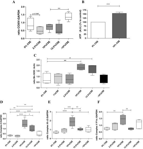
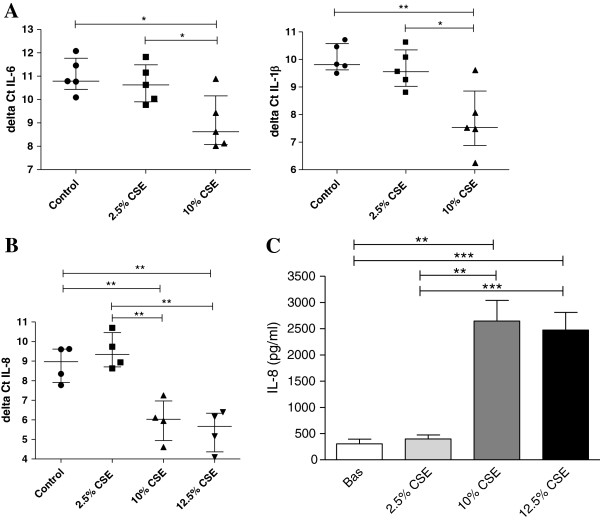
BACKGROUND: Cigarette smoking is the major risk factor for COPD, leading to chronic airway inflammation. We hypothesized that cigarette smoke induces structural and functional changes of airway epithelial mitochondria, with important implications for lung inflammation and COPD pathogenesis. METHODS: We studied changes in mitochondrial morphology and in expression of markers for mitochondrial capacity, damage/biogenesis and fission/fusion in the human bronchial epithelial cell line BEAS-2B upon 6-months from ex-smoking COPD GOLD stage IV patients to age-matched smoking and never-smoking controls. RESULTS: We observed that long-term CSE exposure induces robust changes in mitochondrial structure, including fragmentation, branching and quantity of cristae. The majority of these changes were persistent upon CSE depletion. Furthermore, long-term CSE exposure significantly increased the expression of specific fission/fusion markers (Fis1, Mfn1, Mfn2, Drp1 and Opa1), oxidative phosphorylation (OXPHOS) proteins (Complex II, III and V), and oxidative stress (Mn-SOD) markers. These changes were accompanied by increased levels of the pro-inflammatory mediators IL-6, IL-8, and IL-1β. Importantly, COPD primary bronchial epithelial cells (PBECs) displayed similar changes in mitochondrial morphology as observed in long-term CSE-exposure BEAS-2B cells. Moreover, expression of specific OXPHOS proteins was higher in PBECs from COPD patients than control smokers, as was the expression of mitochondrial stress marker PINK1. CONCLUSION: The observed mitochondrial changes in COPD epithelium are potentially the consequence of long-term exposure to cigarette smoke, leading to impaired mitochondrial function and may play a role in the pathogenesis of COPD.
Overview
The claims and statistics of this paper haven’t been checked yet.
Free: it reads the open-access text and checks the first 5 claims that cite a source we can check. Create a free account to check every claim.
Integrity signals
Registry
Cites
Statistics
Figures




Verification
Claims the paper makes
The claims and statistics of this paper haven’t been checked yet.
Free: it reads the open-access text and checks the first 5 claims that cite a source we can check. Create a free account to check every claim.
Claims other papers make about this one
Each is checked against this paper’s own text: does it say what the citing paper says it does?
The source backs part of the claim, not all of it — or backs it for a different population, condition or comparison.
In the citing paper
“The discrepancies among these studies may be due to different cell culture conditions and the concentration and duration of CSE used (14, 15, 48).”
From Impaired mitophagy leads to cigarette smoke stress‐induced cellular senescence: implications for chronic obstructive pulmonary disease (2015) · cites this paper as [15]
Experimental — accuracy not yet measuredIn the citing paper
“Similarly, elongated mitochondria with increased mitochondrial fusion activity occur during long-term CSE treatment in a human lung epithelial cell line (15).”
From Impaired mitophagy leads to cigarette smoke stress‐induced cellular senescence: implications for chronic obstructive pulmonary disease (2015) · cites this paper as [15]
In the citing paper
“There is a study showing elongated mitochondria with increased mitochondrial fusion activity during long-term CSE treatment in a human lung epithelial cell line (15), which is in agreement with our findings.”
From Impaired mitophagy leads to cigarette smoke stress‐induced cellular senescence: implications for chronic obstructive pulmonary disease (2015) · cites this paper as [15]
In the citing paper
“Recent studies have also shown that smooth muscle and transformed lung epithelial cells are equally sensitive to CS-induced cellular senescence (14, 15).”
From Impaired mitophagy leads to cigarette smoke stress‐induced cellular senescence: implications for chronic obstructive pulmonary disease (2015) · cites this paper as [15]
Self-check
Percentages, sums and reported differences re-computed from the paper’s own numbers.
The self-check needs the paper’s full text.
Citations
Cites
1 retracted
Mitochondrial Localization and Function of Heme Oxygenase-1 in Cigarette Smoke–Induced Cell Death
Slebos, D.-J., Ryter, S. W., van der Toorn, M., Liu, F., Guo, F., Baty, C. J., Karlsson, J. M., Watkins, S. C., Kim, H. P., Wang, X., et al. (2007) · American Journal of Respiratory Cell and Molecular Biology · doi:10.1165/rcmb.2006-0214oc · Cited by 215 · cited 1×
“Eventually, this pro-inflammatory activity of the airway epithelium [2,23,48-51] may contribute to the development of chronic airway inflammation in COPD.”
RetractedVandesompele, J., De Preter, K., Pattyn, F., Poppe, B., Van Roy, N., De Paepe, A., & Speleman, F. (2002) · Genome Biology · doi:10.1186/gb-2002-3-7-research0034 · Cited by 20,038 · cited 1×
“2 RT-qPCR RNA isolation, Primer Sequences, RT-PCR on Tfam, OPA1, Drp1, Fis1, Mfn1, Mfn2 using MyiQ single-color Real-Time thermal cycler (31) and RT-PCR for IL-6, IL-8, IL-1β, Mn-SOD, PINK1, PPARGC1α, OPA1 and TFAM using Taqman® were performed as described in the Supplemental Figure 1.”
MethodMitochondria, Oxidants, and Aging
Balaban, R. S., Nemoto, S., & Finkel, T. (2005) · Cell · doi:10.1016/j.cell.2005.02.001 · Cited by 4,388 · cited 4×
“Of importance, oxidative stress exceeding the anti-oxidant response may also lead to accelerated ageing and thus accelerated lung function decline [10,22,27].”
InfluentialPINK1 Is Selectively Stabilized on Impaired Mitochondria to Activate Parkin
Narendra, D. P., Jin, S. M., Tanaka, A., Suen, D.-F., Gautier, C. A., Shen, J., Cookson, M. R., & Youle, R. J. (2010) · PLoS Biology · doi:10.1371/journal.pbio.1000298 · Cited by 2,944 · cited 1×
“Persistent mitochondrial damage in airway epithelium of COPD patients is further evidenced by the increased expression of PINK1, a sensor for damaged mitochondria, and PPARGC1α, a regulator of mitochondrial biogenesis [20,46].”
Chen, H., Detmer, S. A., Ewald, A. J., Griffin, E. E., Fraser, S. E., & Chan, D. C. (2003) · The Journal of Cell Biology · doi:10.1083/jcb.200211046 · Cited by 2,601 · cited 1×
“Furthermore, it will augment ROS production and cellular apoptosis through cytochrome-C release [8,11-16].”
Oxidative damage and mitochondrial decay in aging.
Shigenaga, M. K., Hagen, T. M., & Ames, B. N. (1994) · Proceedings of the National Academy of Sciences · doi:10.1073/pnas.91.23.10771 · Cited by 2,055 · cited 1×
“Recent studies suggest that due to increased oxidative stress, damaged mtDNA induces premature ageing in human cells (7,27,37).”
BackgroundMitochondria as the central control point of apoptosis
Desagher, S., & Martinou, J.-C. (2000) · Trends in Cell Biology · doi:10.1016/s0962-8924(00)01803-1 · Cited by 1,913 · cited 2×
“Excessive oxidative stress may induce oxidative modifications, including peroxidation of mitochondrial lipids, oxidative damage of mitochondrial proteins including OXPHOS components, cristae remodeling, mutations and severe damage in mitochondrial DNA (mtDNA) [5,6,22,39,40,42,43].”
OPA1 Controls Apoptotic Cristae Remodeling Independently from Mitochondrial Fusion
Frezza, C., Cipolat, S., Martins de Brito, O., Micaroni, M., Beznoussenko, G. V., Rudka, T., Bartoli, D., Polishuck, R. S., Danial, N. N., De Strooper, B., et al. (2006) · Cell · doi:10.1016/j.cell.2006.06.025 · Cited by 1,673 · cited 1×
“Furthermore, it will augment ROS production and cellular apoptosis through cytochrome-C release [8,11-16].”
Olichon, A., Baricault, L., Gas, N., Guillou, E., Valette, A., Belenguer, P., & Lenaers, G. (2003) · Journal of Biological Chemistry · doi:10.1074/jbc.c200677200 · Cited by 1,168 · cited 1×
“Furthermore, it will augment ROS production and cellular apoptosis through cytochrome-C release [8,11-16].”
Mitochondrial Fusion and Fission in Mammals
Chan, D. C. (2006) · Annual Review of Cell and Developmental Biology · doi:10.1146/annurev.cellbio.22.010305.104638 · Cited by 972 · cited 2×
“by producing anti-oxidant scavengers, regulating the oxidative phosphorylation (OXPHOS) process responsible for ATP generation and exchanging mitochondrial DNA (mtDNA) through fusion and fission events [4,7-9].”
Cited by
The Continuum of Aging and Age-Related Diseases: Common Mechanisms but Different Rates
Franceschi, C., Garagnani, P., Morsiani, C., Conte, M., Santoro, A., Grignolio, A., Monti, D., Capri, M., & Salvioli, S. (2018) · Frontiers in Medicine · doi:10.3389/fmed.2018.00061 · Cited by 945
“Age-related pathology Mechanisms shared with aging process Markers References/ reviews Alzheimer’s disease Inflammation Oxidative stress Mitochondrial dysfunction Decreased proteasome activity Cellular senescence Gut microbiota alterations IL-6, TNF-α, IL-1β, TGFβ, IL-12, IL-18, and INFγ 8-hydroxyguanosine, 8-hydroxy-…”
BackgroundInfluentialThe mitophagy pathway and its implications in human diseases
Wang, S., Long, H., Hou, L., Feng, B., Ma, Z., Wu, Y., Zeng, Y., Cai, J., Zhang, D.-W., & Zhao, G. (2023) · Signal Transduction and Targeted Therapy · doi:10.1038/s41392-023-01503-7 · Cited by 883
“234 Nevertheless, the function of mitophagy still remains controversial when it comes to the development of COPD.”
BackgroundMitochondrial fusion, fission, and mitochondrial toxicity
Meyer, J. N., Leuthner, T. C., & Luz, A. L. (2017) · Toxicology · doi:10.1016/j.tox.2017.07.019 · Cited by 522
“cells exposed to 10% CSE for three months, followed by three months of control conditions, exhibited increased branching, suggesting mitochondrial fusion occurs in response to a chronic stress scenario (Hoffmann et al. 2013).”
BackgroundWiegman, C. H., Michaeloudes, C., Haji, G., Narang, P., Clarke, C. J., Russell, K. E., Bao, W., Pavlidis, S., Barnes, P. J., Kanerva, J., et al. (2015) · Journal of Allergy and Clinical Immunology · doi:10.1016/j.jaci.2015.01.046 · Cited by 454
“In addition, other cells can be examined, such as bronchial epithelial cells, in which mitochondrial abnormalities have been recently reported.(32,47) In summary, we have shown mitochondrial dysfunction and excessive mitochondrial ROS in ASM cells of patients with COPD, and this might contribute to their proinflammato…”
BackgroundThe relationship between COPD and lung cancer
Durham, A. L., & Adcock, I. M. (2015) · Lung Cancer · doi:10.1016/j.lungcan.2015.08.017 · Cited by 451
“Mitochondria are the major cellular source of RNOS [26] and the itochondrial dysfunction seen in COPD [81] may link COPD with he development of lung cancer.”
Background
Metadata
- Authors
- Hoffmann, R. F., Zarrintan, S., Brandenburg, S. M., Kol, A., de Bruin, H. G., Jafari, S., Dijk, F., Kalicharan, D., Kelders, M., Gosker, H. R., ten Hacken, N. H., van der Want, J. J., van Oosterhout, A. J., & Heijink, I. H.
- Affiliations
- University Medical Center Groningen (NL)
- Maastricht University Medical Centre (NL)
- Corresponding author
- Hoffmann, Roland F
- ORCID
- Venue
- Respiratory Research
- Published
- 2013
- Volume
- 14
- Issue
- 1
- Pages
- 97
- ISSN
- 1465-9921 (ISSN-L)
- PMC
- PMC3852998
- OpenAlex
- W2154409238
- Cited by
- 316
- References
- 51
- Citations by year
- 2026: 15 · 2025: 31 · 2024: 25 · 2023: 32 · 2022: 32 · 2021: 37 · 2020: 23 · 2019: 26 · 2018: 22 · 2017: 24 · 2016: 18 · 2015: 21 · 2014: 10
- FWCI
- 7.31
- Type
- journal article
- Language
- en
- Open access
- gold
- doi.org
- Locations
- Respiratory Research (publishedVersion)
- Europe PMC (PubMed Central) (publishedVersion, cc-by)
- PubMed Central (publishedVersion, cc-by)
- PubMed (publishedVersion)
- Research Publications (Maastricht University) (publishedVersion, cc-by)
- NORA - Norwegian Open Research Archives (submittedVersion, cc-by)
- University of Groningen research database (University of Groningen / Centre for Information Technology) (submittedVersion, cc-by)
- Research Publications (Maastricht University) (submittedVersion, other-oa)
- Funders
- AstraZeneca
- GlaxoSmithKline
- Danone
- Top Institute Pharma (project T1-201, T1‐201)
- Universitair Medisch Centrum Groningen
- Topics
- Life Sciences › Biochemistry, Genetics and Molecular Biology › Molecular Biology › Mitochondrial Function and Pathology
- Health Sciences › Medicine › Pulmonary and Respiratory Medicine › Chronic Obstructive Pulmonary Disease (COPD) Research
- Health Sciences › Medicine › Public Health, Environmental and Occupational Health › Smoking Behavior and Cessation
- Keywords
- cigarette smoke exposure, chronic obstructive pulmonary disease, mitochondrial morphology, mitochondrial fission and fusion, oxidative phosphorylation, oxidative stress, airway epithelial cells, bronchial epithelium, mitochondrial biogenesis, PINK1, mitochondrial stress, chronic airway inflammation, interleukin-6, interleukin-8, IL-1β, BEAS-2B cells
- MeSH terms
- PTEN-Induced Putative Kinase, Adult, Aged, Bronchi, Cell Line, Cells, Cultured, Epithelial Cells, Female, Humans, Male, Membrane Proteins, Microtubule-Associated Proteins, Middle Aged, Mitochondria, Protein Kinases, Risk Factors, Smoking, Superoxide Dismutase, Time Factors, Case-Control Studies, Cytokines, GTP Phosphohydrolases, Mitochondrial Proteins, Pulmonary Disease, Chronic Obstructive, Mitochondrial Membrane Transport Proteins, Dynamins, Mitochondrial Dynamics, Mitochondrial Turnover, In Vitro Techniques
About these checks
Registry, retraction, Cites and statistics results are computed, not judged, with one exception: a printed reference that no index lists is read once by a model before it counts as a citation error. Claims are extracted by a model and kept only when their quote appears verbatim in the text; each cited source is then checked, with a model’s help, for whether it says what the claim relies on. Those verdicts can be wrong — read the claim’s quote and the cited source before relying on one.
Statistics and arithmetic are recomputed, not judged.
Confidence bands are shown only for fields calibrated on our annotated test set. None is calibrated yet, so no band is shown.
What counts as a claim ›How we measure confidence ›How we check this ›
Are you an author, or do you see a problem with this page? Request a review
How to cite
Cite CoChat for the checks on this page, and the paper for its own findings.
- This page
- CoChat. (2026). Citation check: Prolonged cigarette smoke exposure alters mitochondrial structure and function in airway epithelial cells. https://app.cochat.ai/papers/10.1186/1465-9921-14-97
- The paper
- Hoffmann, R. F., Zarrintan, S., Brandenburg, S. M., Kol, A., de Bruin, H. G., Jafari, S., Dijk, F., Kalicharan, D., Kelders, M., Gosker, H. R., ten Hacken, N. H., van der Want, J. J., van Oosterhout, A. J., & Heijink, I. H. (2013). Prolonged cigarette smoke exposure alters mitochondrial structure and function in airway epithelial cells. Respiratory Research. https://doi.org/10.1186/1465-9921-14-97
Data sources
Bibliographic data: Semantic Scholar, OpenAlex, Crossref.
Contains information from the Semantic Scholar Academic Graph Datasets, made available under the ODC Attribution License (ODC-BY 1.0).
Semantic Scholar’s data is described in: Kinney, R., et al. (2023). The Semantic Scholar Open Data Platform. arXiv:2301.10140.
