

The DOI matches its Crossref record; 54 of the 55 references it cites were found in a registry or an index, and 1 are not checked yet.
Amyloid-β precursor protein promotes cell proliferation and motility of advanced breast cancer
Open accessAbstract
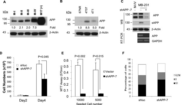
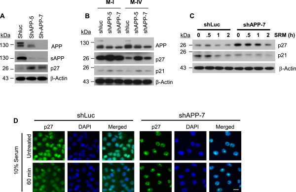
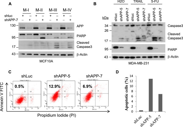
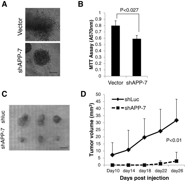
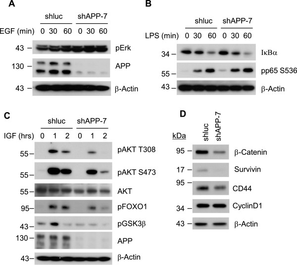
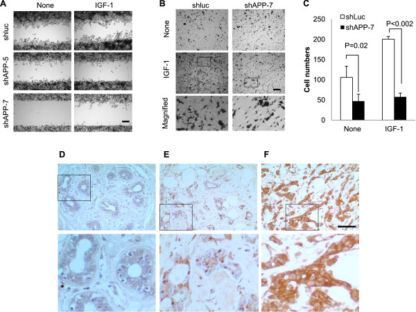
BACKGROUND: Amyloid-β precursor protein (APP) is a highly conserved single transmembrane protein that has been linked to Alzheimer disease. Recently, the increased expression of APP in multiple types of cancers has been reported where it has significant correlation with the cancer cell proliferation. However, the function of APP in the pathogenesis of breast cancer has not previously been determined. In this study, we studied the pathological role of APP in breast cancer and revealed its potential mechanism. METHODS: The expression level of APP in multiple breast cancer cell lines was measured by Western blot analysis and the breast cancer tissue microarray was utilized to analyze the expression pattern of APP in human patient specimens. To interrogate the functional role of APP in cell growth and apoptosis, the effect of APP knockdown in MDA-MB-231 cells were analyzed. Specifically, multiple signal transduction pathways and functional alterations linked to cell survival and motility were examined in in vivo animal model as well as in vitro cell culture with the manipulation of APP expression. RESULTS: We found that the expression of APP is increased in mouse and human breast cancer cell lines, especially in the cell line possessing higher metastatic potential. Moreover, the analysis of human breast cancer tissues revealed a significant correlation between the level of APP and tumor development. Knockdown of APP (APP-kd) in breast cancer cells caused the retardation of cell growth in vitro and in vivo, with both the induction of p27(kip1) and caspase-3-mediated apoptosis. APP-kd cells also had higher sensitivity to treatment of chemotherapeutic agents, TRAIL and 5-FU. Such anti-tumorigenic effects shown in the APP-kd cells partially came from reduced pro-survival AKT activation in response to IGF-1, leading to activation of key signaling regulators for cell growth, survival, and pro-apoptotic events such as GSK3-β and FOXO1. Notably, knock-down of APP in metastatic breast cancer cells limited cell migration and invasion ability upon stimulation of IGF-1. CONCLUSION: The present data strongly suggest that the increase of APP expression is causally linked to tumorigenicity as well as invasion of aggressive breast cancer and, therefore, the targeting of APP may be an effective therapy for breast cancer.
Overview
The claims and statistics of this paper haven’t been checked yet.
Run the check — free, no account needed. It reads the open-access text and checks the sources it cites.
Integrity signals
Registry
Cites
Statistics
Figures
Verification
Claims the paper makes
The claims and statistics of this paper haven’t been checked yet.
Run the check — free, no account needed. It reads the open-access text and checks the sources it cites.
Claims other papers make about this one
Each is checked against this paper’s own text: does it say what the citing paper says it does?
The cited source says what the claim attributes to it.
In the citing paper
“Amyloid precursor protein (APP), for example, is upregulated in advanced breast cancer and has been associated with increased cancer cell proliferation, migration, and invasion4.”
From Oligomeric cystatin C supports the immunosuppressive activity of myeloid cells through interaction with inhibitory receptors (2025) · cites this paper as [4]
Self-check
Percentages, sums and reported differences re-computed from the paper’s own numbers.
The self-check needs the paper’s full text.
Citations
Cites
Jemal, A., Bray, F., Ferlay, J., Ward, E., & Forman, D. (2011) · CA A Cancer Journal for Clinicians · doi:10.3322/caac.20107 · Cited by 55,077
Alzheimer's disease: the amyloid cascade hypothesis
Hardy, J. A., & Higgins, G. A. (1992) · Science · doi:10.1126/science.1566067 · Cited by 7,484
CDK inhibitors: positive and negative regulators of G1-phase progression
Sherr, C. J., & Roberts, J. M. (1999) · Genes & Development · doi:10.1101/gad.13.12.1501 · Cited by 6,115
Genes that mediate breast cancer metastasis to lung
Minn, A. J., Gupta, G. P., Siegel, P. M., Bos, P. D., Shu, W., Giri, D. D., Viale, A., Olshen, A. B., Gerald, W. L., & Massagué, J. (2005) · Nature · doi:10.1038/nature03799 · Cited by 2,922
Amyloid Precursor Protein Processing and Alzheimer's Disease
O'Brien, R. J., & Wong, P. C. (2011) · Annual Review of Neuroscience · doi:10.1146/annurev-neuro-061010-113613 · Cited by 2,061
Three-dimensional culture models of normal and malignant breast epithelial cells
Lee, G. Y., Kenny, P. A., Lee, E. H., & Bissell, M. J. (2007) · Nature Methods · doi:10.1038/nmeth1015 · Cited by 1,336
Larue, L., & Bellacosa, A. (2005) · Oncogene · doi:10.1038/sj.onc.1209091 · Cited by 1,247
Aslakson, C. J., & Miller, F. R. (1992) · PubMed · Cited by 1,234
Kenny, P. A., Lee, G. Y., Myers, C. A., Neve, R. M., Semeiks, J. R., Spellman, P. T., Lorenz, K., Lee, E. H., Barcellos‐Hoff, M. H., PETERSEN, O. W., et al. (2008) · eScholarship (California Digital Library) · Cited by 1,039
Cellular actions of beta-amyloid precursor protein and its soluble and fibrillogenic derivatives
Mattson, M. P. (1997) · Physiological Reviews · doi:10.1152/physrev.1997.77.4.1081 · Cited by 1,006 · cited 1×
“APP protein expression of MDA-MB231 was comparable to that of M-IV cells while MDAMB-231, but not M-IV cells, showed fair amount of soluble APP secretion that is known to enhance cell growth and survival [25,26].”
Result
Cited by
Sorted by citations
Hypoxia-induced alternative splicing: the 11th Hallmark of Cancer
Farina, A. R., Cappabianca, L. A., Sebastiano, M., Zelli, V., Guadagni, S., & Mackay, A. R. (2020) · Journal of Experimental & Clinical Cancer Research · doi:10.1186/s13046-020-01616-9 · Cited by 136
Amyloid-beta and tau protein beyond Alzheimer’s disease
Abyadeh, M., Gupta, V., Paulo, J. A., Mahmoudabad, A. G., Shadfar, S., Mirshahvaladi, S., Gupta, V. B., Nguyen, C. T. O., Finkelstein, D. I., You, Y., et al. (2023) · Neural Regeneration Research · doi:10.4103/1673-5374.386406 · Cited by 126
Amyloid precursor protein and amyloid precursor-like protein 2 in cancer
Pandey, P., Sliker, B. H., Peters, H. L., Tuli, A., Herskovitz, J., Smits, K., Purohit, A., Singh, R. K., Dong, J., Batra, S. K., et al. (2016) · Oncotarget · doi:10.18632/oncotarget.7103 · Cited by 109
Molecular Characteristics of Amyloid Precursor Protein (APP) and Its Effects in Cancer
Lee, H. N., Jeong, M. S., & Jang, S. B. (2021) · International Journal of Molecular Sciences · doi:10.3390/ijms22094999 · Cited by 93
Self-citationFunctions of amyloid precursor protein in metabolic diseases
Guo, Y., Wang, Q., Chen, S., & Xu, C. (2020) · Metabolism · doi:10.1016/j.metabol.2020.154454 · Cited by 89
Metadata
- Authors
- Lim, S., Yoo, B. K., Kim, H.-S., Gilmore, H. L., Lee, Y., Lee, H.-P., Kim, S.-J., Letterio, J., & Lee, H.-G.
- Affiliations
- Case Comprehensive Cancer Center (US)
- Virginia Commonwealth University (US)
- Case Western Reserve University (US)
- CHA University (KR)
- Rainbow Babies & Children's Hospital (US)
- Corresponding author
- Lim, Seunghwan
- Lee, Hyun-pil
- Lee, Hyoung-gon
- Venue
- BMC Cancer
- Published
- 2014-12
- Volume
- 14
- Issue
- 1
- Pages
- 928-928
- ISSN
- 1471-2407 (ISSN-L)
- PMC
- PMC4295427
- OpenAlex
- W2086492959
- Cited by
- 100
- References
- 54
- Citations by year
- 2026: 10 · 2025: 8 · 2024: 9 · 2023: 8 · 2022: 9 · 2021: 9 · 2020: 13 · 2019: 11 · 2018: 10 · 2017: 6 · 2016: 6 · 2015: 1
- FWCI
- 2.89
- Type
- journal article
- Language
- en
- Open access
- gold
- doi.org
- Licence
- Locations
- BMC Cancer (publishedVersion, cc-by)
- PubMed Central (publishedVersion, cc-by)
- PubMed (publishedVersion)
- Funders
- National Institutes of Health (AG028679)
- National Institute on Aging (AG028679, R01AG028679)
- Topics
- Life Sciences › Neuroscience › Cellular and Molecular Neuroscience › Alzheimer's disease research and treatments
- Health Sciences › Medicine › Oncology › Cancer Cells and Metastasis
- Life Sciences › Biochemistry, Genetics and Molecular Biology › Molecular Biology › Mechanisms of cancer metastasis
- Keywords
- amyloid precursor protein, breast cancer, metastasis, cell proliferation, cell migration and invasion, apoptosis, p27, caspase-3, Akt signaling, insulin-like growth factor I, GSK3β, FOXO1, chemosensitivity, TRAIL, 5-fluorouracil, therapeutic target
- MeSH terms
- Animals, Breast Neoplasms, Cell Movement, Cell Survival, Disease Models, Animal, Female, Humans, Insulin-Like Growth Factor I, Neoplasm Staging, Gene Expression, Amyloid beta-Protein Precursor, Apoptosis, Xenograft Model Antitumor Assays, Cell Line, Tumor, Cell Proliferation, Cyclin-Dependent Kinase Inhibitor p27, Proto-Oncogene Proteins c-akt, Mice, Gene Knockdown Techniques
About these checks
Registry, retraction, Cites and statistics results are computed, not judged, with one exception: a printed reference that no index lists is read once by a model before it counts as a citation error. Claims are extracted by a model and kept only when their quote appears verbatim in the text; each cited source is then checked, with a model’s help, for whether it says what the claim relies on. Those verdicts can be wrong — read the claim’s quote and the cited source before relying on one.
Statistics and arithmetic are recomputed, not judged.
Confidence bands are shown only for fields calibrated on our annotated test set. None is calibrated yet, so no band is shown.
What counts as a claim ›How we measure confidence ›How we check this ›
Are you an author, or do you see a problem with this page? Request a review
