

The DOI matches its Crossref record; 60 of the 61 references it cites were found in a registry or an index.
Adaptive Müller cell responses to microglial activation mediate neuroprotection and coordinate inflammation in the retina
Open accessAbstract
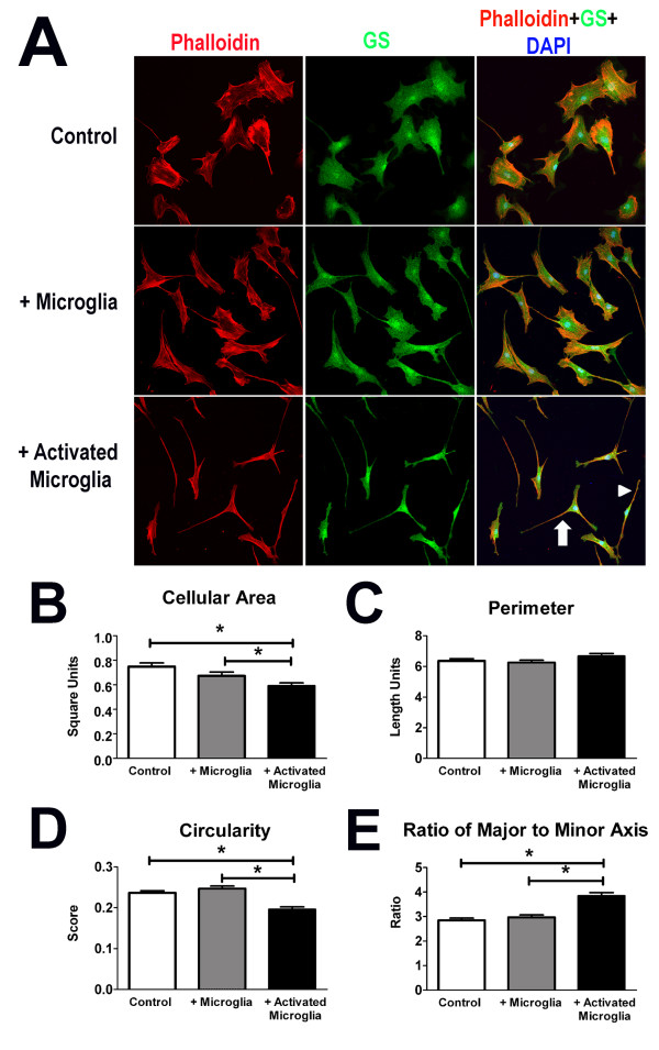
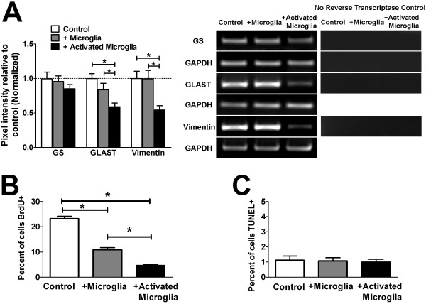
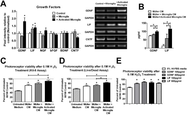
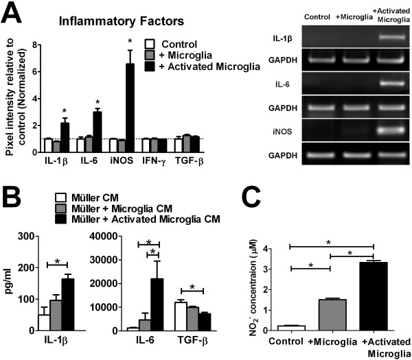
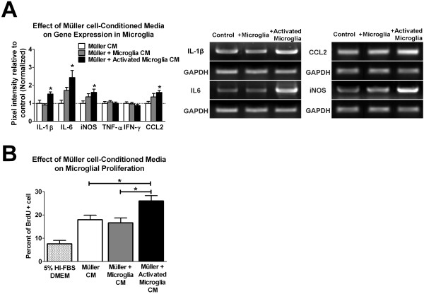
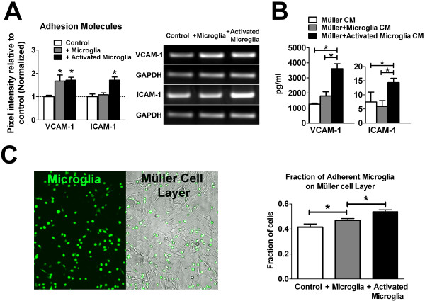
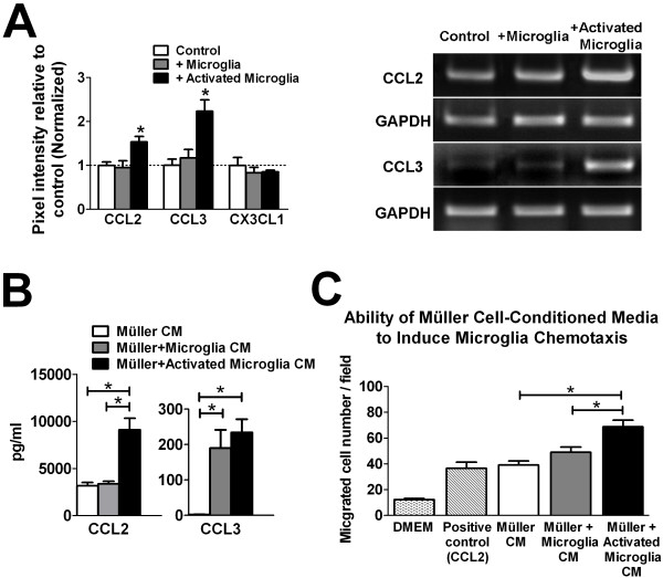
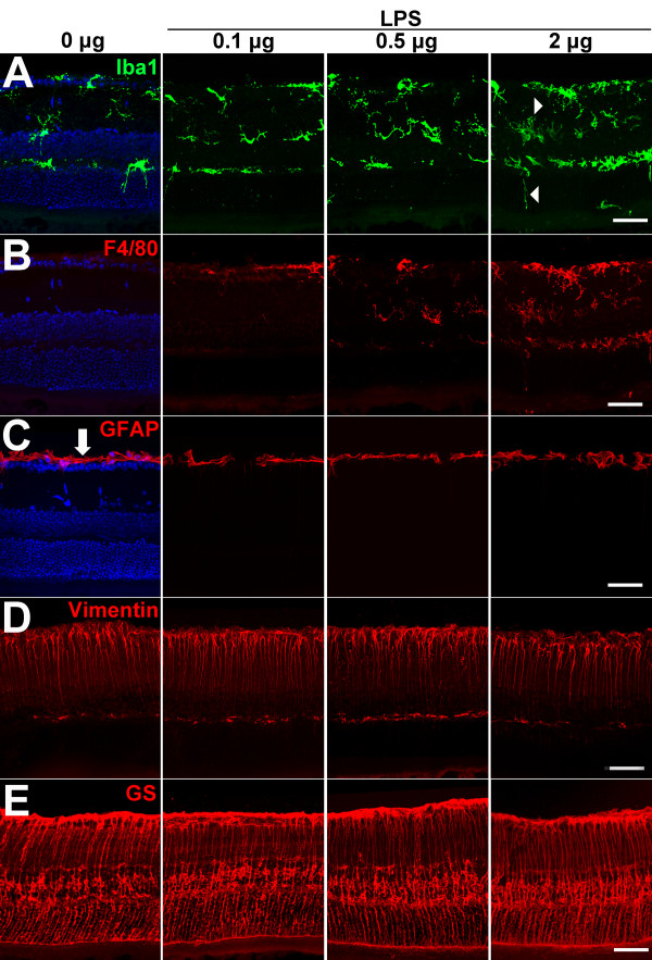
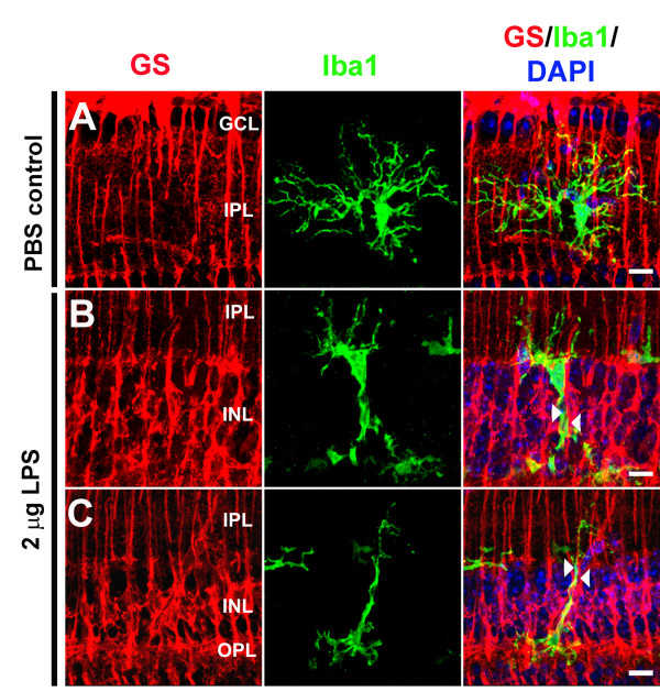
Purpose: Microglia and Müller cells are prominent participants in retinal responses to injury and disease that shape eventual tissue adaptation or damage. This investigation examined how microglia and Müller cells interact with each other following initial microglial activation. Methods: Mouse Müller cells were cultured alone, or co-cultured with activated or unactivated retinal microglia, and their morphological, molecular, and functional responses were evaluated. Müller cell-feedback signaling to microglia was studied using Müller cell-conditioned media. Corroborative in vivo analyses of retinal microglia-Müller cell interactions in the mouse retina were also performed. Results: Our results demonstrate that Müller cells exposed to activated microglia, relative to those cultured alone or with unactivated microglia, exhibit marked alterations in cell morphology and gene expression that differed from those seen in chronic gliosis. These Müller cells demonstrated in vitro (1) an upregulation of growth factors such as GDNF and LIF, and provide neuroprotection to photoreceptor cells, (2) increased pro-inflammatory factor production, which in turn increased microglial activation in a positive feedback loop, and (3) upregulated chemokine and adhesion protein expression, which allowed Müller cells to attract and adhere to microglia. In vivo activation of microglia by intravitreal injection of lipopolysaccharide (LPS) also induced increased Müller cell-microglia adhesion, indicating that activated microglia may translocate intraretinally in a radial direction using Müller cell processes as an adhesive scaffold. Conclusion: Our findings demonstrate that activated microglia are able to influence Müller cells directly, and initiate a program of bidirectional microglia-Müller cell signaling that can mediate adaptive responses within the retina following injury. In the acute aftermath following initial microglia activation, Müller cell responses may serve to augment initial inflammatory responses across retinal lamina and to guide the intraretinal mobilization of migratory microglia using chemotactic cues and adhesive cell contacts. Understanding adaptive microglia-Müller cell interactions in injury responses can help discover therapeutic cellular targets for intervention in retinal disease.
Overview
The claims and statistics of this paper haven’t been checked yet.
Run the check — free, no account needed. It reads the open-access text and checks the sources it cites.
Integrity signals
Registry
Cites
Statistics
Figures
Verification
Claims the paper makes
The claims and statistics of this paper haven’t been checked yet.
Run the check — free, no account needed. It reads the open-access text and checks the sources it cites.
Claims other papers make about this one
Each is checked against this paper’s own text: does it say what the citing paper says it does?
In the citing paper
“Inflammatory immune responses for ocular gene therapies become harder to predict in the clinical setting, as both physiological and barrier functions of immune privilege may already be compromised by aging or the ocular disorder physiopathological conditions such as inflammation or oxidative stress induction [21,66,67].”
From Risk mitigation of immunogenicity: a key to personalized retinal gene therapy (2021) · cites this paper as [21]
Self-check
Percentages, sums and reported differences re-computed from the paper’s own numbers.
The self-check needs the paper’s full text.
Citations
Cites
Resting microglial cells are highly dynamic surveillants of brain parenchyma in vivo
Nimmerjahn, A., Kirchhoff, F., & Helmchen, F. (2005) · Science · doi:10.1126/science.1110647 · Cited by 5,827
ATP mediates rapid microglial response to local brain injury in vivo
Davalos, D., Grutzendler, J., Yang, G., Kim, J. V., Zuo, Y., Jung, S., Littman, D. R., Dustin, M. L., & Gan, W.-B. (2005) · Nature Neuroscience · doi:10.1038/nn1472 · Cited by 3,913
Müller cells in the healthy and diseased retina
Bringmann, A., Pannicke, T., Grosche, J., Francke, M., Wiedemann, P., Skatchkov, S. N., Osborne, N. N., & Reichenbach, A. (2006) · Progress in Retinal and Eye Research · doi:10.1016/j.preteyeres.2006.05.003 · Cited by 1,967
The myeloid cells of the central nervous system parenchyma
Ransohoff, R. M., & Cardona, A. E. (2010) · Nature · doi:10.1038/nature09615 · Cited by 804
Bringmann, A., Iandiev, I., Pannicke, T., Wurm, A., Hollborn, M., Wiedemann, P., Osborne, N. N., & Reichenbach, A. (2009) · Progress in Retinal and Eye Research · doi:10.1016/j.preteyeres.2009.07.001 · Cited by 761
Para-Inflammation in the Aging Retina
Xu, H., Chen, M., & Forrester, J. V. (2009) · Progress in Retinal and Eye Research · doi:10.1016/j.preteyeres.2009.06.001 · Cited by 659 · cited 2×
“An understanding of how the overall injury response, initiated following microglial activation, is subsequently shaped by communications between these two cell types is likely central to elucidating the functional importance of postinjury inflammation and the mechanisms underlying chronic neuroinflammatory changes tho…”
BackgroundAge-related alterations in the dynamic behavior of microglia
Damani, M. R., Zhao, L., Fontainhas, A. M., Amaral, J., Fariss, R. N., & Wong, W. T. (2011) · Aging Cell · doi:10.1111/j.1474-9726.2010.00660.x · Cited by 504
Self-citationMicroglia activation in retinal degeneration
Langmann, T. (2007) · Journal of Leukocyte Biology · doi:10.1189/jlb.0207114 · Cited by 496
Propagation of Intercellular Calcium Waves in Retinal Astrocytes and Müller Cells
Newman, E. A. (2001) · Journal of Neuroscience · doi:10.1523/jneurosci.21-07-02215.2001 · Cited by 449
Assessing Activation States in Microglia
Colton, C. A., & Wilcock, D. M. (2010) · CNS & Neurological Disorders - Drug Targets · doi:10.2174/187152710791012053 · Cited by 430
Cited by
Sorted by citations
Items 101–200 of 228
Alfarhan, M., Liu, F., Shan, S., Pichavaram, P., Somanath, P. R., & Narayanan, S. P. (2022) · International Journal of Molecular Sciences · doi:10.3390/ijms23042133 · Cited by 27
Augustine, J., Pavlou, S., Harkin, K., Stitt, A. W., Xu, H., & Chen, M. (2023) · Disease Models & Mechanisms · doi:10.1242/dmm.050174 · Cited by 27
Santos-Carvalho, A., Aveleira, C. A., Elvas, F., Ambrósio, A. F., & Cavadas, C. (2012) · Investigative Ophthalmology & Visual Science · doi:10.1167/iovs.12-10776 · Cited by 26
Ghosh, F. K., Abdshill, H., Arnér, K. M., Voss, U., & Taylor, L. (2018) · Experimental Eye Research · doi:10.1016/j.exer.2018.02.002 · Cited by 26
Wang, X., Miller, E. B., Goswami, M., Zhang, P., Ronning, K. E., Karlen, S. J., Zawadzki, R. J., Pugh, E. N., & Burns, M. E. (2017) · Journal of Neuroinflammation · doi:10.1186/s12974-017-0886-6 · Cited by 26
Müller cells trophism and pathology as the next therapeutic targets for retinal diseases
Arrigo, A., Cremona, O., Aragona, E., Casoni, F., Consalez, G., Dogru, R. M., Hauck, S. M., Antropoli, A., Bianco, L., Parodi, M. B., et al. (2025) · Progress in Retinal and Eye Research · doi:10.1016/j.preteyeres.2025.101357 · Cited by 27
Mollick, T., Mohlin, C., & Johansson, K. U. I. (2016) · Brain Research · doi:10.1016/j.brainres.2016.06.039 · Cited by 25
Neuroretinal-Derived Caveolin-1 Promotes Endotoxin-Induced Inflammation in the Murine Retina
Gurley, J. M., Gmyrek, G. B., McClellan, M. E., Hargis, E. A., Hauck, S. M., Dozmorov, M. G., Wren, J. D., Carr, D. J. J., & Elliott, M. H. (2020) · Investigative Ophthalmology & Visual Science · doi:10.1167/iovs.61.12.19 · Cited by 25
Mahaling, B., Pandala, N. G., Wang, H.-C. H., & Lavik, E. (2022) · ACS Bio & Med Chem Au · doi:10.1021/acsbiomedchemau.2c00013 · Cited by 25
Ding, X., Sun, Z., Guo, Y., Tang, W., Shu, Q., & Xu, G. (2023) · Annals of Translational Medicine · doi:10.21037/atm-22-2204 · Cited by 25
Zhang, S., Zhang, S., Gong, W., Zhu, G., Wang, S., Wang, Y., Halim, M., Wang, K., Zhou, G., & Liu, Q. (2018) · Frontiers in Neuroscience · doi:10.3389/fnins.2018.00890 · Cited by 23
Systemic Administration of Erythropoietin Inhibits Retinopathy in RCS Rats
Shen, W., Chung, S. H., Irhimeh, M. R., Li, S., Lee, S.-R., & Gillies, M. C. (2014) · PLoS ONE · doi:10.1371/journal.pone.0104759 · Cited by 22
Mechanisms of action of retinal microglia in diabetic retinopathy (Review)
Bai, Y., Wang, X. R., Qi, F., Zuo, X., & Zou, G. (2025) · International Journal of Molecular Medicine · doi:10.3892/ijmm.2025.5643 · Cited by 22
Lu, Y., Fernando, N., Natoli, R., Madigan, M. C., & Valter, K. (2018) · Experimental Eye Research · doi:10.1016/j.exer.2018.01.011 · Cited by 21
Cellular and molecular alterations in neurons and glial cells in inherited retinal degeneration
Martínez‐Gil, N., Maneu, V., Kutsyr, O., Fernández‐Sánchez, L., Sánchez‐Sáez, X., Sánchez‐Castillo, C., Campello, L., Lax, P., Pinilla, I., & Cuenca, N. (2022) · Frontiers in Neuroanatomy · doi:10.3389/fnana.2022.984052 · Cited by 20
Ozawa, Y., Toda, E., Kawashima, H., Homma, K., Osada, H., Nagai, N., Abe, Y., Yasui, M., & Tsubota, K. (2019) · Molecular Neurobiology · doi:10.1007/s12035-019-01661-2 · Cited by 20
Immunology of Retinitis Pigmentosa and Gene Therapy–Associated UveitisPreprint
Yang, P. G., Mustafi, D., & Pepple, K. L. (2023) · Cold Spring Harbor Perspectives in Medicine · doi:10.1101/cshperspect.a041305 · Cited by 20
Risk mitigation of immunogenicity: a key to personalized retinal gene therapy
Varin, J., Morival, C., Maillard, N., Adjali, O., & Cronin, T. (2021) · International Journal of Molecular Sciences · doi:10.3390/ijms222312818 · Cited by 19
Shen, W., Lee, S., Yam, M., Zhu, L., Zhang, T., Pye, V. J., Mathai, A. E., Shibagaki, K., Zhang, J.-Z., Matsugi, T., et al. (2018) · Investigative Ophthalmology & Visual Science · doi:10.1167/iovs.18-25628 · Cited by 19
Andries, L., Groef, L. D., & Moons, L. (2019) · Current Eye Research · doi:10.1080/02713683.2019.1669664 · Cited by 19
Wang, Y., Zhao, X., Gao, M., Wan, X., Guo, Y., Qu, Y., Chen, Y., Li, T., Liu, H., Jiang, M., et al. (2021) · Cell Death and Disease · doi:10.1038/s41419-021-03983-3 · Cited by 19
Norte‐Muñoz, M., Gallego‐Ortega, A., Lucas‐Ruiz, F., González‐Riquelme, M. J., Changa-Espinoza, Y. I., Galindo‐Romero, C., Ponsaerts, P., Vidal‐Sanz, M., García‐Bernal, D., & Agudo‐Barriuso, M. (2022) · Stem Cell Research & Therapy · doi:10.1186/s13287-022-03129-y · Cited by 20
Yu, Y., Bao, Z., Wang, X., Gong, W., Chen, H., Guan, H., Le, Y., Su, S., Chen, K., & Wang, J. M. (2017) · Frontiers in Immunology · doi:10.3389/fimmu.2017.01852 · Cited by 19
Steered migration and changed morphology of human astrocytes by an applied electric field
Yang, C., Wang, L., Weng, W., Wang, S., Ma, Y., Mao, Q., Gao, G., Chen, R., & Feng, J. (2018) · Experimental Cell Research · doi:10.1016/j.yexcr.2018.11.029 · Cited by 19
Biswal, M. R., Wang, Z., Paulson, R. J., Uddin, R. R., Tong, Y., Zhu, P., Li, H., & Lewin, A. S. (2021) · Antioxidants · doi:10.3390/antiox10060842 · Cited by 19
Microglial and macroglial dynamics in a model of retinitis pigmentosa
Díaz‐Lezama, N., Kajtna, J., Wu, J., Ayten, M., & Koch, S. (2023) · Vision Research · doi:10.1016/j.visres.2023.108268 · Cited by 19
Conedera, F. M., Runnels, J. M., Stein, J. V., Alt, C., Enzmann, V., & Lin, C. P. (2023) · Journal of Neuroinflammation · doi:10.1186/s12974-023-02867-x · Cited by 18
Bludau, O., Weber, A., Bosak, V., Kuscha, V., Dietrich, K., Hans, S., & Brand, M. (2024) · Frontiers in Cell and Developmental Biology · doi:10.3389/fcell.2024.1332347 · Cited by 18
Dierschke, S. K., Toro, A. L., Miller, W. P., Sunilkumar, S., & Dennis, M. D. (2020) · Journal of Biological Chemistry · doi:10.1074/jbc.ra120.013711 · Cited by 18
Clock Gene Nr1d1 Alleviates Retinal Inflammation Through Repression of Hmga2 in Microglia
Wang, Z., Huang, Y., Chu, F., Ji, S., Liao, K., Cui, Z., Chen, J., & Tang, S. (2021) · Journal of Inflammation Research · doi:10.2147/jir.s326091 · Cited by 17
Reichenbach, A., & Bringmann, A. (2015) · Colloquium Series on Neuroglia in Biology and Medicine From Physiology to Disease · doi:10.4199/c00122ed1v01y201412ngl003 · Cited by 18
Microglial mediators in autoimmune Uveitis: Bridging neuroprotection and neurotoxicity
Sharma, M., Pal, P., & Gupta, S. K. (2024) · International Immunopharmacology · doi:10.1016/j.intimp.2024.112309 · Cited by 16
Padovani-Claudio, D. A., Morales, M. S., Smith, T. E., Ontko, C. D., Namburu, N. S., Palmer, S. A., Jhala, M. G., Ramos, C. J., Capozzi, M. E., McCollum, G. W., et al. (2024) · Cellular Signalling · doi:10.1016/j.cellsig.2024.111454 · Cited by 16
Vitrectomy for diabetic macular edema and the relevance of external limiting membrane
Ivastinović, D., Haas, A., Weger, M., Seidel, G., Mayer‐Xanthaki, C., Lindner, E., Guttmann, A., & Wedrich, A. (2021) · BMC Ophthalmology · doi:10.1186/s12886-021-02095-y · Cited by 16
Racic, T., Chang, A., Fernando, N., Brandli, A. A., Natoli, R., Penfold, P. L., Provis, J. M., & Rutar, M. (2021) · Experimental Eye Research · doi:10.1016/j.exer.2021.108765 · Cited by 17
Huang, L., Xu, G., Guo, J., Xie, M., Chen, L., & Xu, W. (2015) · Ocular Immunology and Inflammation · doi:10.3109/09273948.2015.1071405 · Cited by 16
Li, H., Chen, D., Sun, W., Chen, J., Luo, C., Xu, H., Hongjie, J., & Tang, S. (2021) · Investigative Ophthalmology & Visual Science · doi:10.1167/iovs.62.2.3 · Cited by 15
Retinal degeneration in mice lacking the cyclic nucleotide‐gated channel subunit CNGA1
Liu, Y., Yf, W., Xiao, Y., Li, X., Ruan, S., Luo, X., Wan, X., Wang, F., & Sun, X. (2021) · The FASEB Journal · doi:10.1096/fj.202101004r · Cited by 15
The immunological link between neonatal lung and eye disease
Wickramasinghe, L. C., van Wijngaarden, P., Tsantikos, E., & Hibbs, M. L. (2021) · Clinical & Translational Immunology · doi:10.1002/cti2.1322 · Cited by 15
Pilotto, E., Torresin, T., Bacelle, M. L., Mojà, G. D., Ferrara, A. M., Zovato, S., Midena, G., & Midena, E. (2022) · PLoS ONE · doi:10.1371/journal.pone.0272318 · Cited by 15
Immunology of age-related macular degeneration
Csaky, K. G., Mettu, P. S., & Cousins, S. W. (2012) · CRC Press eBooks · doi:10.1201/b14548-3 · Cited by 15
Gao, S., Li, N., Wang, Y., Lin, Z., Zhu, Y., Xu, J., Zhang, Q., Zhu, C., Zhou, Y., Zhou, J., et al. (2022) · The FASEB Journal · doi:10.1096/fj.202101294rrr · Cited by 14
Systemic Lipopolysaccharide Exposure Exacerbates Choroidal Neovascularization in Mice
Tsioti, I., Steiner, B. L., Escher, P., Zinkernagel, M. S., Benz, P. M., & Kokona, D. (2022) · Ocular Immunology and Inflammation · doi:10.1080/09273948.2022.2147547 · Cited by 14
Myojin, S., Yoshimura, T., Yoshida, S., Takeda, A., Murakami, Y., Kawano, Y., Oshima, Y., Ishibashi, T., & Sonoda, K.-H. (2016) · PLoS ONE · doi:10.1371/journal.pone.0164355 · Cited by 13
Liu, X., Heitz, P., Roux, M. J., Keller, D., Bourcier, T., Sauer, A., Prévost, G., & Gaucher, D. (2018) · Scientific Reports · doi:10.1038/s41598-018-20590-z · Cited by 13
Venanzi, A. W., McGee, L. D., & Hackam, A. S. (2024) · Molecular Neurobiology · doi:10.1007/s12035-024-04679-3 · Cited by 13
Li, Z., Xiong, Q., Li, Q., & Tang, L. (2025) · International Immunopharmacology · doi:10.1016/j.intimp.2025.114219 · Cited by 12
“Super p53” Mice Display Retinal Astroglial Changes
Salazar, J. J., Gallego‐Pinazo, R., de Hoz, R., Pinazo-Duran, M. D., Rojas, B., Ramirez, A. I., Serrano, M., & Ramirez, J. M. (2013) · PLoS ONE · doi:10.1371/journal.pone.0065446 · Cited by 12
Comparative Anatomy of Glial Cells in Mammals
Reichenbach, A., & Bringmann, A. (2016) · Elsevier eBooks · doi:10.1016/b978-0-12-804042-3.00050-6 · Cited by 12
Schultz, R., Krug, M., Precht, M., Wohl, S. G., Witte, O. W., & Schmeer, C. W. (2018) · Scientific Reports · doi:10.1038/s41598-018-22887-5 · Cited by 13
Automated detection of hyperreflective foci in the outer nuclear layer of the retina
Schmidt, M. F., Christensen, J. L., Dahl, V. A., Toosy, A., Petzold, A., Hanson, J. V. M., Schippling, S., Frederiksen, J. L., & Larsen, M. (2022) · Acta Ophthalmologica · doi:10.1111/aos.15237 · Cited by 11
Lepre, C. C., Russo, M., Trotta, M. C., Petrillo, F., D’Agostino, F. A., Gaudino, G., D’Amico, G., Campitiello, M. R., Crisci, E., Nicoletti, M., et al. (2023) · International Journal of Molecular Sciences · doi:10.3390/ijms24119721 · Cited by 10
Zhang, Y., Ma, Y., Ji, Y., Jiang, Y.-F., Li, D., Mu, W., Yao, M., Yao, J., & Yan, B. (2025) · Cell Communication and Signaling · doi:10.1186/s12964-024-02013-x · Cited by 10
Microglial depletion decreases Müller cell maturation and inner retinal vascular density
Rowthorn-Apel, N., Vridhachalam, N., Connor, K. M., Bonilla, G. M., Sadreyev, R. I., Singh, C., & Gnanaguru, G. (2025) · Cell Communication and Signaling · doi:10.1186/s12964-025-02083-5 · Cited by 10
Zhu, Q., Yu, P. C., Liang, X.-S., Ma, L., Xiao, H.-X., & Yuan, Y. (2020) · International Journal of Ophthalmology · doi:10.18240/ijo.2020.12.16 · Cited by 10
Molecular studies of phenotype variation in canine RPGR-XLPRA1.
Appelbaum, T., Becker, D., Santana, E., & Aguirre, G. K. (2016) · PubMed Central · Cited by 10
Hooper, M. J., Wang, J., Browning, R., & Ash, J. D. (2018) · Scientific Reports · doi:10.1038/s41598-018-27479-x · Cited by 10
Protecting the aging eye with hydrogen sulfide
George, A. K., Homme, R. P., Stanišić, D., Tyagi, S. C., & Singh, M. (2020) · Canadian Journal of Physiology and Pharmacology · doi:10.1139/cjpp-2020-0216 · Cited by 10
Pilotto, E., Parolini, F., Midena, G., Cosmo, E., & Midena, E. (2024) · Experimental Eye Research · doi:10.1016/j.exer.2024.110064 · Cited by 9
Zhou, Z., Feng, Z., Sun, X., Wang, Y., & Dou, G. (2023) · International Journal of Molecular Sciences · doi:10.3390/ijms242115516 · Cited by 9
Ulbricht, E., Pannicke, T., Uhlmann, S., Wiedemann, P., Reichenbach, A., & Francke, M. (2012) · Acta Ophthalmologica · doi:10.1111/j.1755-3768.2012.02527.x · Cited by 9
Martínez‐López, M. A., Rubio‐Casado, S., Felipe, D. S., Martín‐Sánchez, B., Fernández‐Albarral, J. A., Salobrar‐García, E., Matamoros, J. A., Ramirez, J. M., de Hoz, R., Salazar, J. J., et al. (2024) · International Journal of Molecular Sciences · doi:10.3390/ijms25063115 · Cited by 8
Inherited retinal disease-associated uveitis
Hung, J., Jain, T., Khatri, A., Nguyen, T. T., Nguyen, C. D.-T., Yavari, N., Mobasserian, A., Karaca, I., Mohammadi, S. S., Gupta, A., et al. (2025) · Survey of Ophthalmology · doi:10.1016/j.survophthal.2025.03.011 · Cited by 9
Li, S., Zhou, H., Zhao, G., Ding, W., Zhang, Y., Wang, Y., Li, F., Miao, Y., Sun, X., & Wang, Z. (2025) · Neuroscience Bulletin · doi:10.1007/s12264-025-01478-1 · Cited by 8
Glial cells modulate retinal cell survival in rotenone-induced neural degeneration
Tawarayama, H., Inoue-Yanagimachi, M., Himori, N., & Nakazawa, T. (2021) · Scientific Reports · doi:10.1038/s41598-021-90604-w · Cited by 9
Tackling retinal ganglion cell apoptosis in glaucoma: role of adenosine receptors
Agarwal, P., & Agarwal, R. (2021) · Expert Opinion on Therapeutic Targets · doi:10.1080/14728222.2021.1969362 · Cited by 9
Lin, Y., Xu, J., Huang, Q., Heng, X., Yang, Z., & Zhang, H. (2025) · Visual Neuroscience · doi:10.48130/vns-0025-0010 · Cited by 7
Shin, T., Ahn, M., Kim, J., Jung, K., Moon, C., & Kim, M.-D. (2021) · Molecular Neurobiology · doi:10.1007/s12035-021-02355-4 · Cited by 7
Zhang, T., Wei, Q., Li, Z., Meng, W., Zhang, Z., & Zhang, Z. (2023) · Computer Methods and Programs in Biomedicine · doi:10.1016/j.cmpb.2023.107632 · Cited by 7
Elbaz-Hayoun, S., Rinsky, B., Hagbi-Levi, S., Grunin, M. A., & Chowers, I. (2023) · eLife · doi:10.7554/elife.81208 · Cited by 7
Physiology and pathophysiology of the retinal neuroglia
Grosche, A., Grosche, J., & Verkhratsky, A. (2025) · Handbook of clinical neurology · doi:10.1016/b978-0-443-19102-2.00017-x · Cited by 6
Chan‐Ling, T., Hu, P., Calzi, S. L., Warner, J., Uddin, M. N., Dupont, M., Neuringer, M. D., Kievit, P., Renner, L. M., Stoddard, J., et al. (2023) · American Journal Of Pathology · doi:10.1016/j.ajpath.2023.02.019 · Cited by 6
Greferath, U., Huynh, M., Jobling, A. I., Vessey, K. A., Venables, G., Surrao, D. C., O'Neill, H. C., Limnios, I. J., & Fletcher, E. L. (2021) · Frontiers in Cellular Neuroscience · doi:10.3389/fncel.2020.553708 · Cited by 6
Neroev, V. V., Зуева, М. В., Zhuravleva, A. N., & Цапенко, И. В. (2020) · Ophthalmology in Russia · doi:10.18008/1816-5095-2020-3s-533-541 · Cited by 6
Salia, O. I., & Mitchell, D. M. (2020) · BMC Genomics · doi:10.1186/s12864-020-07273-8 · Cited by 6
Neuroprotective Role of Liver Growth Factor “LGF” in an Experimental Model of Cerebellar Ataxia
Calatrava-Ferreras, L., Gonzalo‐Gobernado, R., Reimers, D., Herranz, A. S., Jiménez‐Escrig, A., Díaz-Gil, J. J., Casarejos, M. J., Vega, M. T. M., & Bazán, E. (2014) · International Journal of Molecular Sciences · doi:10.3390/ijms151019056 · Cited by 6
The role of microglia in glaucoma - trigger and potential target
Chen, L., Yang, S., Wang, D., & Huang, P. (2025) · Frontiers in Immunology · doi:10.3389/fimmu.2025.1685495 · Cited by 6
Taylor, L., Arnér, K. M., Blom, A. M., & Ghosh, F. K. (2016) · Journal of Neuroimmunology · doi:10.1016/j.jneuroim.2016.07.014 · Cited by 5
Pfromm, J. K., Bonillo, M., Dauletbekov, D., Bucher, K., & Fischer, M. D. (2022) · Scientific Reports · doi:10.1038/s41598-022-17269-x · Cited by 5
Carrero, L., Antequera, D., Alcalde, I., Megı́as, D., Ordóñez‐Gutiérrez, L., Gutiérrez, C., Merayo-Llovés, J., Wandosell, F. G., Municio, C., & Carro, E. (2023) · International Journal of Molecular Sciences · doi:10.3390/ijms242115679 · Cited by 5
Amato, R., Canovai, A., Melecchi, A., Fenza, M. D., Leone, L., Pavone, V., D’Alonzo, D., Cammalleri, M., & Monte, M. D. (2025) · Investigative Ophthalmology & Visual Science · doi:10.1167/iovs.66.11.29 · Cited by 4
Phenomenology of spontaneous closure in degenerative and mixed type lamellar macular hole
Catania, F., Romano, M. R., Crincoli, E., Allegrini, D., Mière, A., Chehaibou, I., Abdelmassih, Y., Beaumont, W., Chapron, T., Souied, E. H., et al. (2023) · Eye · doi:10.1038/s41433-023-02681-y · Cited by 4
The role of adiponectin and its receptor signaling in ocular inflammation-associated diseases
Zheng, J. G., Zhang, W., Xu, R., & Liu, L. (2024) · Biochemical and Biophysical Research Communications · doi:10.1016/j.bbrc.2024.150041 · Cited by 4
Yang, T., Wang, W., Xie, L., Chen, S., Ye, X., Shen, S., Chen, H., Qi, L., Cui, Z., Xiong, W., et al. (2024) · Journal of Neuroscience Methods · doi:10.1016/j.jneumeth.2024.110181 · Cited by 4
Comparative Anatomy of Glial Cells in Mammals
Reichenbach, A., & Bringmann, A. (2020) · Elsevier eBooks · doi:10.1016/b978-0-12-820584-6.00016-7 · Cited by 4
Quaranta, L., Bruttini, C., Angelis, G. D., Montescani, S., Ardizzone, A., Katsanos, A., Carnevale, C., Oddone, F., & Riva, I. (2021) · Journal of Clinical Medicine · doi:10.3390/jcm10204668 · Cited by 4
Effects of age on retinal macrophage responses to acute elevation of intraocular pressure
Kezic, J. M., Chrysostomou, V., McMenamin, P. G., & Crowston, J. G. (2020) · Experimental Eye Research · doi:10.1016/j.exer.2020.107995 · Cited by 3
Palazzo, I., Todd, L. J., Hoang, T. V., Reh, T. A., Blackshaw, S., & Fischer, A. J. (2021) · bioRxiv (Cold Spring Harbor Laboratory) · doi:10.1101/2021.10.05.463152 · Cited by 3
Fibrogliosis in neovascular age-related macular degeneration: A new mechanistic perspective
Merle, D. A., Beretta, F., Sacconi, R., & Querques, G. (2025) · Survey of Ophthalmology · doi:10.1016/j.survophthal.2025.09.019 · Cited by 2
Parolini, F., Pilotto, E., Midena, E., & Midena, G. (2026) · Journal of Clinical Medicine · doi:10.3390/jcm15041453 · Cited by 2
Kang, S., & Wohl, S. G. (2022) · Journal of Visualized Experiments · doi:10.3791/63651 · Cited by 2
Xu, H., Wang, Q., Chen, X., Huang, Q., Xu, S., & Wu, Z. (2025) · Computational and Structural Biotechnology Journal · doi:10.1016/j.csbj.2025.08.019 · Cited by 2
“Armed in-vitro retina”-generating microglial retinal organoids, where are we now?
Liu, Y., Gao, L., Chen, W., Yan, Y., Ye, Z., & Li, Z. (2025) · Frontiers in Cell and Developmental Biology · doi:10.3389/fcell.2025.1574283 · Cited by 2
Pilotto, E., Midena, G., Torresin, T., Mojà, G. D., Bacelle, M. L., Ferrara, A. M., Zovato, S., & Midena, E. (2021) · Cancers · doi:10.3390/cancers14010170 · Cited by 2
Modification of Müller Glial Cell Fate and Proliferation with the Use of Small Molecules
Hooper, M. J. (2023) · Advances in experimental medicine and biology · doi:10.1007/978-3-031-27681-1_69 · Cited by 1
Isolation and Culture of Primary Retinal Müller Cells from Sprague-Dawley (SD) Rats
Tang, Y., Sun, Y., Mao, Y., Peng, W., Zhang, W., & Zhang, F. (2025) · Journal of Visualized Experiments · doi:10.3791/68129 · Cited by 1
Xin, X., Han, L., Qu, Y., Zhang, L., Zhang, X., Li, J., Lei, B., Wang, Y.-C., & Wang, Z. (2026) · Journal of Neuroinflammation · doi:10.1186/s12974-026-03884-2 · Cited by 1
Las bases: retinopatía diabética y edema macular
García-Rubio, Y. Z., & Lima-Gómez, V. (2014) · Revista del Hospital Juárez de México
Graca, A. B. (2017) · UCL Discovery (University College London)
Metadata
- Authors
- Wang, M., Ma, W., Zhao, L., Fariss, R. N., & Wong, W. T.
- Affiliations
- National Institutes of Health (US)
- Corresponding author
- Wong, Wai T
- Venue
- Journal of Neuroinflammation
- Published
- 2011-12
- Volume
- 8
- Issue
- 1
- Pages
- 173-173
- ISSN
- 1742-2094 (ISSN-L)
- PMC
- PMC3251543
- OpenAlex
- W2077349110
- Cited by
- 228
- References
- 61
- Citations by year
- 2026: 12 · 2025: 17 · 2024: 14 · 2023: 20 · 2022: 20 · 2021: 32 · 2020: 18 · 2019: 11 · 2018: 24 · 2017: 14 · 2016: 11 · 2015: 12 · 2014: 14 · 2013: 5 · 2012: 4
- FWCI
- 4.07
- Type
- journal article
- Language
- en
- Open access
- gold
- doi.org
- Licence
- cc-by
- Locations
- Journal of Neuroinflammation (publishedVersion, cc-by)
- Europe PMC (PubMed Central) (publishedVersion, cc-by)
- PubMed Central (publishedVersion, cc-by)
- PubMed (publishedVersion)
- DOAJ (DOAJ: Directory of Open Access Journals) (submittedVersion)
- Topics
- Life Sciences › Neuroscience › Neurology › Neuroinflammation and Neurodegeneration Mechanisms
- Health Sciences › Medicine › Ophthalmology › Retinal Diseases and Treatments
- Health Sciences › Medicine › Ophthalmology › Retinal Development and Disorders
- Keywords
- Müller cells, retinal microglia, microglial activation, neuroprotection, photoreceptors, glial cell line-derived neurotrophic factor, LiF, pro-inflammatory factors, chemokines, cell adhesion, microglial migration, lipopolysaccharide, retinal inflammation, gliosis, retinal damage
- MeSH terms
- Animals, Cell Adhesion, Cell Communication, Cells, Cultured, Chemotaxis, Gliosis, Inflammation, Mice, Inbred C57BL, Retina, Culture Media, Conditioned, Apoptosis, Microglia, Neuroprotective Agents, Coculture Techniques, Cell Proliferation, Mice
About these checks
Registry, retraction, Cites and statistics results are computed, not judged, with one exception: a printed reference that no index lists is read once by a model before it counts as a citation error. Claims are extracted by a model and kept only when their quote appears verbatim in the text; each cited source is then checked, with a model’s help, for whether it says what the claim relies on. Those verdicts can be wrong — read the claim’s quote and the cited source before relying on one.
Statistics and arithmetic are recomputed, not judged.
Confidence bands are shown only for fields calibrated on our annotated test set. None is calibrated yet, so no band is shown.
What counts as a claim ›How we measure confidence ›How we check this ›
Are you an author, or do you see a problem with this page? Request a review
