

The DOI matches its Crossref record; 60 of the 61 references it cites were found in a registry or an index.
Adaptive Müller cell responses to microglial activation mediate neuroprotection and coordinate inflammation in the retina
Open accessAbstract
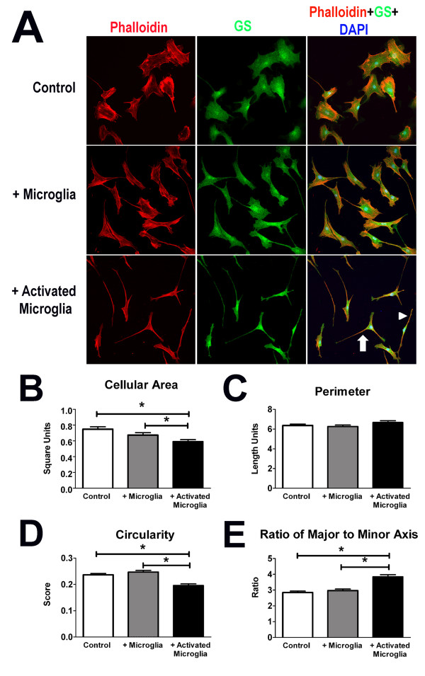
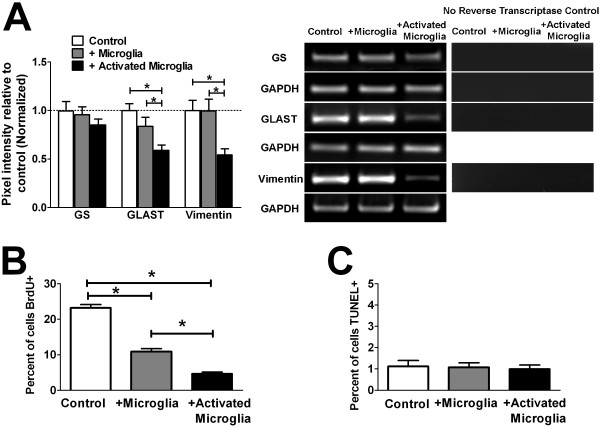
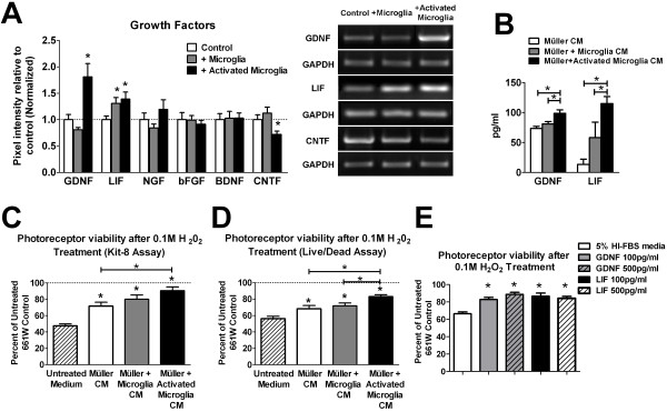
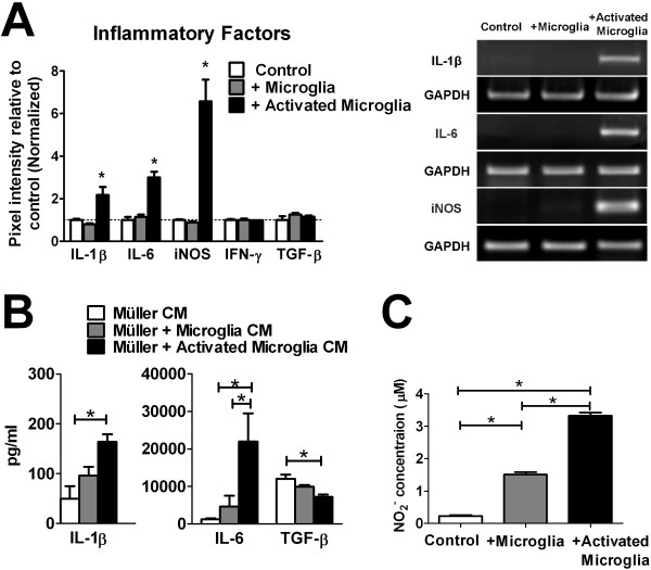
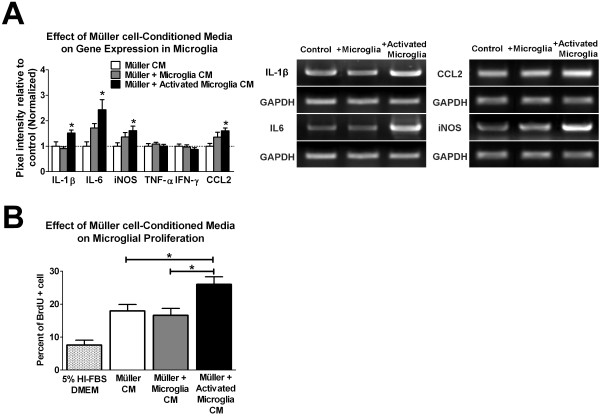
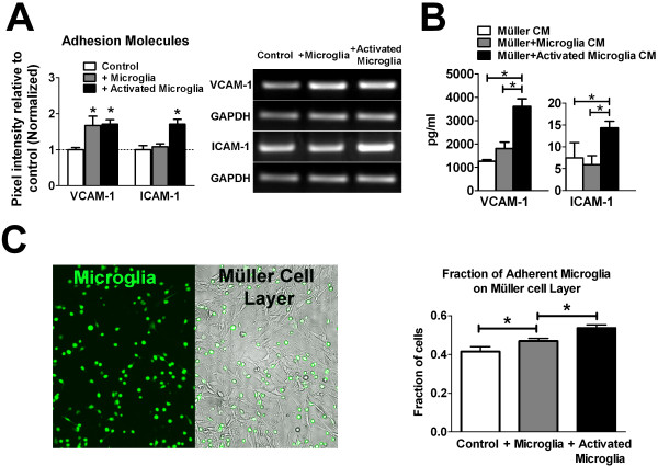
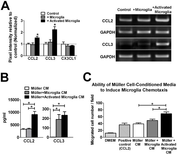
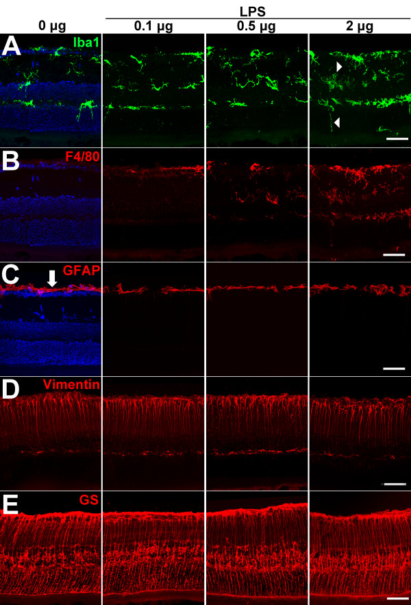
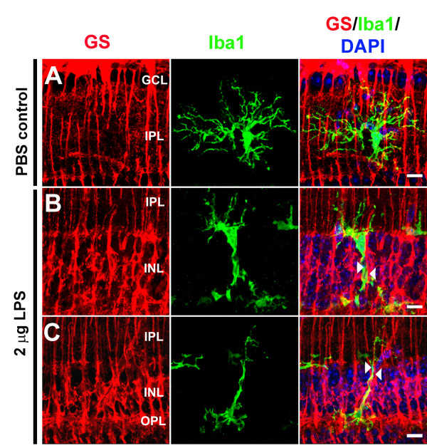
Purpose: Microglia and Müller cells are prominent participants in retinal responses to injury and disease that shape eventual tissue adaptation or damage. This investigation examined how microglia and Müller cells interact with each other following initial microglial activation. Methods: Mouse Müller cells were cultured alone, or co-cultured with activated or unactivated retinal microglia, and their morphological, molecular, and functional responses were evaluated. Müller cell-feedback signaling to microglia was studied using Müller cell-conditioned media. Corroborative in vivo analyses of retinal microglia-Müller cell interactions in the mouse retina were also performed. Results: Our results demonstrate that Müller cells exposed to activated microglia, relative to those cultured alone or with unactivated microglia, exhibit marked alterations in cell morphology and gene expression that differed from those seen in chronic gliosis. These Müller cells demonstrated in vitro (1) an upregulation of growth factors such as GDNF and LIF, and provide neuroprotection to photoreceptor cells, (2) increased pro-inflammatory factor production, which in turn increased microglial activation in a positive feedback loop, and (3) upregulated chemokine and adhesion protein expression, which allowed Müller cells to attract and adhere to microglia. In vivo activation of microglia by intravitreal injection of lipopolysaccharide (LPS) also induced increased Müller cell-microglia adhesion, indicating that activated microglia may translocate intraretinally in a radial direction using Müller cell processes as an adhesive scaffold. Conclusion: Our findings demonstrate that activated microglia are able to influence Müller cells directly, and initiate a program of bidirectional microglia-Müller cell signaling that can mediate adaptive responses within the retina following injury. In the acute aftermath following initial microglia activation, Müller cell responses may serve to augment initial inflammatory responses across retinal lamina and to guide the intraretinal mobilization of migratory microglia using chemotactic cues and adhesive cell contacts. Understanding adaptive microglia-Müller cell interactions in injury responses can help discover therapeutic cellular targets for intervention in retinal disease.
Overview
The claims and statistics of this paper haven’t been checked yet.
Run the check — free, no account needed. It reads the open-access text and checks the sources it cites.
Integrity signals
Registry
Cites
Statistics
Figures
Verification
Claims the paper makes
The claims and statistics of this paper haven’t been checked yet.
Run the check — free, no account needed. It reads the open-access text and checks the sources it cites.
Claims other papers make about this one
Each is checked against this paper’s own text: does it say what the citing paper says it does?
In the citing paper
“Inflammatory immune responses for ocular gene therapies become harder to predict in the clinical setting, as both physiological and barrier functions of immune privilege may already be compromised by aging or the ocular disorder physiopathological conditions such as inflammation or oxidative stress induction [21,66,67].”
From Risk mitigation of immunogenicity: a key to personalized retinal gene therapy (2021) · cites this paper as [21]
Self-check
Percentages, sums and reported differences re-computed from the paper’s own numbers.
The self-check needs the paper’s full text.
Citations
Cites
Resting microglial cells are highly dynamic surveillants of brain parenchyma in vivo
Nimmerjahn, A., Kirchhoff, F., & Helmchen, F. (2005) · Science · doi:10.1126/science.1110647 · Cited by 5,827
ATP mediates rapid microglial response to local brain injury in vivo
Davalos, D., Grutzendler, J., Yang, G., Kim, J. V., Zuo, Y., Jung, S., Littman, D. R., Dustin, M. L., & Gan, W.-B. (2005) · Nature Neuroscience · doi:10.1038/nn1472 · Cited by 3,913
Müller cells in the healthy and diseased retina
Bringmann, A., Pannicke, T., Grosche, J., Francke, M., Wiedemann, P., Skatchkov, S. N., Osborne, N. N., & Reichenbach, A. (2006) · Progress in Retinal and Eye Research · doi:10.1016/j.preteyeres.2006.05.003 · Cited by 1,967
The myeloid cells of the central nervous system parenchyma
Ransohoff, R. M., & Cardona, A. E. (2010) · Nature · doi:10.1038/nature09615 · Cited by 804
Bringmann, A., Iandiev, I., Pannicke, T., Wurm, A., Hollborn, M., Wiedemann, P., Osborne, N. N., & Reichenbach, A. (2009) · Progress in Retinal and Eye Research · doi:10.1016/j.preteyeres.2009.07.001 · Cited by 761
Para-Inflammation in the Aging Retina
Xu, H., Chen, M., & Forrester, J. V. (2009) · Progress in Retinal and Eye Research · doi:10.1016/j.preteyeres.2009.06.001 · Cited by 659 · cited 2×
“An understanding of how the overall injury response, initiated following microglial activation, is subsequently shaped by communications between these two cell types is likely central to elucidating the functional importance of postinjury inflammation and the mechanisms underlying chronic neuroinflammatory changes tho…”
BackgroundAge-related alterations in the dynamic behavior of microglia
Damani, M. R., Zhao, L., Fontainhas, A. M., Amaral, J., Fariss, R. N., & Wong, W. T. (2011) · Aging Cell · doi:10.1111/j.1474-9726.2010.00660.x · Cited by 504
Self-citationMicroglia activation in retinal degeneration
Langmann, T. (2007) · Journal of Leukocyte Biology · doi:10.1189/jlb.0207114 · Cited by 496
Propagation of Intercellular Calcium Waves in Retinal Astrocytes and Müller Cells
Newman, E. A. (2001) · Journal of Neuroscience · doi:10.1523/jneurosci.21-07-02215.2001 · Cited by 449
Assessing Activation States in Microglia
Colton, C. A., & Wilcock, D. M. (2010) · CNS & Neurological Disorders - Drug Targets · doi:10.2174/187152710791012053 · Cited by 430
Tezel, G., & Wax, M. B. (2000) · Journal of Neuroscience · doi:10.1523/jneurosci.20-23-08693.2000 · Cited by 418
Harada, T., Harada, C., Kohsaka, S., Wada, E., Yoshida, K., Ohno, S., Mamada, H., Tanaka, K., Parada, L. F., & Wada, K. (2002) · The Journal of Neuroscience · doi:10.1523/jneurosci.22-21-09228.2002 · Cited by 397
Buffo, A., Rolando, C., & Ceruti, S. (2009) · Biochemical Pharmacology · doi:10.1016/j.bcp.2009.09.014 · Cited by 338
Astrogliosis in CNS Pathologies: Is There A Role for Microglia?
Zhang, D., Hu, X., Li, Q., O'Callaghan, J. P., & Hong, J. (2010) · Molecular Neurobiology · doi:10.1007/s12035-010-8098-4 · Cited by 305
Liu, W., Tang, Y., & Feng, J. (2011) · Life Sciences · doi:10.1016/j.lfs.2011.05.011 · Cited by 303 · cited 2×
“While the astrocytic and microglial responses in these pathogenic contexts have been thought to involve cross-talk between these two cell populations [3], the mechanisms and functional significances underlying these interactions are incompletely understood [4,5].”
BackgroundMonocyte chemoattractant protein 1 mediates retinal detachment-induced photoreceptor apoptosis
Nakazawa, T., Hisatomi, T., Nakazawa, C., Noda, K., Maruyama, K., She, H., Matsubara, A., Miyahara, S., Nakao, S., Yin, Y., et al. (2007) · Proceedings of the National Academy of Sciences · doi:10.1073/pnas.0608167104 · Cited by 272
Microglial cells in astroglial cultures: a cautionary note
Saura, J. (2007) · Journal of Neuroinflammation · doi:10.1186/1742-2094-4-26 · Cited by 233
Hicks, D., & Courtois, Y. (1990) · Experimental Eye Research · doi:10.1016/0014-4835(90)90063-z · Cited by 227
Like cops on the beat: the active role of resting microglia
Raivich, G. (2005) · Trends in Neurosciences · doi:10.1016/j.tins.2005.09.001 · Cited by 225
Tanaka, J., Fujita, H. M., Matsuda, S., Toku, K., Sakanaka, M., & Maeda, N. (1997) · Glia · doi:10.1002/(sici)1098-1136(199705)20:1 · Cited by 223
Ma, W., Zhao, L., Fontainhas, A. M., Fariss, R. N., & Wong, W. T. (2009) · PLoS ONE · doi:10.1371/journal.pone.0007945 · Cited by 222
Self-citationOmri, S., Béhar‐Cohen, F., de Kozak, Y., Sennlaub, F., Veríssimo, L. M., Jonet, L., Savoldelli, M., Omri, B., & Crisanti, P. (2011) · American Journal Of Pathology · doi:10.1016/j.ajpath.2011.04.018 · Cited by 219
Functions of Mature Mammalian Astrocytes: A Current View
Kimelberg, H. K. (2010) · The Neuroscientist · doi:10.1177/1073858409342593 · Cited by 218
Embryonic and postnatal development of microglial cells in the mouse retina
Santos, A. M., Calvente, R., Tassi, M., Carrasco, M., Martín‐Oliva, D., Marín‐Teva, J. L., Navascués, J., & Cuadros, M. A. (2008) · Journal of Comparative Neurology · doi:10.1002/cne.21538 · Cited by 213
Ex Vivo Dynamic Imaging of Retinal Microglia Using Time-Lapse Confocal Microscopy
Lee, J. E., Liang, K. J., Fariss, R. N., & Wong, W. T. (2008) · Investigative Ophthalmology & Visual Science · doi:10.1167/iovs.08-2076 · Cited by 212
Self-citationZeng, H.-Y., Zhu, X. A., Zhang, C., Yang, L.-P., Wu, L.-M., & Tso, M. O. M. (2005) · Investigative Ophthalmology & Visual Science · doi:10.1167/iovs.05-0118 · Cited by 203
Microglia in the healthy and degenerating retina: Insights from novel mouse models
Karlstetter, M., Ebert, S., & Langmann, T. (2010) · Immunobiology · doi:10.1016/j.imbio.2010.05.010 · Cited by 197 · cited 1×
“beneficial and deleterious features [8-10], and the “Janus-faced” nature of these responses may be secondary to the complex system of communications between microglia and macroglia.”
BackgroundDelayed astrocyte reaction following facial nerve axotomy
Graeber, M. B., & Kreutzberg, G. W. (1988) · Journal of Neurocytology · doi:10.1007/bf01674208 · Cited by 183
Intermediate filaments and stress
Pekny, M., & Lane, E. B. (2007) · Experimental Cell Research · doi:10.1016/j.yexcr.2007.04.023 · Cited by 174
Nakazawa, T., Takeda, M., Lewis, G. P., Cho, K.-S., Jiao, J., Wilhelmsson, U., Fisher, S. K., Pekny, M., Chen, D. F., & Miller, J. W. (2007) · Investigative Opthalmology & Visual Science · doi:10.1167/iovs.06-1398 · Cited by 168
The role of Müller cells in fibrocontractive retinal disorders
Guidry, C. (2004) · Progress in Retinal and Eye Research · doi:10.1016/j.preteyeres.2004.07.001 · Cited by 161
Sheng, W., Zong, Y., Mohammad, A., Ajit, D., Cui, J., Han, D., Hamilton, J. L., Simonyi, Á., Sun, A. Y., Gu, Z., et al. (2011) · Journal of Neuroinflammation · doi:10.1186/1742-2094-8-121 · Cited by 161
Lotan, M., & Schwartz, M. (1994) · The FASEB Journal · doi:10.1096/fasebj.8.13.7926367 · Cited by 150
Leukemia Inhibitory Factor Extends the Lifespan of Injured Photoreceptors In Vivo
Joly, S., Lange, C., Thiersch, M., Samardzija, M., & Grimm, C. (2008) · Journal of Neuroscience · doi:10.1523/jneurosci.5114-08.2008 · Cited by 148
Yoshida, S., Yoshida, A., & Ishibashi, T. (2004) · Graefe s Archive for Clinical and Experimental Ophthalmology · doi:10.1007/s00417-004-0874-2 · Cited by 148
Lewis, G. P., Matsumoto, B., & Fisher, S. K. (1995) · PubMed · Cited by 144
Induction and Regulation of Nitric Oxide Synthase in Retinal Müller Glial Cells
Goureau, O., Hicks, D., Courtois, Y., & de Kozak, Y. (1994) · Journal of Neurochemistry · doi:10.1046/j.1471-4159.1994.63010310.x · Cited by 125
Sawada, M., Suzumura, A., & Marunouchi, T. (1995) · International Journal of Developmental Neuroscience · doi:10.1016/0736-5748(94)00076-f · Cited by 119
Ueki, Y., Wang, J., Chollangi, S., & Ash, J. D. (2007) · Journal of Neurochemistry · doi:10.1111/j.1471-4159.2007.05180.x · Cited by 113
Proliferation of Microglia, but not Photoreceptors, in the Outer Nuclear Layer of therd-1Mouse
Zeiss, C. J., & Johnson, E. A. (2004) · Investigative Ophthalmology & Visual Science · doi:10.1167/iovs.03-0301 · Cited by 110
Dalkara, D., Kolstad, K. D., Guerin, K. I., Hoffmann, N., Visel, M., Klimczak, R. R., Schaffer, D. V., & Flannery, J. G. (2011) · Molecular Therapy · doi:10.1038/mt.2011.62 · Cited by 109
Activation of microglia and chemokines in light-induced retinal degeneration.
Zhang, C., Shen, J. K., Lam, T. T., Zeng, H., Chiang, S. K. S., Yang, F. W., & Tso, M. O. M. (2005) · PubMed · Cited by 105
The effect of activated microglia on astrogliosis parameters in astrocyte cultures
Röhl, C., Lucius, R., & Sievers, J. (2007) · Brain Research · doi:10.1016/j.brainres.2006.10.057 · Cited by 98
Microglial response to light‐induced photoreceptor degeneration in the mouse retina
Santos, A. M., Martín‐Oliva, D., Ferrer‐Martín, R. M., Tassi, M., Calvente, R., Sierra, A., Carrasco, M., Marín‐Teva, J. L., Navascués, J., & Cuadros, M. Á. (2009) · The Journal of Comparative Neurology · doi:10.1002/cne.22227 · Cited by 97
Rutar, M., Natoli, R., Valter, K., & Provis, J. M. (2011) · Investigative Ophthalmology & Visual Science · doi:10.1167/iovs.10-6010 · Cited by 97
Inhibition of Reactive Gliosis Attenuates Excitotoxicity-Mediated Death of Retinal Ganglion Cells
Sukka-Ganesh, B., & Chintala, S. K. (2011) · PLoS ONE · doi:10.1371/journal.pone.0018305 · Cited by 82
Generation of Activated Sialoadhesin-Positive Microglia during Retinal Degeneration
Hughes, E. H., Schlichtenbrede, F. C., Murphy, C. C., Sarra, G., Luthert, P. J., Ali, R. R., & Dick, A. D. (2003) · Investigative Ophthalmology & Visual Science · doi:10.1167/iovs.02-0824 · Cited by 81
Induction of basic fibroblast growth factor mRNA by basic fibroblast growth factor in Müller cells.
Cao, W., Wen, R., Li, F., Cheng, T., & Steinberg, R. H. (1997) · PubMed · Cited by 66
Retinal Mueller Glial Cells Trigger the Hallmark Inflammatory Process in Autoimmune Uveitis
Hauck, S. M., Schoeffmann, S., Amann, B., Stangassinger, M., Gerhards, H., Ueffing, M., & Deeg, C. A. (2007) · Journal of Proteome Research · doi:10.1021/pr060668y · Cited by 66
Read, S. P., Cashman, S. M., & Kumar‐Singh, R. (2010) · Molecular Therapy · doi:10.1038/mt.2010.167 · Cited by 62
Role of the chemokine receptor CX3CR1 in the mobilization of phagocytic retinal microglial cells
Raoul, W., Keller, N., Rodéro, M., Behar-Cohen, F., Sennlaub, F., & Combadière, C. (2008) · Journal of Neuroimmunology · doi:10.1016/j.jneuroim.2008.04.014 · Cited by 60
Igarashi, Y., Chiba, H., Utsumi, H., Miyajima, H., Ishizaki, T., Gotoh, T., Kuwahara, K., Tobioka, H., Satoh, M., Mori, M., et al. (2000) · Cell Structure and Function · doi:10.1247/csf.25.237 · Cited by 59
Bürgi, S., Samardzija, M., & Grimm, C. (2009) · PubMed · doi:10.5167/uzh-20288 · Cited by 57
Interleukin-6 (IL-6) production by cytokine-stimulated human Müller cells
Yoshida, S., Sotozono, C., Ikeda, T., & Kinoshita, S. (2001) · Current Eye Research · doi:10.1076/ceyr.22.5.341.5498 · Cited by 49
Desclaux, M., Teigell, M., Amar, L., Vogel, R., y Ribotta, M. G., Privat, A., & Mallet, J. J. (2009) · PLoS ONE · doi:10.1371/journal.pone.0006227 · Cited by 47
Radial migration of developing microglial cells in quail retina: A confocal microscopy study
Sánchez‐López, A. M., Cuadros, M. Á., Calvente, R., Tassi, M., Marín‐Teva, J. L., & Navascués, J. (2004) · Glia · doi:10.1002/glia.20007 · Cited by 40
Differential expression of Kir4.1 and aquaporin 4 in the retina from endotoxin-induced uveitis rat.
Liu, X., Kobayashi, H., Jin, Z., Wada, A., & Nao‐i, N. (2007) · PubMed Central · Cited by 33
ROBERGE, F. G., Caspi, R. R., Chan, C., & Nussenblatt, R. B. (1991) · Journal of Autoimmunity · doi:10.1016/0896-8411(91)90026-9 · Cited by 10
Tassi, M., Calvente, R., Marín‐Teva, J. L., Cuadros, M. Á., Santos, A. M., Carrasco, M., Sánchez‐López, A. M., & Navascués, J. (2006) · Glia · doi:10.1002/glia.20393 · Cited by 9
Cited by
Sorted by citations
Retinal microglia: Just bystander or target for therapy?
Karlstetter, M., Scholz, R., Rutar, M., Wong, W. T., Provis, J. M., & Langmann, T. (2014) · Progress in Retinal and Eye Research · doi:10.1016/j.preteyeres.2014.11.004 · Cited by 570
Self-citationAltmann, C., & Schmidt, M. H. H. (2018) · International Journal of Molecular Sciences · doi:10.3390/ijms19010110 · Cited by 413
Cuenca, N., Fernández-Sánchez, L., Campello, L., Maneu, V., De la Villa, P., Lax, P., & Pinilla, I. (2014) · Progress in Retinal and Eye Research · doi:10.1016/j.preteyeres.2014.07.001 · Cited by 401
Microglia in Retinal Degeneration
Rashid, K., Akhtar‐Schäfer, I., & Langmann, T. (2019) · Frontiers in Immunology · doi:10.3389/fimmu.2019.01975 · Cited by 365
Microglia in the Retina: Roles in Development, Maturity, and Disease
Silverman, S. M., & Wong, W. T. (2018) · Annual Review of Vision Science · doi:10.1146/annurev-vision-091517-034425 · Cited by 364
Self-citation
Metadata
- Authors
- Wang, M., Ma, W., Zhao, L., Fariss, R. N., & Wong, W. T.
- Affiliations
- National Institutes of Health (US)
- Corresponding author
- Wong, Wai T
- Venue
- Journal of Neuroinflammation
- Published
- 2011-12
- Volume
- 8
- Issue
- 1
- Pages
- 173-173
- ISSN
- 1742-2094 (ISSN-L)
- PMC
- PMC3251543
- OpenAlex
- W2077349110
- Cited by
- 228
- References
- 61
- Citations by year
- 2026: 12 · 2025: 17 · 2024: 14 · 2023: 20 · 2022: 20 · 2021: 32 · 2020: 18 · 2019: 11 · 2018: 24 · 2017: 14 · 2016: 11 · 2015: 12 · 2014: 14 · 2013: 5 · 2012: 4
- FWCI
- 4.07
- Type
- journal article
- Language
- en
- Open access
- gold
- doi.org
- Licence
- cc-by
- Locations
- Journal of Neuroinflammation (publishedVersion, cc-by)
- Europe PMC (PubMed Central) (publishedVersion, cc-by)
- PubMed Central (publishedVersion, cc-by)
- PubMed (publishedVersion)
- DOAJ (DOAJ: Directory of Open Access Journals) (submittedVersion)
- Topics
- Life Sciences › Neuroscience › Neurology › Neuroinflammation and Neurodegeneration Mechanisms
- Health Sciences › Medicine › Ophthalmology › Retinal Diseases and Treatments
- Health Sciences › Medicine › Ophthalmology › Retinal Development and Disorders
- Keywords
- Müller cells, retinal microglia, microglial activation, neuroprotection, photoreceptors, glial cell line-derived neurotrophic factor, LiF, pro-inflammatory factors, chemokines, cell adhesion, microglial migration, lipopolysaccharide, retinal inflammation, gliosis, retinal damage
- MeSH terms
- Animals, Cell Adhesion, Cell Communication, Cells, Cultured, Chemotaxis, Gliosis, Inflammation, Mice, Inbred C57BL, Retina, Culture Media, Conditioned, Apoptosis, Microglia, Neuroprotective Agents, Coculture Techniques, Cell Proliferation, Mice
About these checks
Registry, retraction, Cites and statistics results are computed, not judged, with one exception: a printed reference that no index lists is read once by a model before it counts as a citation error. Claims are extracted by a model and kept only when their quote appears verbatim in the text; each cited source is then checked, with a model’s help, for whether it says what the claim relies on. Those verdicts can be wrong — read the claim’s quote and the cited source before relying on one.
Statistics and arithmetic are recomputed, not judged.
Confidence bands are shown only for fields calibrated on our annotated test set. None is calibrated yet, so no band is shown.
What counts as a claim ›How we measure confidence ›How we check this ›
Are you an author, or do you see a problem with this page? Request a review
