

The DOI matches its Crossref record; 60 of the 61 references it cites were found in a registry or an index.
Adaptive Müller cell responses to microglial activation mediate neuroprotection and coordinate inflammation in the retina
Open accessAbstract
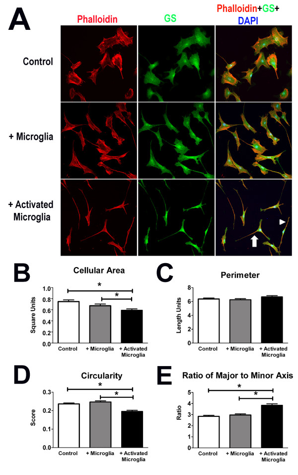
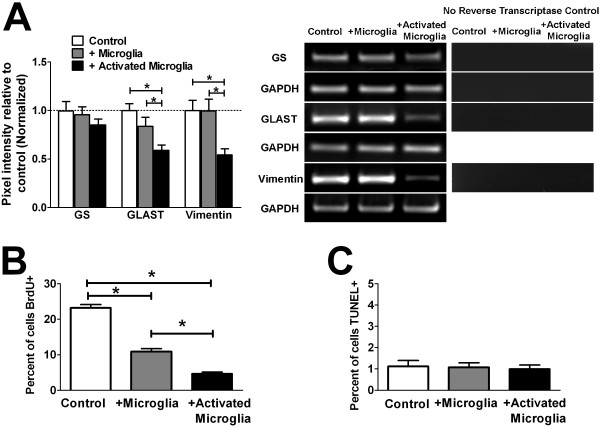
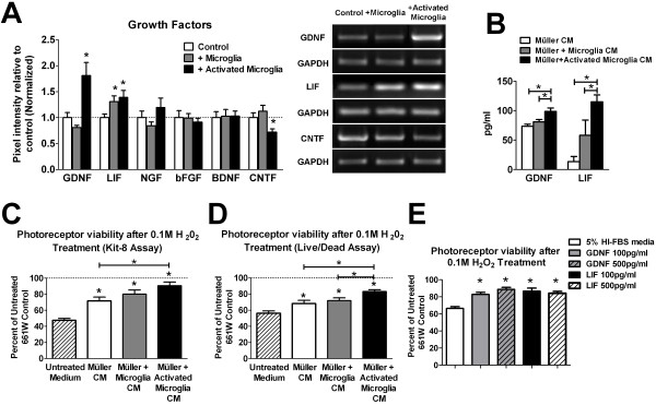
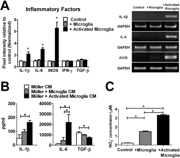
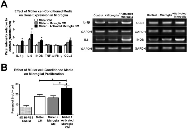
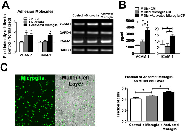
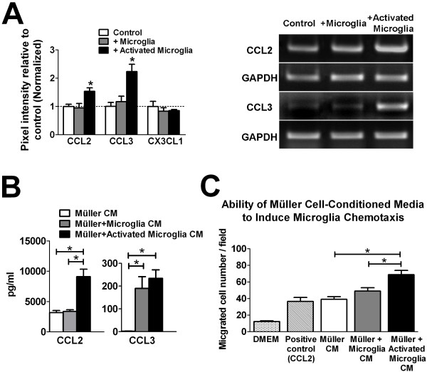
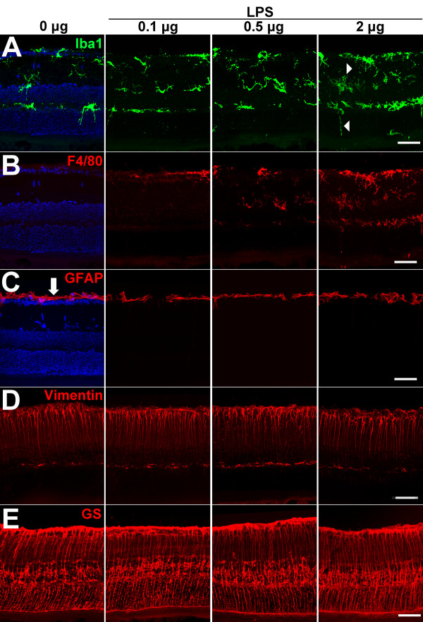
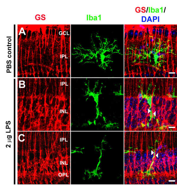
Purpose: Microglia and Müller cells are prominent participants in retinal responses to injury and disease that shape eventual tissue adaptation or damage. This investigation examined how microglia and Müller cells interact with each other following initial microglial activation. Methods: Mouse Müller cells were cultured alone, or co-cultured with activated or unactivated retinal microglia, and their morphological, molecular, and functional responses were evaluated. Müller cell-feedback signaling to microglia was studied using Müller cell-conditioned media. Corroborative in vivo analyses of retinal microglia-Müller cell interactions in the mouse retina were also performed. Results: Our results demonstrate that Müller cells exposed to activated microglia, relative to those cultured alone or with unactivated microglia, exhibit marked alterations in cell morphology and gene expression that differed from those seen in chronic gliosis. These Müller cells demonstrated in vitro (1) an upregulation of growth factors such as GDNF and LIF, and provide neuroprotection to photoreceptor cells, (2) increased pro-inflammatory factor production, which in turn increased microglial activation in a positive feedback loop, and (3) upregulated chemokine and adhesion protein expression, which allowed Müller cells to attract and adhere to microglia. In vivo activation of microglia by intravitreal injection of lipopolysaccharide (LPS) also induced increased Müller cell-microglia adhesion, indicating that activated microglia may translocate intraretinally in a radial direction using Müller cell processes as an adhesive scaffold. Conclusion: Our findings demonstrate that activated microglia are able to influence Müller cells directly, and initiate a program of bidirectional microglia-Müller cell signaling that can mediate adaptive responses within the retina following injury. In the acute aftermath following initial microglia activation, Müller cell responses may serve to augment initial inflammatory responses across retinal lamina and to guide the intraretinal mobilization of migratory microglia using chemotactic cues and adhesive cell contacts. Understanding adaptive microglia-Müller cell interactions in injury responses can help discover therapeutic cellular targets for intervention in retinal disease.
Overview
The claims and statistics of this paper haven’t been checked yet.
Run the check — free, no account needed. It reads the open-access text and checks the sources it cites.
Integrity signals
Registry
Cites
Statistics
Figures
Verification
Claims the paper makes
The claims and statistics of this paper haven’t been checked yet.
Run the check — free, no account needed. It reads the open-access text and checks the sources it cites.
Claims other papers make about this one
Each is checked against this paper’s own text: does it say what the citing paper says it does?
In the citing paper
“Inflammatory immune responses for ocular gene therapies become harder to predict in the clinical setting, as both physiological and barrier functions of immune privilege may already be compromised by aging or the ocular disorder physiopathological conditions such as inflammation or oxidative stress induction [21,66,67].”
From Risk mitigation of immunogenicity: a key to personalized retinal gene therapy (2021) · cites this paper as [21]
Self-check
Percentages, sums and reported differences re-computed from the paper’s own numbers.
The self-check needs the paper’s full text.
Citations
Cites
Resting microglial cells are highly dynamic surveillants of brain parenchyma in vivo
Nimmerjahn, A., Kirchhoff, F., & Helmchen, F. (2005) · Science · doi:10.1126/science.1110647 · Cited by 5,827
ATP mediates rapid microglial response to local brain injury in vivo
Davalos, D., Grutzendler, J., Yang, G., Kim, J. V., Zuo, Y., Jung, S., Littman, D. R., Dustin, M. L., & Gan, W.-B. (2005) · Nature Neuroscience · doi:10.1038/nn1472 · Cited by 3,913
Müller cells in the healthy and diseased retina
Bringmann, A., Pannicke, T., Grosche, J., Francke, M., Wiedemann, P., Skatchkov, S. N., Osborne, N. N., & Reichenbach, A. (2006) · Progress in Retinal and Eye Research · doi:10.1016/j.preteyeres.2006.05.003 · Cited by 1,967
The myeloid cells of the central nervous system parenchyma
Ransohoff, R. M., & Cardona, A. E. (2010) · Nature · doi:10.1038/nature09615 · Cited by 804
Bringmann, A., Iandiev, I., Pannicke, T., Wurm, A., Hollborn, M., Wiedemann, P., Osborne, N. N., & Reichenbach, A. (2009) · Progress in Retinal and Eye Research · doi:10.1016/j.preteyeres.2009.07.001 · Cited by 761
Para-Inflammation in the Aging Retina
Xu, H., Chen, M., & Forrester, J. V. (2009) · Progress in Retinal and Eye Research · doi:10.1016/j.preteyeres.2009.06.001 · Cited by 659 · cited 2×
“An understanding of how the overall injury response, initiated following microglial activation, is subsequently shaped by communications between these two cell types is likely central to elucidating the functional importance of postinjury inflammation and the mechanisms underlying chronic neuroinflammatory changes tho…”
BackgroundAge-related alterations in the dynamic behavior of microglia
Damani, M. R., Zhao, L., Fontainhas, A. M., Amaral, J., Fariss, R. N., & Wong, W. T. (2011) · Aging Cell · doi:10.1111/j.1474-9726.2010.00660.x · Cited by 504
Self-citationMicroglia activation in retinal degeneration
Langmann, T. (2007) · Journal of Leukocyte Biology · doi:10.1189/jlb.0207114 · Cited by 496
Propagation of Intercellular Calcium Waves in Retinal Astrocytes and Müller Cells
Newman, E. A. (2001) · Journal of Neuroscience · doi:10.1523/jneurosci.21-07-02215.2001 · Cited by 449
Assessing Activation States in Microglia
Colton, C. A., & Wilcock, D. M. (2010) · CNS & Neurological Disorders - Drug Targets · doi:10.2174/187152710791012053 · Cited by 430
Cited by
Sorted by citations
Retinal microglia: Just bystander or target for therapy?
Karlstetter, M., Scholz, R., Rutar, M., Wong, W. T., Provis, J. M., & Langmann, T. (2014) · Progress in Retinal and Eye Research · doi:10.1016/j.preteyeres.2014.11.004 · Cited by 570
Self-citationAltmann, C., & Schmidt, M. H. H. (2018) · International Journal of Molecular Sciences · doi:10.3390/ijms19010110 · Cited by 413
Cuenca, N., Fernández-Sánchez, L., Campello, L., Maneu, V., De la Villa, P., Lax, P., & Pinilla, I. (2014) · Progress in Retinal and Eye Research · doi:10.1016/j.preteyeres.2014.07.001 · Cited by 401
Microglia in Retinal Degeneration
Rashid, K., Akhtar‐Schäfer, I., & Langmann, T. (2019) · Frontiers in Immunology · doi:10.3389/fimmu.2019.01975 · Cited by 365
Microglia in the Retina: Roles in Development, Maturity, and Disease
Silverman, S. M., & Wong, W. T. (2018) · Annual Review of Vision Science · doi:10.1146/annurev-vision-091517-034425 · Cited by 364
Self-citationReichenbach, A., & Bringmann, A. (2019) · Glia · doi:10.1002/glia.23727 · Cited by 327
Wang, X., Zhao, L., Zhang, J., Fariss, R. N., Ma, W., Kretschmer, F., Wang, M., Qian, H., Badea, T. C., Diamond, J. S., et al. (2016) · Journal of Neuroscience · doi:10.1523/jneurosci.3575-15.2016 · Cited by 249
Self-citationRetinal microglia – A key player in healthy and diseased retina
Rathnasamy, G., Foulds, W. S., Ling, E., & Kaur, C. (2018) · Progress in Neurobiology · doi:10.1016/j.pneurobio.2018.05.006 · Cited by 250
Age‐Related Macular Degeneration: New Paradigms for Treatment and Management of AMD
Hernández-Zimbrón, L. F., Zamora-Alvarado, R., la Paz, L. D. O.-D., Vélez-Montoya, R., Zenteno, E., Gulías-Cañizo, R., Quiroz–Mercado, H., & González-Salinas, R. (2018) · Oxidative Medicine and Cellular Longevity · doi:10.1155/2018/8374647 · Cited by 247
Wang, M., Wang, X., Zhao, L., Ma, W., Rodríguez, I. R., Fariss, R. N., & Wong, W. T. (2014) · Journal of Neuroscience · doi:10.1523/jneurosci.3153-13.2014 · Cited by 211
Self-citationRetinal Macroglial Responses in Health and Disease
de Hoz, R., Rojas, B., Ramírez, A. I., Salazar, J. J., Gallego, B. I., Triviño, A., & Ramírez, J. M. (2016) · BioMed Research International · doi:10.1155/2016/2954721 · Cited by 187
Au, N. P. B., & Ma, C. H. E. (2022) · Frontiers in Immunology · doi:10.3389/fimmu.2022.860070 · Cited by 192
White, D. T., Sengupta, S., Saxena, M. T., Xu, Q., Hanes, J. S., Ding, D., Ji, H., & Mumm, J. S. (2017) · Proceedings of the National Academy of Sciences · doi:10.1073/pnas.1617721114 · Cited by 167
Todd, L. J., Palazzo, I., Suarez, L., Liu, X., Volkov, L. I., Hoang, T. V., Campbell, W. A., Blackshaw, S., Quan, N., & Fischer, A. J. (2019) · Journal of Neuroinflammation · doi:10.1186/s12974-019-1505-5 · Cited by 164
Okunuki, Y., Mukai, R., Pearsall, E., Klokman, G., Husain, D., Park, D.-H., Korobkina, E. D., Weiner, H. L., Butovsky, O., Ksander, B. R., et al. (2018) · Proceedings of the National Academy of Sciences · doi:10.1073/pnas.1719601115 · Cited by 165
Schnichels, S., Paquet‐Durand, F., Löscher, M., Tsai, T., Hurst, J., Joachim, S. C., & Klettner, A. (2020) · Progress in Retinal and Eye Research · doi:10.1016/j.preteyeres.2020.100880 · Cited by 152
Natoli, R., Fernando, N., Madigan, M. C., Chu‐Tan, J. A., Valter, K., Provis, J. M., & Rutar, M. (2017) · Molecular Neurodegeneration · doi:10.1186/s13024-017-0175-y · Cited by 146
Microglia-Müller Cell Interactions in the Retina
Wang, M., & Wong, W. T. (2014) · Advances in experimental medicine and biology · doi:10.1007/978-1-4614-3209-8_42 · Cited by 136
Self-citationHu, X. T., Zhao, G., Xu, M.-X., Zhou, H., Li, F., Miao, Y., Lei, B., Yang, X.-L., & Wang, Z. (2021) · Journal of Neuroinflammation · doi:10.1186/s12974-021-02366-x · Cited by 131
Microglia: Key Players in Retinal Ageing and Neurodegeneration
Guo, L., Choi, S., Bikkannavar, P., & Cordeiro, M. F. (2022) · Frontiers in Cellular Neuroscience · doi:10.3389/fncel.2022.804782 · Cited by 134
Rutar, M., Natoli, R., Chia, R., Valter, K., & Provis, J. M. (2015) · Journal of Neuroinflammation · doi:10.1186/s12974-014-0224-1 · Cited by 127
Mitchell, D. M., Lovel, A. G., & Stenkamp, D. L. (2018) · Journal of Neuroinflammation · doi:10.1186/s12974-018-1185-6 · Cited by 126
Miao, Y., Zhao, G., Cheng, S., Wang, Z., & Yang, X.-L. (2023) · Progress in Retinal and Eye Research · doi:10.1016/j.preteyeres.2023.101169 · Cited by 126
Maternal immune activation induces sustained changes in fetal microglia motility
Ozaki, K., Kato, D., Ikegami, A., Hashimoto, A., Sugio, S., Guo, Z., Shibushita, M., Tatematsu, T., Haruwaka, K., Moorhouse, A. J., et al. (2020) · Scientific Reports · doi:10.1038/s41598-020-78294-2 · Cited by 123
The innate immune system in diabetic retinopathy
Pan, W. W., Lin, F., & Fort, P. E. (2021) · Progress in Retinal and Eye Research · doi:10.1016/j.preteyeres.2021.100940 · Cited by 117
Glial Cells in Glaucoma: Friends, Foes, and Potential Therapeutic Targets
García-Bermúdez, M. Y., Freude, K., Mouhammad, Z. A., van Wijngaarden, P., Martin, K., & Kolko, M. (2021) · Frontiers in Neurology · doi:10.3389/fneur.2021.624983 · Cited by 114
Fischer, A. J., Zelinka, C. P., Gallina, D., Scott, M. A., & Todd, L. J. (2014) · Glia · doi:10.1002/glia.22703 · Cited by 100
Müller Glia in Retinal Innate Immunity: A Perspective on Their Roles in Endophthalmitis
Kumar, A., Pandey, R. K., Miller, L. J., Singh, P. K., & Kanwar, M. (2013) · Critical Reviews in Immunology · doi:10.1615/critrevimmunol.2013006618 · Cited by 99
A journey into the retina: Müller glia commanding survival and death
Subirada, P. V., Paz, M. C., Ridano, M. E., Lorenc, V. E., Vaglienti, M. V., Barcelona, P. F., Luna, J. D., & Sanchez, M. C. (2018) · European Journal of Neuroscience · doi:10.1111/ejn.13965 · Cited by 99
Glial cell interactions and glaucoma
Chong, R. S., & Martin, K. R. G. (2014) · Current Opinion in Ophthalmology · doi:10.1097/icu.0000000000000125 · Cited by 99
Conedera, F. M., Pousa, A. M. Q., Mercader, N., Tschopp, M., & Enzmann, V. (2019) · Glia · doi:10.1002/glia.23601 · Cited by 94
Aging Changes in Retinal Microglia and their Relevance to Age-related Retinal Disease
Ma, W., & Wong, W. T. (2015) · Advances in experimental medicine and biology · doi:10.1007/978-3-319-17121-0_11 · Cited by 93
Self-citationThe role of Müller glia and microglia in glaucoma
Seitz, R., Ohlmann, A., & Tamm, E. R. (2013) · Cell and Tissue Research · doi:10.1007/s00441-013-1666-y · Cited by 91
Todd, L. J., Squires, N. A., Suarez, L., & Fischer, A. J. (2016) · Scientific Reports · doi:10.1038/srep35703 · Cited by 87
Ma, W., Silverman, S. M., Zhao, L., Villasmil, R., Campos, M. M., Amaral, J., & Wong, W. T. (2019) · eLife · doi:10.7554/elife.42049 · Cited by 88
Self-citationMüller Glia-Mediated Retinal Regeneration
Gao, H., Luodan, A., Huang, X., Chen, X., & Xu, H. (2021) · Molecular Neurobiology · doi:10.1007/s12035-020-02274-w · Cited by 84
Palazzo, I., Todd, L. J., Hoang, T. V., Reh, T. A., Blackshaw, S., & Fischer, A. J. (2022) · Glia · doi:10.1002/glia.24181 · Cited by 82
Molecular Mechanisms Related to Oxidative Stress in Retinitis Pigmentosa
Gallenga, C. E., Lonardi, M., Pacetti, S., Violanti, S. S., Tassinari, P., Virgilio, F. D., Tognon, M. G., & Perri, P. (2021) · Antioxidants · doi:10.3390/antiox10060848 · Cited by 79
Regeneration associated transcriptional signature of retinal microglia and macrophages
Mitchell, D. M., Sun, C. C., Hunter, S. S., New, D. D., & Stenkamp, D. L. (2019) · Scientific Reports · doi:10.1038/s41598-019-41298-8 · Cited by 80
Müller Glia Reactivity and Development of Gliosis in Response to Pathological Conditions
Graca, A. B., Hippert, C., & Pearson, R. A. (2018) · Advances in experimental medicine and biology · doi:10.1007/978-3-319-75402-4_37 · Cited by 77
Deliyanti, D., & Wilkinson‐Berka, J. L. (2015) · Journal of Neuroinflammation · doi:10.1186/s12974-015-0363-z · Cited by 78
Huang, Y., He, J., Liang, H., Hu, K., Jiang, S., Yang, L., Mei, S., Zhu, X. M., Yu, J. H., Kijlstra, A., et al. (2018) · Frontiers in Immunology · doi:10.3389/fimmu.2018.01713 · Cited by 75
Müller glia and phagocytosis of cell debris in retinal tissue
Bejarano‐Escobar, R., Sánchez‐Calderón, H., Otero‐Arenas, J., Martín‐Partido, G., & Francisco‐Morcillo, J. (2017) · Journal of Anatomy · doi:10.1111/joa.12653 · Cited by 77
Roche, S. L., Ruiz‐Lopez, A. M., Moloney, J. N., Byrne, A. M., & Cotter, T. G. (2017) · Glia · doi:10.1002/glia.23243 · Cited by 73
Ocular Vascular Diseases: From Retinal Immune Privilege to Inflammation
Wang, X., Wang, T., Lam, E., Álvarez, D., & Sun, Y. (2023) · International Journal of Molecular Sciences · doi:10.3390/ijms241512090 · Cited by 71
Appelbaum, T., Santana, E., & Aguirre, G. D. (2017) · PLoS ONE · doi:10.1371/journal.pone.0177224 · Cited by 67
Neuroinflammation in retinitis pigmentosa: Therapies targeting the innate immune system
Zhao, L., Hou, C., & Yan, N. (2022) · Frontiers in Immunology · doi:10.3389/fimmu.2022.1059947 · Cited by 65
Self-citationMacrophage/microglia polarization for the treatment of diabetic retinopathy
Yao, Y., Li, J., Zhou, Y., Wang, S., Zhang, Z., Jiang, Q., & Li, K. (2023) · Frontiers in Endocrinology · doi:10.3389/fendo.2023.1276225 · Cited by 60
Reactive retinal microglia, neuronal survival, and the formation of retinal folds and detachments
Fischer, A. J., Zelinka, C. P., & Milani‐Nejad, N. (2014) · Glia · doi:10.1002/glia.22752 · Cited by 60
Palazzo, I., Deistler, K., Hoang, T. V., Blackshaw, S., & Fischer, A. J. (2020) · Development · doi:10.1242/dev.183418 · Cited by 60
Pilotto, E., Leonardi, F., Stefanon, F. G., Longhin, E., Torresin, T., Deganello, D., Cavarzeran, F., Miglionico, G., Parrozzani, R., & Midena, E. (2018) · British Journal of Ophthalmology · doi:10.1136/bjophthalmol-2018-312461 · Cited by 58
Loss of XBP1 accelerates age-related decline in retinal function and neurodegeneration
McLaughlin, T., Falkowski, M., Park, J. W., Keegan, S., Elliott, M. H., Wang, J. J., & Zhang, S. X. (2018) · Molecular Neurodegeneration · doi:10.1186/s13024-018-0250-z · Cited by 57
Keep an eye on adenosine: Its role in retinal inflammation
Santiago, A. R., Madeira, M. H., Boia, R., Aires, I. D., Rodrigues‐Neves, A. C., Santos, P. F., & Ambrósio, A. F. (2020) · Pharmacology & Therapeutics · doi:10.1016/j.pharmthera.2020.107513 · Cited by 58
The Interaction Between Microglia and Macroglia in Glaucoma
Zhao, X., Sun, R., Luo, X., Wang, F., & Sun, X. (2021) · Frontiers in Neuroscience · doi:10.3389/fnins.2021.610788 · Cited by 58
Regulations of Retinal Inflammation: Focusing on Müller Glia
Chen, Y., Xia, Q., Zeng, Y., Zhang, Y., & Zhang, M. (2022) · Frontiers in Cell and Developmental Biology · doi:10.3389/fcell.2022.898652 · Cited by 56
Gao, L., Chen, X., Tang, Y., Zhao, J., Li, Q., Fan, X., Xu, H., & Yin, Z. Q. (2015) · Experimental Eye Research · doi:10.1016/j.exer.2015.04.013 · Cited by 53
Feng, C., Wang, X., Liu, T., Zhang, M., Xu, G., & Ni, Y. (2017) · Europe PMC (PubMed Central) · Cited by 53
Green tea extract attenuates LPS-induced retinal inflammation in rats
Ren, J. L., Yu, Q. X., Liang, W. C., Leung, P. Y., Ng, T. K., Chu, W. K., Pang, C. P., & Chan, S. O. (2018) · Scientific Reports · doi:10.1038/s41598-017-18888-5 · Cited by 51
Szabó, K., Énzsöly, A., Dékány, B., Szabó, A., Hajdú, R. I., Radovits, T., Mátyás, C., Oláh, A., Laurik, L. K., Somfai, G. M., et al. (2017) · Scientific Reports · doi:10.1038/s41598-017-09068-6 · Cited by 50
Choi, S., Keun-young Kim, A., Perkins, G. A., Phan, S., Edwards, G., Xia, Y., Kim, J., Skowronska‐Krawczyk, D., Weinreb, R. N., Ellisman, M. H., et al. (2020) · Redox Biology · doi:10.1016/j.redox.2020.101703 · Cited by 50
Coordinated Intervention of Microglial and Müller Cells in Light-Induced Retinal Degeneration
Pierdomenico, J. D., Martínez-Vacas, A., Hernández-Muñoz, D., Gómez-Ramírez, A. M., Valiente‐Soriano, F. J., Agudo‐Barriuso, M., Vidal‐Sanz, M., Villegas‐Pérez, M. P., & García‐Ayuso, D. (2020) · Investigative Ophthalmology & Visual Science · doi:10.1167/iovs.61.3.47 · Cited by 49
Xu, M.-X., Zhao, G., Hu, X., Zhou, H., Li, S., Li, F., Miao, Y., Lei, B., & Wang, Z. (2022) · Neuroscience Bulletin · doi:10.1007/s12264-022-00833-w · Cited by 48
Müller Glia in Retinal Development: From Specification to Circuit Integration
Tworig, J. M., & Feller, M. B. (2022) · Frontiers in Neural Circuits · doi:10.3389/fncir.2021.815923 · Cited by 52
Lieffrig, S. A., Gyimesi, G., Mao, Y., & Finnemann, S. C. (2023) · Immunological Reviews · doi:10.1111/imr.13264 · Cited by 50
The role of lipopolysaccharides in diabetic retinopathy
Qin, X., & Zou, H. (2022) · BMC Ophthalmology · doi:10.1186/s12886-022-02296-z · Cited by 48
Bray, A., Cevallos, R. R., Gazarian, K. G., & Lamas, M. (2014) · Neuroscience · doi:10.1016/j.neuroscience.2014.09.023 · Cited by 45
Massengill, M. T., Ahmed, C. M. I., Lewin, A. S., & Ildefonso, C. J. (2018) · Advances in experimental medicine and biology · doi:10.1007/978-3-319-75402-4_23 · Cited by 45
Proliferation Potential of Müller Glia after Retinal Damage Varies between Mouse Strains
Suga, A., Sadamoto, K., Fujii, M., Mandai, M., & Takahashi, M. (2014) · PLoS ONE · doi:10.1371/journal.pone.0094556 · Cited by 45
Microglia Reactivity Entails Microtubule Remodeling From Acentrosomal to Centrosomal Arrays
Rosito, M., Sanchini, C., Gosti, G., Moreno, M., Panfilis, S. D., Giubettini, M., Debellis, D., Catalano, F., Peruzzi, G., Marotta, R., et al. (2022) · Cell Reports · doi:10.1016/j.celrep.2023.112104 · Cited by 43
Salkar, A., Wall, R. V., Basavarajappa, D. K., Chitranshi, N., Parrilla, G. E., Mirzaei, M., Yan, P., Graham, S. L., & You, Y. (2024) · Aging and Disease · doi:10.14336/ad.2024.0103 · Cited by 44
Navneet, S., Wilson, K., & Rohrer, B. (2024) · Investigative Ophthalmology & Visual Science · doi:10.1167/iovs.65.2.42 · Cited by 42
Resident Immunity in Tissue Repair and Maintenance: The Zebrafish Model Coming of Age
Martins, R. R., Ellis, P. S., MacDonald, R. B., Richardson, R. J., & Henriques, C. M. (2019) · Frontiers in Cell and Developmental Biology · doi:10.3389/fcell.2019.00012 · Cited by 44
Adelman, R. A., Parnes, A. J., Michalewska, Z., Parolini, B., Boscher, C., & Ducournau, D. H. (2015) · BioMed Research International · doi:10.1155/2015/352487 · Cited by 41
Monte, M. D., Rezzola, S., Cammalleri, M., Belleri, M., Locri, F., Morbidelli, L., Corsini, M., Paganini, G., Semeraro, F., Cancarini, A., et al. (2015) · Investigative Ophthalmology & Visual Science · doi:10.1167/iovs.14-16323 · Cited by 41
Involvement of NT3 and P75NTR in photoreceptor degeneration following selective Müller cell ablation
Shen, W., Zhu, L., Lee, S.-R., Chung, S. H., & Gillies, M. C. (2013) · Journal of Neuroinflammation · doi:10.1186/1742-2094-10-137 · Cited by 41
Gallina, D., Zelinka, C. P., Cebulla, C. M., & Fischer, A. J. (2015) · Experimental Neurology · doi:10.1016/j.expneurol.2015.08.007 · Cited by 41
Angiotensin II and aldosterone activate retinal microglia
Rana, I., Suphapimol, V., Jerome, J. R., Talia, D. M., Deliyanti, D., & Wilkinson‐Berka, J. L. (2019) · Experimental Eye Research · doi:10.1016/j.exer.2019.107902 · Cited by 41
Harnessing the Neuroprotective Behaviors of Müller Glia for Retinal Repair
Peña, J. S., & Vázquez, M. (2022) · Frontiers in Bioscience-Landmark · doi:10.31083/j.fbl2706169 · Cited by 40
Microglia-derived TNF-α mediates Müller cell activation by activating the TNFR1-NF-κB pathway
Ji, M., Sun, Q., Zhang, G., Huang, Z., Zhang, Y., Shen, Q., & Guan, H. (2021) · Experimental Eye Research · doi:10.1016/j.exer.2021.108852 · Cited by 38
IL-10 Is Significantly Involved in HSP70-Regulation of Experimental Subretinal Fibrosis
Yang, Y., Takeda, A., Yoshimura, T., Oshima, Y., Sonoda, K.-H., & Ishibashi, T. (2013) · PLoS ONE · doi:10.1371/journal.pone.0080288 · Cited by 38
Incorporating microglia‐like cells in human induced pluripotent stem cell‐derived retinal organoids
Chichagova, V., Georgiou, M., Carter, M., Dorgau, B., Hilgen, G., Collin, J., Queen, R., Chung, G., Ajeian, J., Moya‐Molina, M., et al. (2023) · Journal of Cellular and Molecular Medicine · doi:10.1111/jcmm.17670 · Cited by 37
Inflammation-Induced Photoreceptor Cell Death
Sène, A., & Apte, R. S. (2018) · Advances in experimental medicine and biology · doi:10.1007/978-3-319-75402-4_25 · Cited by 37
Müller glia–myeloid cell crosstalk accelerates optic nerve regeneration in the adult zebrafish
Dyck, A. V., Bollaerts, I., Beckers, A., Vanhunsel, S., Glorian, N., houcke, J. V., van Ham, T. J., Groef, L. D., Andries, L., & Moons, L. (2021) · Glia · doi:10.1002/glia.23972 · Cited by 39
Garces, K. N., Carmy, T., Illiano, P., Brambilla, R., & Hackam, A. S. (2020) · Journal of Molecular Neuroscience · doi:10.1007/s12031-020-01503-0 · Cited by 36
Deliyanti, D., Lee, J., Petratos, S., Meyer, C. J., Ward, K. W., Wilkinson‐Berka, J. L., & de Haan, J. B. (2016) · Clinical Science · doi:10.1042/cs20160068 · Cited by 36
Culture and Characterization of Microglia from the Adult Murine Retina
Devarajan, G., Chen, M., Muckersie, E., & Xu, H. (2014) · The Scientific World JOURNAL · doi:10.1155/2014/894368 · Cited by 35
Schmalen, A., Lorenz, L. M., Grosche, A., Pauly, D., Deeg, C. A., & Hauck, S. M. (2021) · Frontiers in Pharmacology · doi:10.3389/fphar.2021.771571 · Cited by 38
Carotenoids in the Management of Glaucoma: A Systematic Review of the Evidence
Lem, D. W., Gierhart, D. L., & Davey, P. G. (2021) · Nutrients · doi:10.3390/nu13061949 · Cited by 35
Elevated Glucose and Interleukin-1β Differentially Affect Retinal Microglial Cell Proliferation
Baptista, F. I., Aveleira, C. A., Castilho, Á., & Ambrósio, A. F. (2017) · Mediators of Inflammation · doi:10.1155/2017/4316316 · Cited by 34
Hyperreflective Foci in the Retina of Active Relapse-Onset Multiple Sclerosis
Pilotto, E., Miante, S., Torresin, T., Puthenparampil, M., Frizziero, L., Federle, L., Gallo, P., & Midena, E. (2020) · Ophthalmology · doi:10.1016/j.ophtha.2020.03.024 · Cited by 33
The expression of Toll-like receptors in murine Müller cells, the glial cells in retina
Lin, X., Fang, D., Zhou, H., & Su, S. B. (2012) · Neurological Sciences · doi:10.1007/s10072-012-1236-1 · Cited by 33
Effects of Lycium barbarum on the Visual System
Manthey, A. L., Chiu, K., & So, K. (2017) · International review of neurobiology · doi:10.1016/bs.irn.2017.02.002 · Cited by 33
Ferrer‐Martín, R. M., Martín‐Oliva, D., Sierra, A., Carrasco, M., Martín‐Estebané, M., Calvente, R., Martín‐Guerrero, S. M., Marín‐Teva, J. L., Navascués, J., & Cuadros, M. Á. (2015) · PLoS ONE · doi:10.1371/journal.pone.0135238 · Cited by 32
Puthenparampil, M., Torresin, T., Franciotta, S., Marin, A., Napoli, F. D., Mauceri, V. A., Miante, S., Pilotto, E., Midena, E., & Gallo, P. (2022) · Frontiers in Immunology · doi:10.3389/fimmu.2022.852183 · Cited by 32
Zhao, N., Hao, X.-N., Huang, J.-M., Song, Z., & Tao, Y. (2024) · Aging and Disease · doi:10.14336/ad.2023.0823-3 · Cited by 32
Microglia activation is essential for BMP7-mediated retinal reactive gliosis
Dharmarajan, S., Fisk, D. L., Sorenson, C. M., Sheibani, N., & Belecky‐Adams, T. L. (2017) · Journal of Neuroinflammation · doi:10.1186/s12974-017-0855-0 · Cited by 31
Tumor necrosis factor-alpha aggravates gliosis and inflammation of activated retinal Müller cells
Hu, X., Xu, M.-X., Zhou, H., Cheng, S., Li, F., Miao, Y., & Wang, Z. (2020) · Biochemical and Biophysical Research Communications · doi:10.1016/j.bbrc.2020.07.102 · Cited by 31
Who let the dogs out?: detrimental role of Galectin-3 in hypoperfusion-induced retinal degeneration
Manouchehrian, O., Arnér, K. M., Deierborg, T., & Taylor, L. (2015) · Journal of Neuroinflammation · doi:10.1186/s12974-015-0312-x · Cited by 30
Lebecetin, a C‐type lectin, inhibits choroidal and retinal neovascularization
Montassar, F., Darche, M., Blaizot, A., Augustin, S., Conart, J., Millet, A., Ayeb, M. E., Sahel, J., Goazigo, A. R.-L., Sennlaub, F., et al. (2016) · The FASEB Journal · doi:10.1096/fj.201600351r · Cited by 30
Natural plant medications for the treatment of retinal diseases: The blood-retinal barrier as a clue
Ren, Y., Liang, H., Xie, M., & Zhang, M. (2024) · Phytomedicine · doi:10.1016/j.phymed.2024.155568 · Cited by 33
Metadata
- Authors
- Wang, M., Ma, W., Zhao, L., Fariss, R. N., & Wong, W. T.
- Affiliations
- National Institutes of Health (US)
- Corresponding author
- Wong, Wai T
- Venue
- Journal of Neuroinflammation
- Published
- 2011-12
- Volume
- 8
- Issue
- 1
- Pages
- 173-173
- ISSN
- 1742-2094 (ISSN-L)
- PMC
- PMC3251543
- OpenAlex
- W2077349110
- Cited by
- 228
- References
- 61
- Citations by year
- 2026: 12 · 2025: 17 · 2024: 14 · 2023: 20 · 2022: 20 · 2021: 32 · 2020: 18 · 2019: 11 · 2018: 24 · 2017: 14 · 2016: 11 · 2015: 12 · 2014: 14 · 2013: 5 · 2012: 4
- FWCI
- 4.07
- Type
- journal article
- Language
- en
- Open access
- gold
- doi.org
- Licence
- cc-by
- Locations
- Journal of Neuroinflammation (publishedVersion, cc-by)
- Europe PMC (PubMed Central) (publishedVersion, cc-by)
- PubMed Central (publishedVersion, cc-by)
- PubMed (publishedVersion)
- DOAJ (DOAJ: Directory of Open Access Journals) (submittedVersion)
- Topics
- Life Sciences › Neuroscience › Neurology › Neuroinflammation and Neurodegeneration Mechanisms
- Health Sciences › Medicine › Ophthalmology › Retinal Diseases and Treatments
- Health Sciences › Medicine › Ophthalmology › Retinal Development and Disorders
- Keywords
- Müller cells, retinal microglia, microglial activation, neuroprotection, photoreceptors, glial cell line-derived neurotrophic factor, LiF, pro-inflammatory factors, chemokines, cell adhesion, microglial migration, lipopolysaccharide, retinal inflammation, gliosis, retinal damage
- MeSH terms
- Animals, Cell Adhesion, Cell Communication, Cells, Cultured, Chemotaxis, Gliosis, Inflammation, Mice, Inbred C57BL, Retina, Culture Media, Conditioned, Apoptosis, Microglia, Neuroprotective Agents, Coculture Techniques, Cell Proliferation, Mice
About these checks
Registry, retraction, Cites and statistics results are computed, not judged, with one exception: a printed reference that no index lists is read once by a model before it counts as a citation error. Claims are extracted by a model and kept only when their quote appears verbatim in the text; each cited source is then checked, with a model’s help, for whether it says what the claim relies on. Those verdicts can be wrong — read the claim’s quote and the cited source before relying on one.
Statistics and arithmetic are recomputed, not judged.
Confidence bands are shown only for fields calibrated on our annotated test set. None is calibrated yet, so no band is shown.
What counts as a claim ›How we measure confidence ›How we check this ›
Are you an author, or do you see a problem with this page? Request a review
