

The DOI matches its Crossref record; 21 of the 21 references it cites were found in a registry or an index.
CX3CL1 reduces neurotoxicity and microglial activation in a rat model of Parkinson’s disease
Open accessAbstract
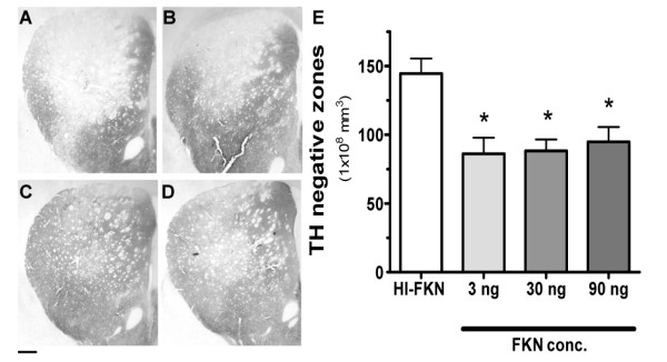
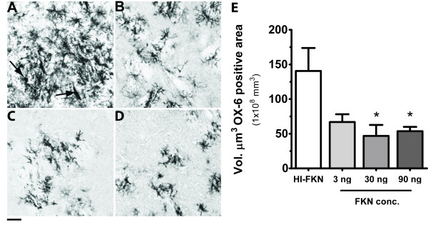
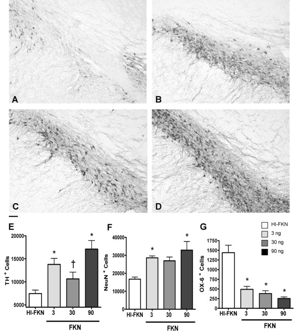
BACKGROUND: Parkinson's disease is characterized by a progressive loss of dopaminergic neurons in the substantia nigra. The cause of the neurodegeneration is unknown. Neuroinflammation has been clearly shown in Parkinson's disease and may be involved in the progressive nature of the disease. Microglia are capable of producing neuronal damage through the production of bioactive molecules such as cytokines, as well as reactive oxygen species (ROS), and nitric oxide (NO). The inflammatory response in the brain is tightly regulated at multiple levels. One form of immune regulation occurs via neurons. Fractalkine (CX3CL1), produced by neurons, suppresses the activation of microglia. CX3CL1 is constitutively expressed. It is not known if addition of exogenous CX3CL1 beyond otherwise physiologically normal levels could decrease microglia activation and thereby minimize the secondary neurodegeneration following a neurotoxic insult. METHODS: The intrastriatal 6-hydroxydopamine (6-OHDA) rat model of Parkinson disease, was used to test the hypothesis that exogenous CX3CL1 could be neuroprotective. Treatment with recombinant CX3CL1 was delivered to the striatum by an osmotic minipump for 28 days beginning 7 days after the initial insult. Unbiased stereological methods were used to quantify the lesion size in the striatum, the amount of neuronal loss in the substantia nigra, and the amount of microglia activation. RESULTS: As hypothesized, CX3CL1 was able to suppress this microglia activation. The reduced microglia activation was found to be neuroprotective as the CX3CL1 treated rats had a smaller lesion volume in the striatum and importantly significantly fewer neurons were lost in the CX3CL1 treated rats. CONCLUSION: These findings demonstrated that CX3CL1 plays a neuroprotective role in 6-OHDA-induced dopaminergic lesion and it might be an effective therapeutic target for many neurodegenerative diseases, including Parkinson disease and Alzheimer disease, where inflammation plays an important role.
Overview
The claims and statistics of this paper haven’t been checked yet.
Run the check — free, no account needed. It reads the open-access text and checks the sources it cites.
Integrity signals
Registry
Cites
Statistics
Figures
Verification
Claims the paper makes
The claims and statistics of this paper haven’t been checked yet.
Run the check — free, no account needed. It reads the open-access text and checks the sources it cites.
Claims other papers make about this one
Each is checked against this paper’s own text: does it say what the citing paper says it does?
In the citing paper
“The reduction in microglia activation may be caused, in part, by Spirulina’s capacity to enhance expression on the microglia of the fractalkin receptor (CX3CR1), which is neuroprotective in a 6-OHDA model of PD [126].”
From Beneficial Effects of Spirulina Consumption on Brain Health (2022) · cites this paper as [126]
In the citing paper
“The authors showed that Spirulina was neuroprotective in this α-Syn model of PD, observing a major number of TH+ and NeuN+ cells and a decrease in the number of activated microglial cells as evaluated by MHC II expression.”
From Beneficial Effects of Spirulina Consumption on Brain Health (2022) · cites this paper as [126]
Self-check
Percentages, sums and reported differences re-computed from the paper’s own numbers.
The self-check needs the paper’s full text.
Citations
Cites
The Danger Model: A Renewed Sense of Self
Matzinger, P. (2002) · Science · doi:10.1126/science.1071059 · Cited by 4,205
Microglia-mediated neurotoxicity: uncovering the molecular mechanisms
Block, M. L., Zecca, L., & Hong, J.-S. (2007) · Nature Reviews Neuroscience · doi:10.1038/nrn2038 · Cited by 4,110
McGeer, P. L., Itagaki, S., Boyes, B. E., & McGeer, E. G. (1988) · Neurology · doi:10.1212/wnl.38.8.1285 · Cited by 2,990
Jung, S., Aliberti, J., Graemmel, P., Sunshine, M. J., Kreutzberg, G. W., Sher, A., & Littman, D. R. (2000) · Molecular and Cellular Biology · doi:10.1128/mcb.20.11.4106-4114.2000 · Cited by 2,632
A new class of membrane-bound chemokine with a CX3C motif
Bazan, J. F., Bacon, K. B., Hardiman, G., Wang, W., Soo, K., Rossi, D., Greaves, D. R., Zlotnik, A., & Schall, T. J. (1997) · Nature · doi:10.1038/385640a0 · Cited by 2,013
Neuroinflammation in Parkinson’s disease: A target for neuroprotection?
Hirsch, E. C., & Hunot, S. (2009) · The Lancet Neurology · doi:10.1016/s1474-4422(09)70062-6 · Cited by 1,984
Control of microglial neurotoxicity by the fractalkine receptor
Cardona, A. E., Pioro, E. P., Sasse, M. E., Kostenko, V., Cardona, S. M., Dijkstra, I. M., Huang, D., Kidd, G., Dombrowski, S., Dutta, R., et al. (2006) · Nature Neuroscience · doi:10.1038/nn1715 · Cited by 1,531
Harrison, J. K., Jiang, Y., Chen, S., Xia, Y., Maciejewski, D., McNamara, R. K., Streit, W. J., Salafranca, M. N., Adhikari, S., Thompson, D. A., et al. (1998) · Proceedings of the National Academy of Sciences · doi:10.1073/pnas.95.18.10896 · Cited by 1,176
Hundhausen, C., Misztela, D., Berkhout, T. A., BROADWAY, N. M., Säftig, P., Reiß, K., Hartmann, D., Fahrenholz, F., Postina, R., Matthews, V. B., et al. (2003) · Blood · doi:10.1182/blood-2002-12-3775 · Cited by 731
Garton, K. J., Gough, P. J., Blobel, C., Murphy, G., Greaves, D. R., Dempsey, P. J., & Raines, E. W. (2001) · Journal of Biological Chemistry · doi:10.1074/jbc.m106434200 · Cited by 653
Cited by
Sorted by citations
Items 101–200 of 240
Zhai, S., Cui, Q., Simmons, D. V., & Surmeier, D. J. (2023) · Current Opinion in Neurobiology · doi:10.1016/j.conb.2023.102798 · Cited by 47
Haas, S. J.-P., Zhou, X., Machado, V., Wree, A., KRIEGLSTEIN, K. G., & Spittau, B. (2016) · Frontiers in Molecular Neuroscience · doi:10.3389/fnmol.2016.00007 · Cited by 44
Seo, Y., Kim, H., Kang, I., Choi, S. W., Shin, T., Shin, J., Lee, B., Lee, J. Y., Kim, J.-J., Kook, M. G., et al. (2016) · Glia · doi:10.1002/glia.23077 · Cited by 43
Kōyama, Y., Kotani, M., Sawamura, T., Kuribayashi, M., Konishi, R., & Michinaga, S. (2013) · Journal of Neuroinflammation · doi:10.1186/1742-2094-10-51 · Cited by 44
Compta, Y., Dias, S. P., Giraldo, D. M., Pérez‐Soriano, A., Muñoz, E., Saura, J., Fernández, M., Bravo, P., Cámara, A., Pulido-Salgado, M., et al. (2019) · Parkinsonism & Related Disorders · doi:10.1016/j.parkreldis.2019.05.040 · Cited by 42
Effect of Inhibition of Colony-Stimulating Factor 1 Receptor on Choroidal Neovascularization in Mice
Schwarzer, P., Kokona, D., Ebneter, A., & Zinkernagel, M. S. (2019) · American Journal Of Pathology · doi:10.1016/j.ajpath.2019.10.011 · Cited by 40
Ślusarczyk, J., Trojan, E., Wydra, K., Głombik, K., Chamera, K., Kucharczyk, M. W., Budziszewska, B., Kubera, M., Lasoń, W., Filip, M., et al. (2016) · Biochemical Pharmacology · doi:10.1016/j.bcp.2016.05.008 · Cited by 41
Enam, S. F., Krieger, J. R., Saxena, T. K., Watts, B. E., Olingy, C. E., Botchwey, E. A., & Bellamkonda, R. V. (2017) · Biomaterials · doi:10.1016/j.biomaterials.2017.07.013 · Cited by 39
Fu, Y., Zhen, J., & Lu, Z. (2017) · DNA and Cell Biology · doi:10.1089/dna.2017.3643 · Cited by 38
Szymura, J., Kubica, J., Więcek, M., & Pera, J. (2020) · Journal of Clinical Medicine · doi:10.3390/jcm9010184 · Cited by 38
Rodriguez, D., Church, K. A., Pietramale, A. N., Cardona, S. M., Vanegas, D., Rorex, C., Leary, M. C., Muzzio, I. A., Nash, K. R., & Cardona, A. E. (2024) · Journal of Neuroinflammation · doi:10.1186/s12974-023-02983-8 · Cited by 37
Fractalkine enhances oligodendrocyte regeneration and remyelination in a demyelination mouse model
de Almeida, M. M. A., Watson, A. E. S., Bibi, S., Dittmann, N. L., Goodkey, K., Sharafodinzadeh, P., Galleguillos, D., Nakhaei‐Nejad, M., Kosaraju, J., Steinberg, N., et al. (2023) · Stem Cell Reports · doi:10.1016/j.stemcr.2022.12.001 · Cited by 36
CNS-Wide over Expression of Fractalkine Improves Cognitive Functioning in a Tauopathy Model
Finneran, D. J., Morgan, D., Gordon, M. N., & Nash, K. R. (2019) · Journal of Neuroimmune Pharmacology · doi:10.1007/s11481-018-9822-5 · Cited by 34
Desforges, N. M., Hebron, M. L., Algarzae, N. K., Lonskaya, I., & Moussa, C. (2012) · International Journal of Alzheimer s Disease · doi:10.1155/2012/345472 · Cited by 36
HIV-1 Tat Disrupts CX3CL1-CX3CR1 Axis in Microglia via the NF-κBYY1 Pathway
Duan, M., Yao, H., Cai, Y. C., Liao, K., Seth, P., & Buch, S. J. (2014) · Current HIV Research · doi:10.2174/1570162x12666140526123119 · Cited by 34
Briones, T. L., Woods, J., & Wadowska, M. (2014) · Journal of Neuroinflammation · doi:10.1186/1742-2094-11-13 · Cited by 35
RetractedRole of chemokines in Parkinson's disease
Liu, J., Chu, S., Zhou, X., Zhang, D., & Chen, N. (2019) · Brain Research Bulletin · doi:10.1016/j.brainresbull.2019.05.020 · Cited by 37
Astrocyte subtype-specific approach to Alzheimer's disease treatment
Sarkar, S., & Biswas, S. C. (2021) · Neurochemistry International · doi:10.1016/j.neuint.2021.104956 · Cited by 35
Fractalkine suppression during hepatic encephalopathy promotes neuroinflammation in mice
McMillin, M., Grant, S., Frampton, G., Andry, S., Brown, A., & DeMorrow, S. (2016) · Journal of Neuroinflammation · doi:10.1186/s12974-016-0674-8 · Cited by 32
The Fine Balance of Chemokines During Disease: Trafficking, Inflammation, and Homeostasis
Cardona, S. M., Garcia, J. A., & Cardona, A. E. (2013) · Methods in molecular biology · doi:10.1007/978-1-62703-426-5_1 · Cited by 34
Fractalkine signaling regulates oligodendroglial cell genesis from SVZ precursor cells
Watson, A. E. S., de Almeida, M. M. A., Dittmann, N. L., Li, Y., Torabi, P., Footz, T. K., Vetere, G., Galleguillos, D., Sipione, S., Cardona, A. E., et al. (2021) · Stem Cell Reports · doi:10.1016/j.stemcr.2021.06.010 · Cited by 34
Huntington, T. E., & Srinivasan, R. (2021) · Aging and disease · doi:10.14336/ad.2021.0517 · Cited by 34
Chen, M., Luo, C., Peñalva, R. G., & Xu, H. (2013) · Investigative Ophthalmology & Visual Science · doi:10.1167/iovs.12-10888 · Cited by 33
The Cytokine CX3CL1 and ADAMs/MMPs in Concerted Cross-Talk Influencing Neurodegenerative Diseases
Iemmolo, M., Ghersi, G., & Bivona, G. (2023) · International Journal of Molecular Sciences · doi:10.3390/ijms24098026 · Cited by 35
Monocyte Subsets and Related Chemokines in Carotid Artery Stenosis and Ischemic Stroke
Große, G. M., Schulz‐Schaeffer, W., Teebken, O. E., Schuppner, R., Dirks, M., Worthmann, H., Lichtinghagen, R., Maye, G., Limbourg, F. P., & Weißenborn, K. (2016) · International Journal of Molecular Sciences · doi:10.3390/ijms17040433 · Cited by 33
More, S. V., Kumar, H., Kim, I., Koppula, S., Kim, B.-W., & Choi, D. (2013) · CNS & Neurological Disorders - Drug Targets · doi:10.2174/18715273113129990059 · Cited by 31
Regulation of cyclic AMP response element‐binding protein during neuroglial interactions
Qin, L., Bouchard, R. J., & Pugazhenthi, S. (2015) · Journal of Neurochemistry · doi:10.1111/jnc.13497 · Cited by 31
Role of MSK1 in the Induction of NF-κB by the Chemokine CX3CL1 in Microglial Cells
Galán-Ganga, M., García‐Yagüe, Á. J., & Lastres‐Becker, I. (2019) · Cellular and Molecular Neurobiology · doi:10.1007/s10571-019-00664-w · Cited by 30
Aurelian, L., & Balan, I. S. (2019) · Psychopharmacology · doi:10.1007/s00213-019-05220-4 · Cited by 31
Microglial Cx3cr1knockout reduces prion disease incubation time in mice
Grizenkova, J., Akhtar, S., Brandner, S., Collinge, J., & Lloyd, S. E. (2014) · BMC Neuroscience · doi:10.1186/1471-2202-15-44 · Cited by 29
Chrysanthou, M., Estruch, I. M., Rietjens, I. M. C. M., Wichers, H. J., & Hoppenbrouwers, T. (2022) · Nutrients · doi:10.3390/nu14020363 · Cited by 31
Huang, M., Xu, L., Liu, J., Huang, P., Tan, Y., & Chen, S. (2022) · Frontiers in Aging Neuroscience · doi:10.3389/fnagi.2022.828457 · Cited by 31
Zhu, Y., Li, L., Jin, X., Li, Z., Wang, C., Teng, L., Li, Y., Zhang, Y., & Wang, D. (2023) · International Journal of Biological Macromolecules · doi:10.1016/j.ijbiomac.2023.128819 · Cited by 29
Yang, S.-Q., Tian, Q., Li, D., He, S.-Q., Hu, M., Liu, S.-Y., Zou, W., Chen, Y.-J., Zhang, P., & Tang, X.-Q. (2020) · Neurochemistry International · doi:10.1016/j.neuint.2020.104692 · Cited by 25
Modulators of microglia: a patent review
Papa, S., Caron, I., Rossi, F., & Veglianese, P. (2016) · Expert Opinion on Therapeutic Patents · doi:10.1517/13543776.2016.1135901 · Cited by 26
Große, G. M., Tryc, A. B., Dirks, M., Schuppner, R., Pflugrad, H., Lichtinghagen, R., Weißenborn, K., & Worthmann, H. (2014) · Journal of Neuroinflammation · doi:10.1186/1742-2094-11-74 · Cited by 26
Murai, N., Mitalipova, M., & Jaenisch, R. (2020) · European Journal of Neuroscience · doi:10.1111/ejn.14879 · Cited by 33
Wu, J., Zou, W., Hu, M., Tian, Q., Xiao, F., Li, M., Zhang, P., Chen, Y., & Jiang, J. (2021) · Clinical and Experimental Pharmacology and Physiology · doi:10.1111/1440-1681.13587 · Cited by 25
Banerjee, J., Dorfman, M. D., Fasnacht, R. D., Douglass, J. D., Wyse‐Jackson, A. C., Barría, A., & Thaler, J. P. (2022) · International Journal of Molecular Sciences · doi:10.3390/ijms23126380 · Cited by 26
Parkinson's disease is positively associated with periodontal inflammation
Yılmaz, M., Yay, E., Balcı, N., Toygar, H. U., Kılıç, B. B., Zirh, A. T., Rivas, C. A., & Kantarcı, A. (2023) · Journal of Periodontology · doi:10.1002/jper.23-0274 · Cited by 28
Chandrasekaran, S., & Bonchev, D. (2016) · Computational and Structural Biotechnology Journal · doi:10.1016/j.csbj.2016.02.001 · Cited by 27
Jiang, L., Zhang, X., Wang, S., Zhang, J., Chen, J., Lu, J., Yao, L., Jin, W., Li, N., & Li, Q. (2024) · Advanced Science · doi:10.1002/advs.202410070 · Cited by 22
Nahum, K., Todder, D., Zohar, J., & Cohen, H. (2022) · International Journal of Molecular Sciences · doi:10.3390/ijms23137185 · Cited by 24
Tristão, F. S. M., Lazzarini, M., Martin, S., Amar, M., Stühmer, W., Kirchhoff, F., Gomes, L. C., Lanfumey, L., Prediger, R. D., Sepulveda-Diaz, J. E., et al. (2015) · Neurotoxicity Research · doi:10.1007/s12640-015-9557-5 · Cited by 22
Immunomodulators as Therapeutic Agents in Mitigating the Progression of Parkinson’s Disease
Grimmig, B., Morganti, J., Nash, K., & Bickford, P. (2016) · Brain Sciences · doi:10.3390/brainsci6040041 · Cited by 17
Rodríguez, M. J., & Mahy, N. (2016) · Current Medicinal Chemistry · doi:10.2174/0929867324666161123091314 · Cited by 22
Role of Peripheral and Brain-Derived Dopamine (DA) in Immune Regulation
Tóth, B. E., Vecsernyés, M., Zelles, T., Kádár, K., & Nagy, G. M. (2012) · Advances in Neuroimmune Biology · doi:10.3233/nib-2012-012044 · Cited by 21
Ruchaya, P. J., Antunes, V. R., Paton, J. F. R., Murphy, D., & Yao, S. T. (2013) · Experimental Physiology · doi:10.1113/expphysiol.2013.075432 · Cited by 20
Age-dependent changes on fractalkine forms and their contribution to neurodegenerative diseases
Eugenín, J. L., Bernhardi, L. E.-V., & Rommy von Bernhardi, M. (2023) · Frontiers in Molecular Neuroscience · doi:10.3389/fnmol.2023.1249320 · Cited by 19
CX3CL1 Regulation of Gliosis in Neuroinflammatory and Neuroprotective Processes
Gutiérrez, I. L., Martín‐Hernández, D., MacDowell, K. S., García‐Bueno, B., Caso, J. R., Leza, J. C., & Madrigal, J. L. M. (2025) · International Journal of Molecular Sciences · doi:10.3390/ijms26030959 · Cited by 20
Fractalkine Attenuates Microglial Cell Activation Induced by Prenatal Stress
Ślusarczyk, J., Trojan, E., Głombik, K., Chamera, K., Roman, A., Budziszewska, B., & Basta‐Kaim, A. (2016) · Neural Plasticity · doi:10.1155/2016/7258201 · Cited by 17
RetractedTiwari, R. K., Moin, A., Rizvi, S. M. D., Shahid, S. M. A., & Bajpai, P. (2021) · Metabolic Brain Disease · doi:10.1007/s11011-021-00696-6 · Cited by 19
Chemokines in neurodegenerative diseases
Vida, H., Sahar, M., Nikdouz, A., & Hosseini, A. (2024) · Immunology and Cell Biology · doi:10.1111/imcb.12843 · Cited by 22
Hao, F., Zhang, N., Zhang, D., Bai, H., Piao, H. Z., Yuan, B., Zhu, H.-Y., Yu, H., Xiao, C.-S., & Li, A.-P. (2013) · Neurochemical Research · doi:10.1007/s11064-013-1010-7 · Cited by 17
Toedebusch, C. M., Snyder, J. C., Jones, M. R., Garcia, V. B., Johnson, G. C., Villalón, E., Coates, J. R., & Garcia, M. L. (2018) · Molecular and Cellular Neuroscience · doi:10.1016/j.mcn.2018.01.009 · Cited by 16
Ahn, J., Kim, D., Park, J. H., Lee, T., Lee, H., Won, M., & Lee, C. H. (2019) · International Journal of Molecular Medicine · doi:10.3892/ijmm.2019.4273 · Cited by 16
Neuronal-Glial Mechanisms of Exercise-Evoked Stress Robustness
Fleshner, M., Greenwood, B. N., & Yirmiya, R. (2014) · Current topics in behavioral neurosciences · doi:10.1007/7854_2014_277 · Cited by 16
Li, Z., Cao, X., Ma, H., Cui, Y., Li, X., Wang, N., & Zhou, Y. (2018) · Journal of Neuropathology & Experimental Neurology · doi:10.1093/jnen/nly051 · Cited by 15
Stothert, A. R., & Kaur, T. (2021) · Frontiers in Cellular Neuroscience · doi:10.3389/fncel.2021.694292 · Cited by 15
Kim, B., Vasanthakumar, A., Li, Q. S., Nudelman, K. N. H., Risacher, S. L., Davis, J. W., Idler, K. B., Lee, J., Seo, S. W., Waring, J. F., et al. (2022) · Alzheimer s & Dementia Diagnosis Assessment & Disease Monitoring · doi:10.1002/dad2.12354 · Cited by 13
Rethinking the network determinants of motor disability in Parkinson’s disease
Surmeier, D. J., Zhai, S., Cui, Q., & Simmons, D. V. (2023) · Frontiers in Synaptic Neuroscience · doi:10.3389/fnsyn.2023.1186484 · Cited by 17
Chemokine CX3CL1 (Fractalkine) Signaling and Diabetic Encephalopathy
Wątroba, M., Grabowska, A. D., & Szukiewicz, D. (2024) · International Journal of Molecular Sciences · doi:10.3390/ijms25147527 · Cited by 13
Liu, J., Wang, Y., Sun, H., Lei, D., Liu, J.-F., Fei, Y., Wang, C., & Han, C. (2024) · Neuroscience Letters · doi:10.1016/j.neulet.2024.138089 · Cited by 13
Zhang, Q. C., Cao, X., Yang, H., Liu, X., Lou, S., Kong, L., Rong, L., Shan, J.-J., & Xu, Y. (2022) · Neural Regeneration Research · doi:10.4103/1673-5374.355759 · Cited by 13
Microglia in the crosstalk between peripheral and central nervous systems in Parkinson’s disease
Li, T., Qiu, T., Jiang, F., Cai, H., & Le, W. (2025) · Translational Neurodegeneration · doi:10.1186/s40035-025-00531-3 · Cited by 15
Zhan, L., Qiu, M., Zheng, J., Lai, M., Lin, K., Dai, J., Sun, W., & Xu, E. (2024) · Cell Communication and Signaling · doi:10.1186/s12964-024-01830-4 · Cited by 11
The Role of Fractalkine in Diabetic Retinopathy: Pathophysiology and Clinical Implications
Lee, C.-Y., & Yang, C. (2025) · International Journal of Molecular Sciences · doi:10.3390/ijms26010378 · Cited by 11
Marcondes, M. C. G., Ojakian, R., Bortell, N., Flynn, C. T., Conti, B., & Fox, H. S. (2014) · Mediators of Inflammation · doi:10.1155/2014/358218 · Cited by 12
Ageing-related changes in the regulation of microglia and their interaction with neurons
Rommy von Bernhardi, M., & Eugenín, J. L. (2024) · Neuropharmacology · doi:10.1016/j.neuropharm.2024.110241 · Cited by 10
Prieto, I., Segarra, A. B., Villarejo, A. B., de Gasparo, M., Martínez-Cañamero, M. M., & Ramírez‐Sánchez, M. (2018) · Journal of Hypertension · doi:10.1097/hjh.0000000000001884 · Cited by 10
Research and progress on biomarkers of neuromyelitis optica spectrum disorders
Wang, J., Liu, J., Li, R., & Wang, C. (2020) · Journal of Receptors and Signal Transduction · doi:10.1080/10799893.2020.1830109 · Cited by 10
Aguilar-Peralta, A. K., Gonzalez-Vazquez, A., Tomás-Sanchez, C., Blanco-Álvarez, V. M., Martínez‐Fong, D., González-Barrios, J. A., Limón, I. D., Peña, L. M.-P., Flores, G., Soto-Rodríguez, G., et al. (2022) · Behavioural Neurology · doi:10.1155/2022/5388944 · Cited by 9
Kandeil, M. A., Mohammed, E. T., Ibrahim, M. A., Radi, R. A., Fouad, A. G., Abdel‐Razik, A. H., Khalıl, F., & Sabry, D. (2025) · Molecular Neurobiology · doi:10.1007/s12035-025-05241-5 · Cited by 9
The Advances of Nanozyme in Brain Disease
Zhang, R., Yan, X., & Fan, K. (2019) · Springer eBooks · doi:10.1007/978-981-13-8731-9_6 · Cited by 8
Distinct systemic cytokine networks in symptomatic and asymptomatic carotid stenosis
Stauss, R. D., Große, G. M., Neubert, L., Falk, C. S., Jonigk, D. D., Kühnel, M., Gabriel, M. M., Schuppner, R., Lichtinghagen, R., Wilhelmi, M. H., et al. (2020) · Scientific Reports · doi:10.1038/s41598-020-78941-8 · Cited by 8
Fractalkine is a key player in skeletal muscle metabolism and pathophysiology
Swalsingh, G., Pani, P., Sadayappan, S., & Bal, N. C. (2025) · FEBS Journal · doi:10.1111/febs.70267 · Cited by 12
Manickam, V., Maity, S., Murali, S. V., Gawande, D. Y., Stothert, A. R., Batalkina, L., Cardona, A. E., & Kaur, T. (2024) · Frontiers in Cellular Neuroscience · doi:10.3389/fncel.2024.1486740 · Cited by 7
Swan, M. (2011) · Rejuvenation Research · doi:10.1089/rej.2011.1216 · Cited by 7
Balan, I. S., Grusca, A., Chéry, S. L., Materia, B. R., O’Buckley, T. K., & Morrow, A. L. (2024) · Life · doi:10.3390/life14070860 · Cited by 6
Viral-based animal models in polyglutamine disorders
Henriques, C., Lopes, M. M., Silva, A. C., Lobo, D. D., Badin, R. A., Hantraye, P., de Almeida, L. P., & Nobre, R. J. (2024) · Brain · doi:10.1093/brain/awae012 · Cited by 6
Soni, D., Jamwal, S., Chawla, R., Singh, S. K., Singh, D., Singh, T. G., Khurana, N., Kanwal, A., Dureja, H., Patil, U. K., et al. (2024) · Food Reviews International · doi:10.1080/87559129.2024.2337766 · Cited by 6
Neuroinflammation During Parkinson’s Disease: Key Cells and Molecules Involved in It
Sinha, P., Ghosh, N., Mitra, S., & Bhattacharyya, A. (2016) · Springer eBooks · doi:10.1007/978-981-10-1711-7_7 · Cited by 6
Fractalkine, sICAM-1 and Kynurenine Pathway in Restrictive Anorexia Nervosa–Exploratory Study
Dudzińska, E., Szymona, K., Kloc, R., Kocki, T., Gil-Kulik, P., Bogucki, J., Kocki, J., Paduch, R., & Urbańska, E. M. (2021) · Nutrients · doi:10.3390/nu13020339 · Cited by 6
Jong, E. D., Hacibekiroglu, S., Guo, L., Sawula, E., Li, B., Li, C., Ho, M. T., Shoichet, M. S., Wallace, V. A., & Nagy, A. (2023) · Stem Cell Research & Therapy · doi:10.1186/s13287-023-03434-0 · Cited by 6
An Update on CSF Biomarkers of Parkinson’s Disease
LeWitt, P. A., Huber, B. R., & Zhang, J. (2012) · Advances in predictive, preventive and personalised medicine · doi:10.1007/978-94-007-5866-7_8 · Cited by 6
Paul, D., & Basavan, D. (2022) · Future Journal of Pharmaceutical Sciences · doi:10.1186/s43094-022-00446-0 · Cited by 5
Chu, Y., Harms, A. S., Boehringer, A. V., & Kordower, J. H. (2025) · Frontiers in Cellular Neuroscience · doi:10.3389/fncel.2025.1557645 · Cited by 5
The therapeutic use of clonal neural stem cells in experimental Parkinson´s disease
Nelke, A., García‐López, S., Caso, J. R., & Pereira, M. P. (2024) · Stem Cell Research & Therapy · doi:10.1186/s13287-024-03965-0 · Cited by 4
Anti-inflammatory microglial cell function in the light of the latest scientific research
Łabuzek, K., Skrudlik, E., Gabryel, B., & Okopień, B. (2015) · Annales Academiae Medicae Silesiensis · doi:10.18794/aams/32608 · Cited by 4
Kalinderi, K., Bostantjopoulou, S., Katsarou, Z., Clarimón, J., & Fidani, L. (2012) · Genetic Testing and Molecular Biomarkers · doi:10.1089/gtmb.2011.0330 · Cited by 4
Sun, Q., Sun, X., Zhao, M., Wen, W., & Wei, F. (2025) · ORL · doi:10.1159/000545134 · Cited by 3
Kato, J., Murase, R., Minoshima, R., Lu, F., Toramaru, T., Niki, Y., Kosugi, S., & Morisaki, H. (2023) · Acta Anaesthesiologica Scandinavica · doi:10.1111/aas.14278 · Cited by 4
Neuroprotection Versus Neurotoxicity
Biber, K. P. H., & Block, M. L. (2014) · Springer eBooks · doi:10.1007/978-1-4939-1429-6_6 · Cited by 3
Briones, T. L., Woods, J., & Wadowska, M. (2014) · The FASEB Journal · doi:10.1096/fasebj.28.1_supplement.877.1 · Cited by 3
Yang, J., Yang, J., Hu, H., Jiang, W., Zhang, X., Wang, G., Zhao, F., Liao, Y., & Jin, Y. (2025) · Ecotoxicology and Environmental Safety · doi:10.1016/j.ecoenv.2025.119322 · Cited by 2
Therapeutic potential of exerkines in neurodegenerative and mental disorders: a narrative review
Yang, S., Sang, H.-W., Kim, S., Cho, E., Choi, Y., Kang, D., Jang, Y. C., Park, D.-H., Kwak, H., & Yook, J. S. (2026) · Frontiers in Physiology · doi:10.3389/fphys.2026.1793043 · Cited by 2
Immunity in Parkinson's disease – The role of innate responses
Iyengar, A. S., Dunkley, P. R., & Dickson, P. W. (2025) · Journal of Neuroimmunology · doi:10.1016/j.jneuroim.2025.578716 · Cited by 4
Briones, T. L., Woods, J., & Wadowska, M. (2015) · Journal of Neuroinflammation · doi:10.1186/s12974-015-0442-1 · Cited by 2
RetractedParkinson's Disease Involving Glial Cell Dysfunction
Oukhrib, M., Tamegart, L., Ghachi, H. E., Haj-Khlifa, A., Hamdan, Y. A., Maloui, A. B., Gamrani, H., & Chraa, M. (2023) · Advances in bioinformatics and biomedical engineering book series · doi:10.4018/978-1-6684-9675-6.ch009 · Cited by 1
Role of chemokines in depression: Highlights on CX 3 CL1/CX 3 CR1 signaling
Yin, Y., & Li, Y. (2025) · Journal of Psychopharmacology · doi:10.1177/02698811251389532 · Cited by 1
Metadata
- Authors
- Pabon, M. M., Bachstetter, A. D., Hudson, C. E., Gemma, C., & Bickford, P. C.
- Affiliations
- University of South Florida (US)
- University of Kentucky (US)
- James A. Haley Veterans' Hospital (US)
- Corresponding author
- Gemma, Carmelina
- Bickford, Paula C
- Venue
- Journal of Neuroinflammation
- Published
- 2011-01-25
- Volume
- 8
- Issue
- 1
- Pages
- 9-9
- ISSN
- 1742-2094 (ISSN-L)
- PMC
- PMC3039584
- OpenAlex
- W2163743819
- Cited by
- 241
- References
- 21
- Citations by year
- 2026: 9 · 2025: 13 · 2024: 24 · 2023: 13 · 2022: 17 · 2021: 14 · 2020: 13 · 2019: 21 · 2018: 14 · 2017: 11 · 2016: 20 · 2015: 16 · 2014: 20 · 2013: 17 · 2012: 13
- FWCI
- 9.89
- Type
- journal article
- Language
- en
- Open access
- gold
- doi.org
- Licence
- cc-by
- Locations
- Journal of Neuroinflammation (publishedVersion, cc-by)
- PubMed Central (publishedVersion, cc-by)
- PubMed (publishedVersion)
- UKnowledge (University of Kentucky) (submittedVersion, cc-by)
- DOAJ (DOAJ: Directory of Open Access Journals) (submittedVersion)
- Funders
- National Institutes of Health
- National Institute on Aging (R21AG024165, P01 AG04418, F32AG037280, P01AG004418)
- Topics
- Life Sciences › Neuroscience › Neurology › Neuroinflammation and Neurodegeneration Mechanisms
- Health Sciences › Medicine › Clinical Neurology › Parkinson's Disease Mechanisms and Treatments
- Life Sciences › Biochemistry, Genetics and Molecular Biology › Molecular Biology › Nuclear Receptors and Signaling
- Keywords
- fractalkine, Parkinson's disease, microglial activation, neuroinflammation, neuroprotection, 6-hydroxydopamine, dopaminergic neuron loss, substantia nigra, striatum, neurodegeneration, recombinant protein therapy
- MeSH terms
- Animals, Corpus Striatum, Disease Models, Animal, Humans, Male, Nerve Degeneration, Neurons, Parkinson Disease, Rats, Inbred F344, Substantia Nigra, Oxidopamine, Microglia, Rats, Chemokine CX3CL1
About these checks
Registry, retraction, Cites and statistics results are computed, not judged, with one exception: a printed reference that no index lists is read once by a model before it counts as a citation error. Claims are extracted by a model and kept only when their quote appears verbatim in the text; each cited source is then checked, with a model’s help, for whether it says what the claim relies on. Those verdicts can be wrong — read the claim’s quote and the cited source before relying on one.
Statistics and arithmetic are recomputed, not judged.
Confidence bands are shown only for fields calibrated on our annotated test set. None is calibrated yet, so no band is shown.
What counts as a claim ›How we measure confidence ›How we check this ›
Are you an author, or do you see a problem with this page? Request a review
