

The DOI matches its Crossref record; 21 of the 21 references it cites were found in a registry or an index.
CX3CL1 reduces neurotoxicity and microglial activation in a rat model of Parkinson’s disease
Open accessAbstract
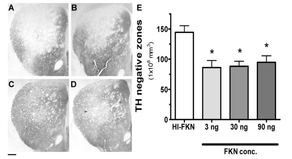
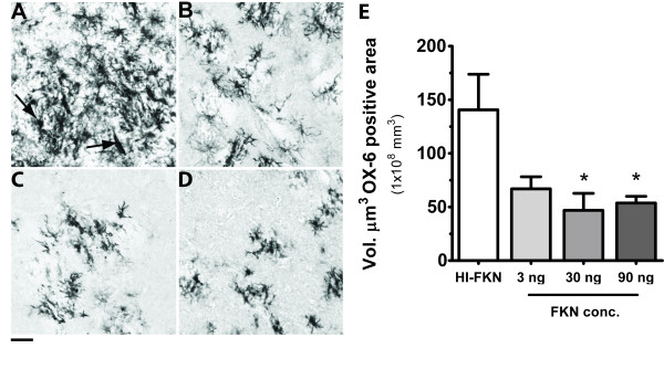
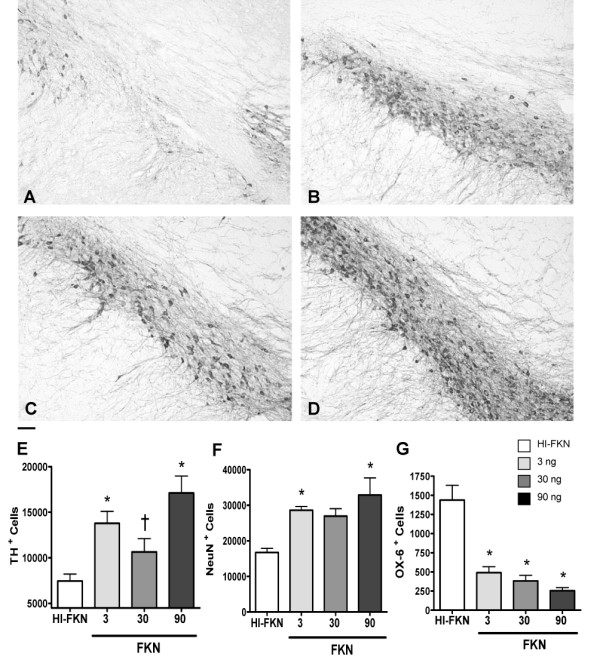
BACKGROUND: Parkinson's disease is characterized by a progressive loss of dopaminergic neurons in the substantia nigra. The cause of the neurodegeneration is unknown. Neuroinflammation has been clearly shown in Parkinson's disease and may be involved in the progressive nature of the disease. Microglia are capable of producing neuronal damage through the production of bioactive molecules such as cytokines, as well as reactive oxygen species (ROS), and nitric oxide (NO). The inflammatory response in the brain is tightly regulated at multiple levels. One form of immune regulation occurs via neurons. Fractalkine (CX3CL1), produced by neurons, suppresses the activation of microglia. CX3CL1 is constitutively expressed. It is not known if addition of exogenous CX3CL1 beyond otherwise physiologically normal levels could decrease microglia activation and thereby minimize the secondary neurodegeneration following a neurotoxic insult. METHODS: The intrastriatal 6-hydroxydopamine (6-OHDA) rat model of Parkinson disease, was used to test the hypothesis that exogenous CX3CL1 could be neuroprotective. Treatment with recombinant CX3CL1 was delivered to the striatum by an osmotic minipump for 28 days beginning 7 days after the initial insult. Unbiased stereological methods were used to quantify the lesion size in the striatum, the amount of neuronal loss in the substantia nigra, and the amount of microglia activation. RESULTS: As hypothesized, CX3CL1 was able to suppress this microglia activation. The reduced microglia activation was found to be neuroprotective as the CX3CL1 treated rats had a smaller lesion volume in the striatum and importantly significantly fewer neurons were lost in the CX3CL1 treated rats. CONCLUSION: These findings demonstrated that CX3CL1 plays a neuroprotective role in 6-OHDA-induced dopaminergic lesion and it might be an effective therapeutic target for many neurodegenerative diseases, including Parkinson disease and Alzheimer disease, where inflammation plays an important role.
Overview
The claims and statistics of this paper haven’t been checked yet.
Run the check — free, no account needed. It reads the open-access text and checks the sources it cites.
Integrity signals
Registry
Cites
Statistics
Figures
Verification
Claims the paper makes
The claims and statistics of this paper haven’t been checked yet.
Run the check — free, no account needed. It reads the open-access text and checks the sources it cites.
Claims other papers make about this one
Each is checked against this paper’s own text: does it say what the citing paper says it does?
In the citing paper
“The reduction in microglia activation may be caused, in part, by Spirulina’s capacity to enhance expression on the microglia of the fractalkin receptor (CX3CR1), which is neuroprotective in a 6-OHDA model of PD [126].”
From Beneficial Effects of Spirulina Consumption on Brain Health (2022) · cites this paper as [126]
In the citing paper
“The authors showed that Spirulina was neuroprotective in this α-Syn model of PD, observing a major number of TH+ and NeuN+ cells and a decrease in the number of activated microglial cells as evaluated by MHC II expression.”
From Beneficial Effects of Spirulina Consumption on Brain Health (2022) · cites this paper as [126]
Self-check
Percentages, sums and reported differences re-computed from the paper’s own numbers.
The self-check needs the paper’s full text.
Citations
Cites
The Danger Model: A Renewed Sense of Self
Matzinger, P. (2002) · Science · doi:10.1126/science.1071059 · Cited by 4,205
Microglia-mediated neurotoxicity: uncovering the molecular mechanisms
Block, M. L., Zecca, L., & Hong, J.-S. (2007) · Nature Reviews Neuroscience · doi:10.1038/nrn2038 · Cited by 4,110
McGeer, P. L., Itagaki, S., Boyes, B. E., & McGeer, E. G. (1988) · Neurology · doi:10.1212/wnl.38.8.1285 · Cited by 2,990
Jung, S., Aliberti, J., Graemmel, P., Sunshine, M. J., Kreutzberg, G. W., Sher, A., & Littman, D. R. (2000) · Molecular and Cellular Biology · doi:10.1128/mcb.20.11.4106-4114.2000 · Cited by 2,632
A new class of membrane-bound chemokine with a CX3C motif
Bazan, J. F., Bacon, K. B., Hardiman, G., Wang, W., Soo, K., Rossi, D., Greaves, D. R., Zlotnik, A., & Schall, T. J. (1997) · Nature · doi:10.1038/385640a0 · Cited by 2,013
Neuroinflammation in Parkinson’s disease: A target for neuroprotection?
Hirsch, E. C., & Hunot, S. (2009) · The Lancet Neurology · doi:10.1016/s1474-4422(09)70062-6 · Cited by 1,984
Control of microglial neurotoxicity by the fractalkine receptor
Cardona, A. E., Pioro, E. P., Sasse, M. E., Kostenko, V., Cardona, S. M., Dijkstra, I. M., Huang, D., Kidd, G., Dombrowski, S., Dutta, R., et al. (2006) · Nature Neuroscience · doi:10.1038/nn1715 · Cited by 1,531
Harrison, J. K., Jiang, Y., Chen, S., Xia, Y., Maciejewski, D., McNamara, R. K., Streit, W. J., Salafranca, M. N., Adhikari, S., Thompson, D. A., et al. (1998) · Proceedings of the National Academy of Sciences · doi:10.1073/pnas.95.18.10896 · Cited by 1,176
Hundhausen, C., Misztela, D., Berkhout, T. A., BROADWAY, N. M., Säftig, P., Reiß, K., Hartmann, D., Fahrenholz, F., Postina, R., Matthews, V. B., et al. (2003) · Blood · doi:10.1182/blood-2002-12-3775 · Cited by 731
Garton, K. J., Gough, P. J., Blobel, C., Murphy, G., Greaves, D. R., Dempsey, P. J., & Raines, E. W. (2001) · Journal of Biological Chemistry · doi:10.1074/jbc.m106434200 · Cited by 653
Neurotactin, a membrane-anchored chemokine upregulated in brain inflammation
Pan, Y., Lloyd, C. M., Zhou, H., Dolich, S., Deeds, J., Gonzalo, J.-Á., Vath, J., Gosselin, M., Ma, J., Dussault, B. J., et al. (1997) · Nature · doi:10.1038/42491 · Cited by 643
Combadière, C., Feumi, C., Raoul, W., Keller, N., Rodéro, M., Pézard, A., Lavalette, S., Houssier, M., Jonet, L., Picard, E., et al. (2007) · Journal of Clinical Investigation · doi:10.1172/jci31692 · Cited by 618 · cited 3×
“Since microglia are the predominant cells in the CNS that express the CX3CL1 receptor, the effects of CX3CL1 are most likely a direct effect on microglia activity [12,13, 17 ].”
InfluentialFractalkine and CX3CR1 regulate hippocampal neurogenesis in adult and aged rats
Bachstetter, A. D., Morganti, J. M., Jernberg, J., Schlunk, A., Mitchell, S. H., Brewster, K. W., Hudson, C. E., Cole, M. J., Harrison, J. K., Bickford, P. C., et al. (2011) · Neurobiology of Aging · doi:10.1016/j.neurobiolaging.2009.11.022 · Cited by 364
Production and neuroprotective functions of fractalkine in the central nervous system
Mizuno, T., Kawanokuchi, J., Numata, K., & Suzumura, A. (2003) · Brain Research · doi:10.1016/s0006-8993(03)02867-1 · Cited by 342
Lyons, A., Lynch, A. M., Downer, E. J., Hanley, R., O’Sullivan, J. B., Smith, A., & Lynch, M. A. (2009) · Journal of Neurochemistry · doi:10.1111/j.1471-4159.2009.06253.x · Cited by 202
Strömberg, I., Gemma, C., Vila, J., & Bickford, P. C. (2005) · Experimental Neurology · doi:10.1016/j.expneurol.2005.08.013 · Cited by 110
Zujovic, V., Schussler, N., Jourdain, D., Duverger, D., & Taupin, V. (2001) · Journal of Neuroimmunology · doi:10.1016/s0165-5728(01)00259-4 · Cited by 102
Shan, S., Hong-Min, T., Yi, F., Jun-Peng, G., Yue, F. L., Yan-hong, T., Yun-ke, Y., Li, W., Wang, X., Ma, J., et al. (2009) · Neurobiology of Aging · doi:10.1016/j.neurobiolaging.2009.03.004 · Cited by 82
Early inhibition of TNFα increases 6-hydroxydopamine-induced striatal degeneration
Gemma, C., Catlow, B. J., Cole, M., Hudson, C., Samec, A. M., Shah, N. K., Vila, J., Bachstetter, A. D., & Bickford, P. C. (2007) · Brain Research · doi:10.1016/j.brainres.2007.02.003 · Cited by 21
Langston, J. W., Forno, L. S., Tetrud, J., Reeves, A. G., Kaplan, J. A., & Karluk, D. (1999) · Annals of Neurology · doi:10.1002/1531-8249(199910)46:4 · Cited by 1
Zujovic (2000) · doi:10.1002/(sici)1098-1136(20000215)29:4
Cited by
Sorted by citations
Microglia in neurodegenerative diseases: mechanism and potential therapeutic targets
Gao, C., Jiang, J., Tan, Y., & Chen, S. (2023) · Signal Transduction and Targeted Therapy · doi:10.1038/s41392-023-01588-0 · Cited by 1,616
Haroon, E., Raison, C. L., & Miller, A. H. (2012) · Neuropsychopharmacology · doi:10.1038/npp.2011.205 · Cited by 1,000
Neuroinflammation in Parkinson's disease and its potential as therapeutic target
Wang, Q., Liu, Y., & Zhou, J. (2015) · Translational Neurodegeneration · doi:10.1186/s40035-015-0042-0 · Cited by 884
Neurotoxin-based models of Parkinson's disease
Bové, J., & Perier, C. (2011) · Neuroscience · doi:10.1016/j.neuroscience.2011.10.057 · Cited by 534
Bidirectional Microglia–Neuron Communication in Health and Disease
Szepesi, Z., Manouchehrian, O., Bachiller, S., & Deierborg, T. (2018) · Frontiers in Cellular Neuroscience · doi:10.3389/fncel.2018.00323 · Cited by 459
Metadata
- Authors
- Pabon, M. M., Bachstetter, A. D., Hudson, C. E., Gemma, C., & Bickford, P. C.
- Affiliations
- University of South Florida (US)
- University of Kentucky (US)
- James A. Haley Veterans' Hospital (US)
- Corresponding author
- Gemma, Carmelina
- Bickford, Paula C
- Venue
- Journal of Neuroinflammation
- Published
- 2011-01-25
- Volume
- 8
- Issue
- 1
- Pages
- 9-9
- ISSN
- 1742-2094 (ISSN-L)
- PMC
- PMC3039584
- OpenAlex
- W2163743819
- Cited by
- 241
- References
- 21
- Citations by year
- 2026: 9 · 2025: 13 · 2024: 24 · 2023: 13 · 2022: 17 · 2021: 14 · 2020: 13 · 2019: 21 · 2018: 14 · 2017: 11 · 2016: 20 · 2015: 16 · 2014: 20 · 2013: 17 · 2012: 13
- FWCI
- 9.89
- Type
- journal article
- Language
- en
- Open access
- gold
- doi.org
- Licence
- cc-by
- Locations
- Journal of Neuroinflammation (publishedVersion, cc-by)
- PubMed Central (publishedVersion, cc-by)
- PubMed (publishedVersion)
- UKnowledge (University of Kentucky) (submittedVersion, cc-by)
- DOAJ (DOAJ: Directory of Open Access Journals) (submittedVersion)
- Funders
- National Institutes of Health
- National Institute on Aging (R21AG024165, P01 AG04418, F32AG037280, P01AG004418)
- Topics
- Life Sciences › Neuroscience › Neurology › Neuroinflammation and Neurodegeneration Mechanisms
- Health Sciences › Medicine › Clinical Neurology › Parkinson's Disease Mechanisms and Treatments
- Life Sciences › Biochemistry, Genetics and Molecular Biology › Molecular Biology › Nuclear Receptors and Signaling
- Keywords
- fractalkine, Parkinson's disease, microglial activation, neuroinflammation, neuroprotection, 6-hydroxydopamine, dopaminergic neuron loss, substantia nigra, striatum, neurodegeneration, recombinant protein therapy
- MeSH terms
- Animals, Corpus Striatum, Disease Models, Animal, Humans, Male, Nerve Degeneration, Neurons, Parkinson Disease, Rats, Inbred F344, Substantia Nigra, Oxidopamine, Microglia, Rats, Chemokine CX3CL1
About these checks
Registry, retraction, Cites and statistics results are computed, not judged, with one exception: a printed reference that no index lists is read once by a model before it counts as a citation error. Claims are extracted by a model and kept only when their quote appears verbatim in the text; each cited source is then checked, with a model’s help, for whether it says what the claim relies on. Those verdicts can be wrong — read the claim’s quote and the cited source before relying on one.
Statistics and arithmetic are recomputed, not judged.
Confidence bands are shown only for fields calibrated on our annotated test set. None is calibrated yet, so no band is shown.
What counts as a claim ›How we measure confidence ›How we check this ›
Are you an author, or do you see a problem with this page? Request a review
