

The DOI matches its Crossref record; 54 of the 56 references it cites were found in a registry or an index.
Influenza A penetrates host mucus by cleaving sialic acids with neuraminidase
Open accessAbstract
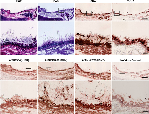
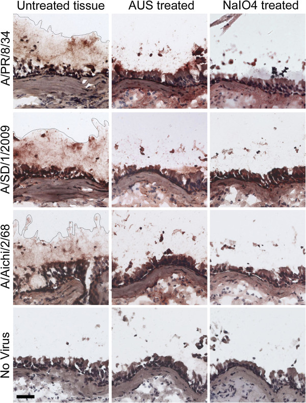
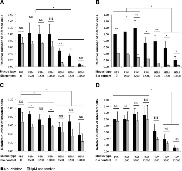
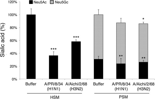
BACKGROUND: Influenza A virus (IAV) neuraminidase (NA) cleaves sialic acids (Sias) from glycans. Inhibiting NA with oseltamivir suppresses both viral infection, and viral release from cultured human airway epithelial cells. The role of NA in viral exit is well established: it releases budding virions by cleaving Sias from glycoconjugates on infected cells and progeny virions. The role of NA in viral entry remains unclear. Host respiratory epithelia secrete a mucus layer rich in heavily sialylated glycoproteins; these could inhibit viral entry by mimicking sialylated receptors on the cell surface. It has been suggested that NA allows influenza to penetrate the mucus by cleaving these sialylated decoys, but the exact mechanism is not yet established. METHODS: We tested IAV interaction with secreted mucus using frozen human trachea/bronchus tissue sections, and bead-bound purified human salivary mucins (HSM) and purified porcine submaxillary mucins (PSM). The protective effect of mucus was analyzed using MDCK cells coated with purified HSM and PSM with known Sia content. Oseltamivir was used to inhibit NA activity, and the fluorescent reporter substrate, 4MU-Neu5Ac, was used to quantify NA activity. RESULTS: IAV binds to the secreted mucus layer of frozen human trachea/bronchus tissues in a Sia dependent manner. HSM inhibition of IAV infection is Sia dose-dependent, but PSM cannot inhibit infection of underlying cells. HSM competitively inhibits NA cleavage of 4MU-Neu5Ac, reporter substrate. Human IAV effectively cleaves Sias from HSM but not from PSM, and binds to HSM but not to PSM. CONCLUSION: IAV interacts with human mucus on frozen tissue sections and mucus-coated beads. Inhibition of IAV infection by sialylated human mucus is dose-dependent, and enhanced when NA is inhibited with oseltamivir. Thus NA cleaves sialylated decoys during initial stages of infection. Understanding IAV interactions with host mucins is a promising new avenue for drug development.
Overview
The claims and statistics of this paper haven’t been checked yet.
Run the check — free, no account needed. It reads the open-access text and checks the sources it cites.
Integrity signals
Registry
Cites
Statistics
Figures
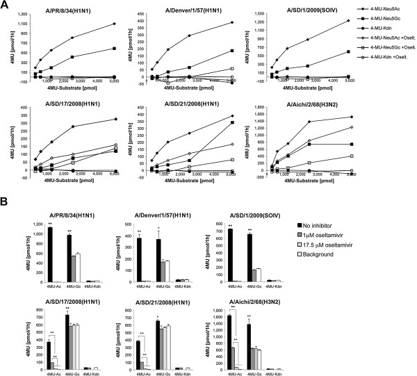
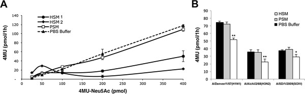
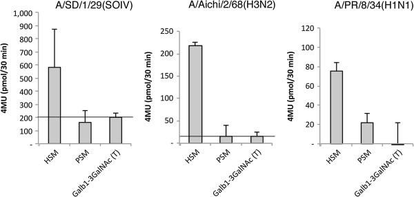
Verification
Claims the paper makes
The claims and statistics of this paper haven’t been checked yet.
Run the check — free, no account needed. It reads the open-access text and checks the sources it cites.
Self-check
Percentages, sums and reported differences re-computed from the paper’s own numbers.
The self-check needs the paper’s full text.
Citations
Cites
Wiederschain, G. Y. (2009) · Biochemistry (Moscow) · doi:10.1134/s0006297909090156 · Cited by 4,089
Varki, A. P., Cummings, R. D., Esko, J. D., Freeze, H. H., Stanley, P., Bertozzi, C. R., Hart, G. W., & Etzler, M. E. (1999) · Cited by 2,384
Airway Mucus Function and Dysfunction
Fahy, J. V., & Dickey, B. F. (2010) · New England Journal of Medicine · doi:10.1056/nejmra0910061 · Cited by 1,804
Cone, R. A. (2008) · Advanced Drug Delivery Reviews · doi:10.1016/j.addr.2008.09.008 · Cited by 1,459
Chemical Diversity in the Sialic Acids and Related α-Keto Acids: An Evolutionary Perspective
Angata, T., & Varki, A. P. (2002) · Chemical Reviews · doi:10.1021/cr000407m · Cited by 1,254
Mucus clearance as a primary innate defense mechanism for mammalian airways
Knowles, M. R., & Boucher, R. C. (2002) · Journal of Clinical Investigation · doi:10.1172/jci0215217 · Cited by 1,054
Structure and Function of the Polymeric Mucins in Airways Mucus
Thornton, D. J., Rousseau, K., & McGuckin, M. A. (2007) · Annual Review of Physiology · doi:10.1146/annurev.physiol.70.113006.100702 · Cited by 858
Human and avian influenza viruses target different cell types in cultures of human airway epithelium
Matrosovich, M. N., Matrosovich, T. Y., Gray, T. E., Roberts, N. A., & Klenk, H. (2004) · Proceedings of the National Academy of Sciences · doi:10.1073/pnas.0308001101 · Cited by 777
The airway epithelium: soldier in the fight against respiratory viruses
Vareille, M., Kieninger, E., Edwards, M. R., & Regamey, N. (2011) · Clinical Microbiology Reviews · doi:10.1128/cmr.00014-10 · Cited by 655
Matrosovich, M. N., Matrosovich, T. Y., Gray, T., Roberts, N. A., & Klenk, H.-D. (2004) · Journal of Virology · doi:10.1128/jvi.78.22.12665-12667.2004 · Cited by 606
Cited by
Sorted by citations
Items 201–290 of 290
Pohl, M. O., Violaki, K., Liu, L., Gaggioli, E., Glas, I., von Kempis, J., Messerli, C., Lin, C., Terrettaz, C., David, S. C., et al. (2025) · mSphere · doi:10.1128/msphere.00365-25 · Cited by 7
Purification of viral neuraminidase from inclusion bodies produced by recombinant Escherichia coli
Lipničanová, S., Chmelová, D., Godány, A., Ondrejovič, M., & Miertuš, S. (2020) · Journal of Biotechnology · doi:10.1016/j.jbiotec.2020.04.005 · Cited by 6
Antibody Neutralization of an Influenza Virus that Uses Neuraminidase for Receptor Binding
Gentles, L. E., Wan, H., Eichelberger, M. C., & Bloom, J. D. (2020) · Viruses · doi:10.3390/v12060597 · Cited by 7
Kannan, S., Shankar, R., & Kolandaivel, P. (2019) · Journal of Molecular Modeling · doi:10.1007/s00894-019-3982-y · Cited by 7
Liu, M., Wu, X., van de Garde, M. D. B., Narimatsu, Y., van Kuppeveld, F. J. M., Clausen, H., de Haan, C. A. M., & de Vries, E. (2025) · iScience · doi:10.1016/j.isci.2025.112549 · Cited by 6
Fuchs, J., Hübner, J., Schmidt, A. P., Irrgang, P., Maier, C., Antão, A. V., Oltmanns, F., Thirion, C., Lapuente, D., & Tenbusch, M. (2024) · npj Vaccines · doi:10.1038/s41541-024-01001-z · Cited by 7
The Application of Pseudotypes to Influenza Pandemic Preparedness
Ferrara, F., Molesti, E., & Temperton, N. J. (2015) · Future Virology · doi:10.2217/fvl.15.36 · Cited by 6
Küchler, J., Opitz, P., Jordan, I., Genzel, Y., Benndorf, D., & Reichl, U. (2025) · Applied Microbiology and Biotechnology · doi:10.1007/s00253-025-13423-3 · Cited by 5
Gattani, A., Kumar, A., Agrawal, A., Khan, M. H., Mahawar, M., Bag, S., Rajak, K. K., Singh, R. K., & Singh, P. (2020) · Microchemical Journal · doi:10.1016/j.microc.2020.105229 · Cited by 5
Harless, W. W., Lewis, B., Qorri, B., Abdulkhalek, S., & Szewczuk, M. R. (2023) · Cells · doi:10.3390/cells12091332 · Cited by 5
The sliding motility of the bacilliform virions of Influenza A viruses
Stevens, L. A., de Buyl, S., & Mognetti, B. M. (2023) · Soft Matter · doi:10.1039/d3sm00371j · Cited by 5
Bridging Viral Glycobiology and Lectin Biotechnology for Antiviral and Diagnostic Strategies
Cavada, B. S., Osterne, V. J. S., Oliveira, M. V., Ferreira, W. P., Neto, C. C., Nascimento, K. S., & Pinto‐Junior, V. R. (2025) · ACS Bio & Med Chem Au · doi:10.1021/acsbiomedchemau.5c00115 · Cited by 4
Combating respiratory diseases with mucosal vaccines
Tang, P. C. H., Ling, Z., Lee, M., King, N. J. C., & Mahalingam, S. (2026) · Journal of Virology · doi:10.1128/jvi.00146-26 · Cited by 5
Song, H., Liu, X., Gao, X., Li, J., Shang, Y., Gao, W., Li, Y., & Zhang, Z. (2022) · Infection Genetics and Evolution · doi:10.1016/j.meegid.2022.105332 · Cited by 5
MUC5AC: A potential biomarker of severity in pediatric patients infected with influenza
López, E. L., Ferolla, F. M., Meza, S. N., Mascardi, N., Neira, P., Chiormi, A., Reyero, M., Retta, A., Contrini, M. M., & Acosta, P. L. (2024) · Journal of Medical Virology · doi:10.1002/jmv.29715 · Cited by 4
Evaluation of catalytic activity of human and animal origin viral neuraminidase: Current prospect
Gattani, A., Agrawal, A., Khan, M. H., Gupta, R., & Singh, P. (2023) · Analytical Biochemistry · doi:10.1016/j.ab.2023.115157 · Cited by 4
Li, K., Bartlett, J. A., Wohlford-Lenane, C., Xue, B., Thurman, A. L., Gallagher, T. M., Pezzulo, A. A., & McCray, P. B. (2023) · The Journal of Infectious Diseases · doi:10.1093/infdis/jiad383 · Cited by 4
Substrate specificity of avian influenza H5N1 neuraminidase
Onsirisakul, N. (2014) · World Journal of Virology · doi:10.5501/wjv.v3.i4.30 · Cited by 4
Phlorotannin-Rich Ascophyllum nodosum Seaweed Extract Inhibits Influenza Infection
Mega, D. F., Sharma, P., Kipar, A., Hetzel, U., Bramwell, C., Merritt, A., Wright, S., Plummer, C., Urbanowicz, R. A., & Stewart, J. P. (2024) · Viruses · doi:10.3390/v16121919 · Cited by 3
Rather, M. A., Gul, I., Aman, M., Hassan, A., Nazki, S., Koul, P. A., Potdar, V. A., Ganai, N. A., Khan, A. A., Chikan, N. A., et al. (2025) · Archives of Microbiology · doi:10.1007/s00203-025-04371-7 · Cited by 3
Singh, K., Malik, Y. S., & Hemida, M. G. (2025) · Pathogens · doi:10.3390/pathogens14090864 · Cited by 3
Human Innate Lymphoid Cells in Influenza Infection and Vaccination
Kumar, A., Kumari, R., Liu, T., Cao, W., Davidson, B. A., Knight, P. R., & Sambhara, S. R. (2021) · Critical Reviews in Immunology · doi:10.1615/critrevimmunol.2021040801 · Cited by 3
Do, T. H. T., Wille, M., Wheatley, A. K., & Koutsakos, M. (2024) · Journal of Virology · doi:10.1128/jvi.01186-24 · Cited by 3
Sriwilaijaroen, N., Hanamatsu, H., Yokota, I., Nishikaze, T., Ijichi, T., Takahashi, T., Sakoda, Y., Furukawa, J., & Suzuki, Y. (2024) · Antiviral Research · doi:10.1016/j.antiviral.2024.106040 · Cited by 3
Enzymatic Cellular Nanoparticles Deliver Payloads across Mucosal Barriers
Kubiatowicz, L. J., Pourafzal, N. N., Krishnan, N., Bao, L., Mohapatra, A., Gao, W., Fang, R. H., & Zhang, L. (2025) · Bioconjugate Chemistry · doi:10.1021/acs.bioconjchem.5c00262 · Cited by 3
Airway mucus, infection, and therapeutic strategies
NandyMazumdar, M. (2023) · Elsevier eBooks · doi:10.1016/b978-0-323-85730-7.00056-4 · Cited by 3
Septer, K. M., Heinly, T. A., Sim, D. G., Patel, D., Roder, A. E., Wang, W., Chung, M. C., Johnson, K., Ghedin, E., & Sutton, T. C. (2024) · mBio · doi:10.1128/mbio.02161-24 · Cited by 3
Butnarasu, C., Safferthal, M., Thomas, J., Povolotsky, T. L., Diehn, R., Fentker, K., Mertins, P., Pagel, K., & Lauster, D. C. (2025) · International Journal of Biological Macromolecules · doi:10.1016/j.ijbiomac.2025.142357 · Cited by 4
Microrheology of gel-forming airway mucins isolated from porcine trachea
Engle, E. M., Yang, S., Boboltz, A., Kumar, S., Stern, A., & Duncan, G. A. (2025) · Soft Matter · doi:10.1039/d4sm01343c · Cited by 2
Implications of morphological variation in influenza viruses
Uchechukwu, C. F., & Robb, N. C. (2025) · Microbiology and Molecular Biology Reviews · doi:10.1128/mmbr.00015-25 · Cited by 2
The receptor landscape of influenza A viruses
Ciminski, K., Reuther, P., Rijkers, R., de Vries, R. P., & Schwemmle, M. (2026) · Journal of Virology · doi:10.1128/jvi.01443-25 · Cited by 2
Knockin’ on Cell’s Door: Influenza A Virus Adsorption and Its Pharmacological Inhibition
Gorshkov, A. N., & Анатольевна, В. Е. (2025) · Microbiology Research · doi:10.3390/microbiolres16020037 · Cited by 2
Shultz, P. K., Crofts, K. F., Holbrook, B. C., & Alexander‐Miller, M. A. (2020) · JCI Insight · doi:10.1172/jci.insight.141655 · Cited by 2
Wautlet, A. J., Patel, P., Chávez, P., & Codispoti, C. D. (2020) · Annals of Allergy Asthma & Immunology · doi:10.1016/j.anai.2020.11.012 · Cited by 2
Song, H., & Gao, G. F. (2022) · Methods in molecular biology · doi:10.1007/978-1-0716-2635-1_15 · Cited by 2
Hewajuli, D. A., & Dharmayanti, N. (2019) · Indonesian Bulletin of Animal and Veterinary Sciences · doi:10.14334/wartazoa.v29i2.1951 · Cited by 2
TRANSMISSION AND PATHOGENESIS OF HANTAVIRUS
Pettersson, L. (2015) · KTH Publication Database DiVA (KTH Royal Institute of Technology) · Cited by 2
Global evolution of influenza A viruses in swine
Vincent, A. L., Lewis, N. S., & Webby, R. J. (2016) · doi:10.1002/9781118924341.ch18 · Cited by 2
Smirnov, I. V., Besavilla, D. F., Schön, K., Axelsson, H., & Angeletti, D. (2025) · npj Viruses · doi:10.1038/s44298-025-00123-y · Cited by 1
Prediction of Host Tropism and Cross-Species Infection of Viruses
Son, H. S., & Cho, M. (2025) · Springer eBooks · doi:10.1007/978-981-96-5757-5_7 · Cited by 1
Sornberger, T. A., Wolters, R. M., Gilchuk, I. M., Myers, L., Binshtein, E., Irving, R. P., Chen, E. C., Gilchuk, P., Nargi, R. S., Sutton, R. E., et al. (2025) · Journal of Clinical Investigation · doi:10.1172/jci188887 · Cited by 1
Ellingson, M. O., & Bevan, M. A. (2026) · Soft Matter · doi:10.1039/d5sm01176k · Cited by 1
Du, X.-Y., Xu, L., Liu, D., Zhang, Z.-J., & Li, R.-T. (2026) · Journal of Ethnopharmacology · doi:10.1016/j.jep.2026.121332 · Cited by 1
Sialic acids are a barrier to the entry of non-influenza orthomyxoviruses
Dupré, G., Moreno‐García, J., Bendl, E., Kochs, G., Dufloo, J., & Sanjuán, R. (2026) · PLoS Pathogens · doi:10.1371/journal.ppat.1013902 · Cited by 1
Host cell entry and neutralisation sensitivity of H3N2 influenza A virus subclade K
Moor, N., Chen, N., Behrens, G. M. N., Zhang, L., Moldenhauer, A.-S., Winkler, M., Happle, C., Steffens, S., Stankov, M. V., Lürken, K., et al. (2026) · npj Vaccines · doi:10.1038/s41541-026-01573-y · Cited by 1
Reshaping and enzymatic activity may allow viruses to move through the mucus
Ziebert, F., Dokonon, K. G., & Kulić, I. M. (2024) · Soft Matter · doi:10.1039/d4sm00592a · Cited by 1
Lai, L., Li, R., Chen, Y., Deng, J., Yu, S., Lin, Q., Chen, L., & Ren, T. (2025) · Veterinary Research · doi:10.1186/s13567-025-01577-x · Cited by 1
Influenza A virus-host interactions and their control by viral non-structural protein NS1
Anastasina, M. (2015) · Työväentutkimus Vuosikirja · Cited by 1
Interactions of pseudorabies virus and swine influenza virus with porcine respiratory mucus
Yang, X. (2015) · Ghent University Academic Bibliography (Ghent University) · Cited by 1
Development of Influenza Virus Sialidase Inhibitors
Pascolutti, M., Thomson, R. J., & von Itzstein, M. (2016) · Methods and principles in medicinal chemistry · doi:10.1002/9783527677047.ch22 · Cited by 1
Barnard, K. N., Alford-Lawrence, B. K., Buchholz, D. W., Wasik, B. R., LaClair, J. R., Yu, H., Honce, R., Rühl, S., Pajic, P., Daugherity, E. K., et al. (2019) · bioRxiv (Cold Spring Harbor Laboratory) · doi:10.1101/800300 · Cited by 1
Immunological studies on Swine Influenza Virus in pigs: from gene to epitopes
Baratelli, M. (2015) · Tesis Doctorals en Xarxa (Consorci de Serveis Universitaris de Catalunya)
Coating the Flu with Sticky Polymers to Look for New Drugs
Gibson, M. I. (2016) · ACS Central Science · doi:10.1021/acscentsci.6b00301
Hoffmann, R. F. (2016) · University of Groningen research database (University of Groningen / Centre for Information Technology)
Tuning of influenza A virus neuraminidase activity
Dai, M. (2017) · Utrecht University Repository (Utrecht University)
Is the Neuraminidase Inhibitor Tamiflu Effective in the Treatment of Influenza
Hershkop, E. (2015) · Touro Scholar (Touro College)
Lung-resident, M2e-specific CD4 T cells critically protect during influenza infection
Omokanye, A. (2018) · Gothenburg University Publications Electronic Archive (Gothenburg University)
PARK, J. E. (2019) · Applied Ecology and Environmental Research · doi:10.15666/aeer/1705_1101711022
The glycobiology of human adenovirus infections : implications for tropism and treatment
Chandra, N. (2019) · DiVA at Umeå University (Umeå University)
Understanding mucus selective permeability across length scales
Samad, T. S. (2019) · DSpace@MIT (Massachusetts Institute of Technology)
da Costa, K. A. S., Rosario, J. M. M. D., Ferrari, M., Vishwanath, S., Asbach, B., Kinsley, R., Wagner, R., Heeney, J. L., Carnell, G., & Temperton, N. J. (2021) · bioRxiv (Cold Spring Harbor Laboratory) · doi:10.1101/2021.09.19.460966
Veerman, E. C. I., & Amerongen, A. V. N. (2014) · Bohn Stafleu van Loghum eBooks · doi:10.1007/978-90-368-0387-8_7
Universal influenza vaccine technologies and recombinant virosome production
Khalaj‐Hedayati, A., Chua, C. L. L., Smooker, P. M., & Lee, K. W. (2022) · Methods in microbiology · doi:10.1016/bs.mim.2022.04.001
Ativação do inflamassoma: a importância na leishmaniose, na influenza e na COVID-19
de Sá, K. S. G. (2022) · Digital Library of Theses and Dissertations (Universidade de São Paulo) · doi:10.11606/t.17.2022.tde-03022023-110423
Influenza management with new therapies
Donadello, K., Gottin, L., BOETTI, R. P., & Martín‐Loeches, I. (2023) · Minerva Respiratory Medicine · doi:10.23736/s2784-8477.23.02064-8
Polmoniti virali gravi dell’adulto
Delmotte, P.-R., & Monsel, A. (2024) · EMC - Anestesia-Rianimazione · doi:10.1016/s1283-0771(23)48694-x
Neumonía viral grave en adultos
Delmotte, P.-R., & Monsel, A. (2024) · EMC - Anestesia-Reanimación · doi:10.1016/s1280-4703(23)48663-x · Cited by 2
Do, T. H. T., Wille, M., Wheatley, A. K., & Koutsakos, M. (2024) · bioRxiv (Cold Spring Harbor Laboratory) · doi:10.1101/2024.07.09.602673
Scalable Extraction of Airway Mucins from Porcine TracheaPreprint
Yang, S., Engle, E. M., Boboltz, A., Kumar, S., Stern, A., & Duncan, G. A. (2024) · bioRxiv (Cold Spring Harbor Laboratory) · doi:10.1101/2024.10.08.617223
Li, J., Lin, X., Wang, H., Zhao, N., & Guo, X. (2025) · Journal of the American Society for Mass Spectrometry · doi:10.1021/jasms.4c00446
Mechanisms for Physicochemical Interaction between Mucus and Respiratory Viruses
Hiremath, I. S., & Wagner, C. E. (2025) · ACS Biomaterials Science & Engineering · doi:10.1021/acsbiomaterials.5c00425
Role of the host genetic variability in the influenza-A virus susceptibility; a reviewPreprint
Arcanjo, A. C., Mazzocco, G., de Oliveira, S. F., Plewczyński, D., & Radomski, J. P. (2014) · arXiv (Cornell University) · doi:10.48550/arxiv.1409.2031
Онколитический потенциал вируса гриппа в терапии глиом
Мустафаева, А., Shurygina, A.-P. S., Stukova, M., & Егоров, А. (2025) · Microbiology Independent Research Journal (MIR Journal) · doi:10.18527/202512180103.ru
Oncolytic potential of the influenza virus in glioma therapy
Мустафаева, А., Shurygina, A.-P. S., Stukova, M., & Egorov, A. (2025) · Microbiology Independent Research Journal (MIR Journal) · doi:10.18527/202512180103
INTERACTION BETWEEN THE COMPOSITION OF THE MICROBIOME AND MUCOPOLYSACCHARIDES OF THE PHARYNX
Deyeva, J. V., Kyiv, U., & Koshel, O. (2025) · OTORHINOLARYNGOLOGY · doi:10.37219/2528-8253-2025-6-58
Mucosal Wound Repair: Reinforcement of Respiratory Mucus Barrier Function by Inorganic Polyphosphate
Wang, X., Schröder, H. C., Neufurth, M., & Müller, W. E. G. (2026) · Progress in molecular and subcellular biology · doi:10.1007/978-3-032-17771-1_6
Decoding sialidase: physiological roles, pathological pathways, and clinical opportunities
Li, R., Liu, Y. C., Zou, R., & Li, M. (2026) · Frontiers in Cellular and Infection Microbiology · doi:10.3389/fcimb.2026.1814721
Lauster, D. C., Sieben, C., Ballauff, M., & Herrmann, A. (2026) · npj Viruses · doi:10.1038/s44298-026-00236-y
From viral infection to emission: host barriers shaping airborne transmission
MARTINEZ, P., Rohde, M., Yeasin, M., Basu, S., Sieben, C., & Port, J. R. (2026) · Journal of Virology · doi:10.1128/jvi.00753-26
Madeira, D., Correia, V., dos Santos, L. J., Rebelo‐de‐Andrade, H., & Branco, L. C. (2026) · Pharmaceutics · doi:10.3390/pharmaceutics18101340
Sun, Y., Xie, Y., Liu, L., Yang, H., Nie, J., Wang, W., Zhang, S., Quan, Y., & Li, C. (2026) · Pathogens · doi:10.3390/pathogens15111120
Immunogenicity testing of a recombinant N1 vaccine candidate in vivo
Krammer, F. (2024) · ImmPort · doi:10.21430/m32jmcqp66
BindingDB (2016) · UC San Diego · doi:10.7270/q2gx4db7
Metadata
- Authors
- Cohen, M., Zhang, X.-Q., Senaati, H. P., Chen, H.-W., Varki, N. M., Schooley, R. T., & Gagneux, P.
- Affiliations
- University of California San Diego (US)
- National Taiwan University (TW)
- Corresponding author
- Gagneux, Pascal
- Venue
- Virology Journal
- Published
- 2013-11-22
- Volume
- 10
- Issue
- 1
- Pages
- 321-321
- ISSN
- 1743-422X (ISSN-L)
- PMC
- PMC3842836
- OpenAlex
- W2119621200
- Cited by
- 288
- References
- 56
- Citations by year
- 2026: 12 · 2025: 25 · 2024: 23 · 2023: 27 · 2022: 23 · 2021: 26 · 2020: 26 · 2019: 31 · 2018: 21 · 2017: 21 · 2016: 25 · 2015: 17 · 2014: 11
- FWCI
- 13.12
- Type
- journal article
- Language
- en
- Open access
- gold
- doi.org
- Licence
- cc-by
- Locations
- Virology Journal (publishedVersion, cc-by)
- Europe PMC (PubMed Central) (publishedVersion, cc-by)
- PubMed Central (publishedVersion, cc-by)
- PubMed (publishedVersion)
- eScholarship (California Digital Library) (submittedVersion)
- Funders
- University of California, San Diego (#NS047101, P30 CA23100, P30 NS047101)
- National Cancer Institute (P30-CA-23100, P30CA023100)
- National Institute of Allergy and Infectious Diseases (U01AI074521)
- National Institute of Neurological Disorders and Stroke (NS047101, P30NS047101)
- Topics
- Health Sciences › Medicine › Epidemiology › Influenza Virus Research Studies
- Life Sciences › Biochemistry, Genetics and Molecular Biology › Molecular Biology › Glycosylation and Glycoproteins Research
- Physical Sciences › Chemistry › Organic Chemistry › Carbohydrate Chemistry and Synthesis
- Keywords
- influenza A virus, neuraminidase, sialic acid, mucus, viral entry, salivary mucins, submaxillary mucin, oseltamivir, respiratory epithelium
- MeSH terms
- Animals, Bronchi, Cell Line, Dogs, Humans, Mucus, Neuraminidase, Influenza A virus, Saliva, Sialic Acids, Trachea, Viral Proteins, Virus Internalization
About these checks
Registry, retraction, Cites and statistics results are computed, not judged, with one exception: a printed reference that no index lists is read once by a model before it counts as a citation error. Claims are extracted by a model and kept only when their quote appears verbatim in the text; each cited source is then checked, with a model’s help, for whether it says what the claim relies on. Those verdicts can be wrong — read the claim’s quote and the cited source before relying on one.
Statistics and arithmetic are recomputed, not judged.
Confidence bands are shown only for fields calibrated on our annotated test set. None is calibrated yet, so no band is shown.
What counts as a claim ›How we measure confidence ›How we check this ›
Are you an author, or do you see a problem with this page? Request a review
