

The DOI matches its Crossref record; 59 of the 59 references it cites were found in a registry or an index.
Genomic scars as biomarkers of homologous recombination deficiency and drug response in breast and ovarian cancers
Open accessAbstract
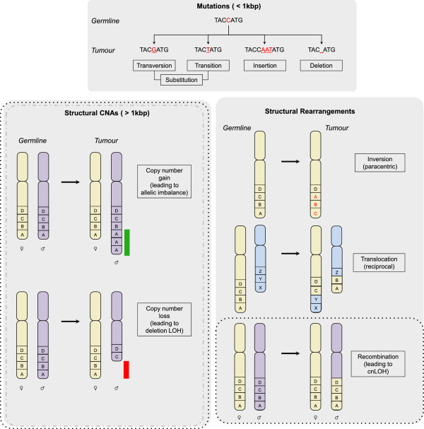
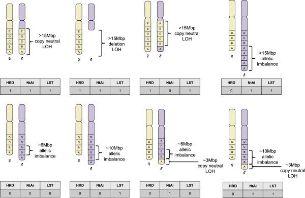
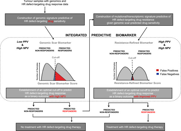
Poly (ADP-ribose) polymerase (PARP) inhibitors and platinum-based chemotherapies have been found to be particularly effective in tumors that harbor deleterious germline or somatic mutations in the BRCA1 or BRCA2 genes, the products of which contribute to the conservative homologous recombination repair of DNA double-strand breaks. Nonetheless, several setbacks in clinical trial settings have highlighted some of the issues surrounding the investigation of PARP inhibitors, especially the identification of patients who stand to benefit from such drugs. One potential approach to finding this patient subpopulation is to examine the tumor DNA for evidence of a homologous recombination defect. However, although the genomes of many breast and ovarian cancers are replete with aberrations, the presence of numerous factors able to shape the genomic landscape means that only some of the observed DNA abnormalities are the outcome of a cancer cell's inability to faithfully repair DNA double-strand breaks. Consequently, recently developed methods for comprehensively capturing the diverse ways in which homologous recombination deficiencies may arise beyond BRCA1/2 mutation have used DNA microarray and sequencing data to account for potentially confounding features in the genome. Scores capturing telomeric allelic imbalance, loss of heterozygosity (LOH) and large scale transition score, as well as the total number of coding mutations are measures that summarize the total burden of certain forms of genomic abnormality. By contrast, other studies have comprehensively catalogued different types of mutational pattern and their relative contributions to a given tumor sample. Although at least one study to explore the use of the LOH scar in a prospective clinical trial of a PARP inhibitor in ovarian cancer is under way, limitations that result in a relatively low positive predictive value for these biomarkers remain. Tumors whose genome has undergone one or more events that restore high-fidelity homologous recombination are likely to be misclassified as double-strand break repair-deficient and thereby sensitive to PARP inhibitors and DNA damaging chemotherapies as a result of prior repair deficiency and its genomic scarring. Therefore, we propose that integration of a genomic scar-based biomarker with a marker of resistance in a high genomic scarring burden context may improve the performance of any companion diagnostic for PARP inhibitors.
Overview
The claims and statistics of this paper haven’t been checked yet.
Run the check — free, no account needed. It reads the open-access text and checks the sources it cites.
Integrity signals
Registry
Cites
Statistics
Figures
Verification
Claims the paper makes
The claims and statistics of this paper haven’t been checked yet.
Run the check — free, no account needed. It reads the open-access text and checks the sources it cites.
Claims other papers make about this one
Each is checked against this paper’s own text: does it say what the citing paper says it does?
In the citing paper
“Defects in HRR can lead to the presence of “genomic scars” caused by the cancer cell’s inability to accurately repair sites of double strand breaks (DSBs).”
From Three-dimensional modelling of ovarian cancer: from cell lines to organoids for discovery and personalized medicine (2022) · cites this paper as Watkins et al., 2014
Self-check
Percentages, sums and reported differences re-computed from the paper’s own numbers.
The self-check needs the paper’s full text.
Citations
Cites
Signatures of mutational processes in human cancer
Initiative, A. P. C. G., Alexandrov, L. B., ICGC Breast Cancer Consortium, ICGC MMML-Seq Consortium, PedBrain, I., Nik-Zainal, S., Wedge, D. C., Aparicio, S. A. J. R., Behjati, S., Biankin, A. V., et al. (2013) · Nature · doi:10.1038/nature12477 · Cited by 9,660
Self-citationIntegrated genomic analyses of ovarian carcinoma
Perou, C. M. (2019) · Carolina Digital Repository (University of North Carolina at Chapel Hill) · doi:10.17615/hvp3-wg08 · Cited by 7,927
Targeting the DNA repair defect in BRCA mutant cells as a therapeutic strategy
Farmer, H., McCabe, N., Lord, C. J., Tutt, A. N. J., Johnson, D. A., Richardson, T. B., Santarosa, M., Dillon, K. J., Hickson, I., Knights, C., et al. (2005) · Nature · doi:10.1038/nature03445 · Cited by 6,683
Self-citationSpecific killing of BRCA2-deficient tumours with inhibitors of poly(ADP-ribose) polymerase
Bryant, H. E., Schultz, N., Thomas, H. D., Parker, K. M., Flower, D., Lopez, E., Kyle, S., Meuth, M., Curtin, N. J., & Helleday, T. (2005) · Nature · doi:10.1038/nature03443 · Cited by 5,198
Inhibition of Poly(ADP-Ribose) Polymerase in Tumors from BRCA Mutation Carriers
Fong, P. C., Boss, D. S., Yap, T. A., Tutt, A., Wu, P., Mergui-Roelvink, M., Mortimer, P., Swaisland, H., Lau, A., O'Connor, M. J., et al. (2009) · New England Journal of Medicine · doi:10.1056/nejmoa0900212 · Cited by 3,634
Self-citationGreaves, M., & Maley, C. C. (2012) · Nature · doi:10.1038/nature10762 · Cited by 3,162
Massive genomic rearrangement acquired in a single catastrophic event during cancer development
Stephens, P. J., Greenman, C. D., Fu, B., Yang, F., Bignell, G. R., Mudie, L. J., Pleasance, E. D., Lau, K. W., Beare, D., Stebbings, L. A., et al. (2011) · Cell · doi:10.1016/j.cell.2010.11.055 · Cited by 2,414
Audeh, M. W., Carmichael, J., Penson, R. T., Friedlander, M., Powell, B., Bell-McGuinn, K. M., Scott, C., Weitzel, J. N., Oaknin, A., Loman, N., et al. (2010) · The Lancet · doi:10.1016/s0140-6736(10)60893-8 · Cited by 2,279
Self-citationMutational Processes Molding the Genomes of 21 Breast Cancers
Nik-Zainal, S., Alexandrov, L. B., Wedge, D. C., Van Loo, P., Greenman, C. D., Raine, K., Jones, D., Hinton, J., Marshall, J., Stebbings, L. A., et al. (2012) · Cell · doi:10.1016/j.cell.2012.04.024 · Cited by 1,909
Self-citationCancer as an evolutionary and ecological process
Merlo, L. M. F., Pepper, J. W., Reid, B. J., & Maley, C. C. (2006) · Nature reviews. Cancer · doi:10.1038/nrc2013 · Cited by 1,743
Research works printed but not linked: 1
- de Bono JS, Mina LA, Gonzalez M, Curtin NJ, Wang E, Henshaw JW, Chadha M, Sachdev JC, Matei D, Jameson GS, Ong M, Basu B, Wainberg ZA, Byers LA, Chugh R, Dorr A, Kaye SB, Ramanthan RK: First-in human trial of novel oral PARP inhibitor BMN673 in patients with solid tumors. J Clin Oncol. 2013, 31: abstract 2580
Cited by
Sorted by citations
Coleman, R. L., Oza, A. M., Lorusso, D., Aghajanian, C. A., Oaknin, A., Dean, A. P., Colombo, N., Weberpals, J. I., Clamp, A. R., Scambia, G., et al. (2017) · The Lancet · doi:10.1016/s0140-6736(17)32440-6 · Cited by 1,699
Lord, C. J., & Ashworth, A. (2016) · Nature Reviews Cancer · doi:10.1038/nrc.2015.21 · Cited by 1,257
State-of-the-art strategies for targeting the DNA damage response in cancer
Pilié, P. G., Tang, C., Mills, G. B., & Yap, T. A. (2019) · Nature Reviews Clinical Oncology · doi:10.1038/s41571-018-0114-z · Cited by 1,189
Swisher, E. M., Lin, K. K., Oza, A. M., Scott, C. L., Giordano, H., Sun, J., Konecny, G. E., Coleman, R. L., Tinker, A. V., O'Malley, D. M., et al. (2017) · The Lancet Oncology · doi:10.1016/s1470-2045(16)30559-9 · Cited by 1,196
HRDetect is a predictor of BRCA1 and BRCA2 deficiency based on mutational signatures
Davies, H., Glodzik, D., Morganella, S., Yates, L. R., Staaf, J., Zou, X., Ramakrishna, M., Martin, S., Boyault, S., Sieuwerts, A. M., et al. (2017) · Nature Medicine · doi:10.1038/nm.4292 · Cited by 1,086
Self-citation
Metadata
- Authors
- Watkins, J. A., Irshad, S., Grigoriadis, A., & Tutt, A. N.
- Affiliations
- Guy's Hospital (GB)
- Corresponding author
- Grigoriadis, Anita
- Venue
- Breast Cancer Research
- Published
- 2014-06-03
- Volume
- 16
- Issue
- 3
- Pages
- 211-211
- ISSN
- 1465-5411 (ISSN-L)
- 1465-542X
- PMC
- PMC4053155
- OpenAlex
- W2100419317
- Cited by
- 327
- References
- 58
- Citations by year
- 2026: 14 · 2025: 14 · 2024: 21 · 2023: 31 · 2022: 54 · 2021: 42 · 2020: 25 · 2019: 25 · 2018: 23 · 2017: 27 · 2016: 35 · 2015: 15 · 2014: 1
- FWCI
- 2.95
- Type
- journal article
- Language
- en
- Open access
- gold
- doi.org
- Licence
- Locations
- Breast Cancer Research (publishedVersion, cc-by)
- PubMed Central (publishedVersion, cc-by)
- PubMed (publishedVersion)
- Research Portal (King's College London) (publishedVersion, other-oa)
- Funders
- National Institute for Health and Care Research
- King's College London
- Topics
- Health Sciences › Medicine › Oncology › PARP inhibition in cancer therapy
- Life Sciences › Biochemistry, Genetics and Molecular Biology › Genetics › BRCA gene mutations in cancer
- Life Sciences › Biochemistry, Genetics and Molecular Biology › Cancer Research › Cancer Genomics and Diagnostics
- Keywords
- homologous recombination deficiency, PARP inhibitors, BRCA1, BRCA2, loss of heterozygosity, telomeric allelic imbalance, mutational signatures, drug response biomarkers, breast cancer, ovarian cancer, DNA double-strand break repair, platinum-based chemotherapy, companion diagnostics, predictive biomarkers
- MeSH terms
- Poly(ADP-ribose) Polymerase Inhibitors, Breast Neoplasms, Chromosome Aberrations, Female, Humans, Ovarian Neoplasms, Biomarkers, Tumor, BRCA1 Protein, BRCA2 Protein, DNA Breaks, Double-Stranded, Homologous Recombination, Recombinational DNA Repair
About these checks
Registry, retraction, Cites and statistics results are computed, not judged, with one exception: a printed reference that no index lists is read once by a model before it counts as a citation error. Claims are extracted by a model and kept only when their quote appears verbatim in the text; each cited source is then checked, with a model’s help, for whether it says what the claim relies on. Those verdicts can be wrong — read the claim’s quote and the cited source before relying on one.
Statistics and arithmetic are recomputed, not judged.
Confidence bands are shown only for fields calibrated on our annotated test set. None is calibrated yet, so no band is shown.
What counts as a claim ›How we measure confidence ›How we check this ›
Are you an author, or do you see a problem with this page? Request a review
