

The DOI matches its Crossref record; 82 of the 82 references it cites were found in a registry or an index.
Lactobacillus crispatus inhibits growth of Gardnerella vaginalis and Neisseria gonorrhoeae on a porcine vaginal mucosa model
Open accessAbstract
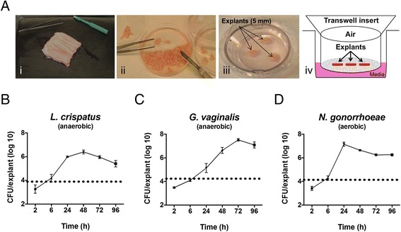
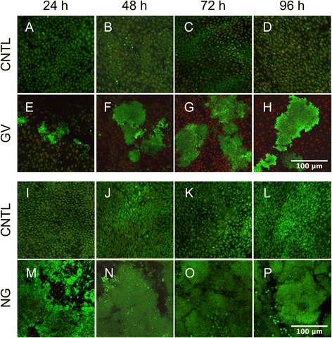
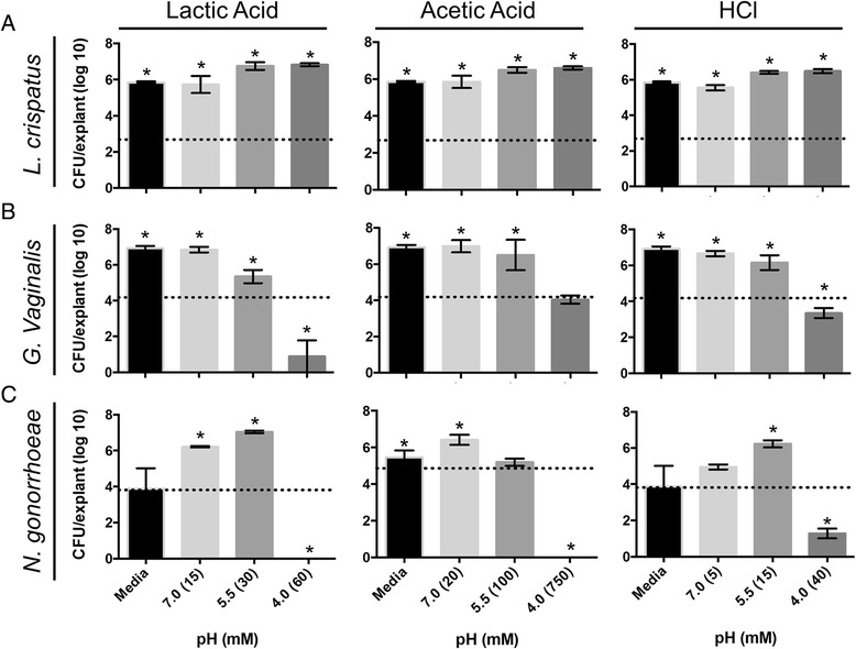
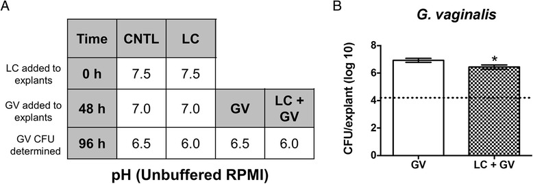
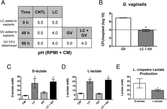
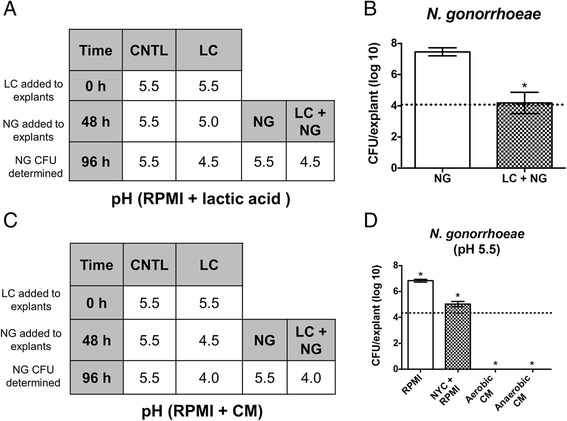
BACKGROUND: The vaginal microbiota can impact the susceptibility of women to bacterial vaginosis (BV) and sexually transmitted infections (STIs). BV is characterized by depletion of Lactobacillus spp., an overgrowth of anaerobes (often dominated by Gardnerella vaginalis) and a pH > 4.5. BV is associated with an increased risk of acquiring STIs such as chlamydia and gonorrhea. While these associations have been identified, the molecular mechanism(s) driving the risk of infections are unknown. An ex vivo porcine vaginal mucosal model (PVM) was developed to explore the mechanistic role of Lactobacillus spp. in affecting colonization by G. vaginalis and Neisseria gonorrhoeae. RESULTS: The data presented here demonstrate that all organisms tested can colonize and grow on PVM to clinically relevant densities. Additionally, G. vaginalis and N. gonorrhoeae form biofilms on PVM. It was observed that lactic acid, acetic acid, and hydrochloric acid inhibit the growth of G. vaginalis on PVM in a pH-dependent manner. N. gonorrhoeae grows best in the presence of lactic acid at pH 5.5, but did not grow well at this pH in the presence of acetic acid. Finally, a clinical Lactobacillus crispatus isolate (24-9-7) produces lactic acid and inhibits growth of both G. vaginalis and N. gonorrhoeae on PVM. CONCLUSIONS: These data reveal differences in the effects of pH, various acids and L. crispatus on the growth of G. vaginalis and N. gonorrhoeae on a live vaginal mucosal surface. The PVM is a useful model for studying the interactions of commensal vaginal microbes with pathogens and the mechanisms of biofilm formation on the vaginal mucosa.
Overview
The claims and statistics of this paper haven’t been checked yet.
Run the check — free, no account needed. It reads the open-access text and checks the sources it cites.
Integrity signals
Registry
Cites
Statistics
Figures
Verification
Claims the paper makes
The claims and statistics of this paper haven’t been checked yet.
Run the check — free, no account needed. It reads the open-access text and checks the sources it cites.
Claims other papers make about this one
Each is checked against this paper’s own text: does it say what the citing paper says it does?
In the citing paper
“This is significant because it then follows that vaginal Lactobacillus spp. (sensu lato) limit colonization of BV-associated microbes and thus by extension are protective against the aforementioned deleterious viral effects through maintenance of a low pH (Breshears et al., 2015), and bacteriocin production (Stoyancheva et al., 2014).”
From Viral Infections, the Microbiome, and Probiotics (2021) · cites this paper as Breshears et al., 2015
Self-check
Percentages, sums and reported differences re-computed from the paper’s own numbers.
The self-check needs the paper’s full text.
Citations
Cites
Vaginal microbiome of reproductive-age women
Ravel, J., Gajer, P., Abdo, Z., Schneider, G. M., Koenig, S. S. K., McCulle, S. L., Karlebach, S., Gorle, R., Russell, J., Tacket, C. O., et al. (2011) · Proceedings of the National Academy of Sciences · doi:10.1073/pnas.1002611107 · Cited by 4,254
Self-citationNugent, R. P., Krohn, M. A., & Hillier, S. L. (1991) · Journal of Clinical Microbiology · doi:10.1128/jcm.29.2.297-301.1991 · Cited by 3,332
Temporal Dynamics of the Human Vaginal Microbiota
Gajer, P., Brotman, R. M., Bai, G., Sakamoto, J. M., Schütte, U. M. E., Zhong, X., Koenig, S. S. K., Fu, L., Ma, Z. S., Zhou, X., et al. (2012) · Science Translational Medicine · doi:10.1126/scitranslmed.3003605 · Cited by 1,583
Self-citationMolecular Identification of Bacteria Associated with Bacterial Vaginosis
Fredricks, D. N., Fiedler, T. L., & Marrazzo, J. M. (2005) · New England Journal of Medicine · doi:10.1056/nejmoa043802 · Cited by 1,303
Eschenbach, D. A., Davick, P., Williams, B. L., Klebanoff, S. J., Young-Smith, K., Critchlow, C. M., & Holmes, K. K. (1989) · Journal of Clinical Microbiology · doi:10.1128/jcm.27.2.251-256.1989 · Cited by 709
HIV-1 infection associated with abnormal vaginal flora morphology and bacterial vaginosis
Sewankambo, N. K., Gray, R. H., Wawer, M. J., Paxton, L. A., McNairn, D., Wabwire‐Mangen, F., Serwadda, D., Li, C., Kiwanuka, N., Hillier, S. L., et al. (1997) · The Lancet · doi:10.1016/s0140-6736(97)01063-5 · Cited by 657
Wiesenfeld, H. C., Hillier, S. L., Krohn, M. A., Landers, D. V., & Sweet, R. L. (2003) · Clinical Infectious Diseases · doi:10.1086/367658 · Cited by 546
Beamer, M. A., Hawes, S. E., & Hillier, S. L. (1999) · The Journal of Infectious Diseases · doi:10.1086/315109 · Cited by 500
Adherent Biofilms in Bacterial Vaginosis
Swidsinski, A. V., Mendling, W., Loening-Baucke, V. A., Ladhoff, A., Swidsinski, S., Hale, L. P., & Lochs, H. H. (2005) · Obstetrics and Gynecology · doi:10.1097/01.aog.0000183594.45524.d2 · Cited by 484
Defense factors of vaginal lactobacilli
Aroutcheva, A. A., Gariti, D., Simon, M., Shott, S., Faro, J. P., Simoes, J. A., Gurguis, A., & Faro, S. (2001) · American Journal of Obstetrics and Gynecology · doi:10.1067/mob.2001.115867 · Cited by 426
Control of the Microbial Flora of the Vagina by H202-Generating Lactobacilli
Klebanoff, S. J., Hillier, S. L., Eschenbach, D. A., & Waltersdorph, A. M. (1991) · The Journal of Infectious Diseases · doi:10.1093/infdis/164.1.94 · Cited by 420
Witkin, S. S., Mendes‐Soares, H., Linhares, I. M., Jayaram, A., Ledger, W. J., & Forney, L. J. (2013) · mBio · doi:10.1128/mbio.00460-13 · Cited by 395
O'Hanlon, D. E., Moench, T. R., & Cone, R. A. (2011) · BMC Infectious Diseases · doi:10.1186/1471-2334-11-200 · Cited by 375
Ravel, J., Brotman, R. M., Gajer, P., Ma, B., Nandy, M., Fadrosh, D. W., Sakamoto, J. M., Koenig, S. S. K., Fu, L., Zhou, X., et al. (2013) · Microbiome · doi:10.1186/2049-2618-1-29 · Cited by 349
Self-citationSwidsinski, A. V., Mendling, W., Loening-Baucke, V. A., Swidsinski, S., Dörffel, Y., Scholze, J., Lochs, H. H., & Verstraelen, H. (2007) · American Journal of Obstetrics and Gynecology · doi:10.1016/j.ajog.2007.06.039 · Cited by 337
Lower genital tract infection and endometritis: insight into subclinical pelvic inflammatory disease
Wiesenfeld, H. (2002) · Obstetrics and Gynecology · doi:10.1016/s0029-7844(02)02118-x · Cited by 306
The microbiology of bacterial vaginosis
Hill, G. B. (1993) · American Journal of Obstetrics and Gynecology · doi:10.1016/0002-9378(93)90339-k · Cited by 294
Bacterial vaginosis as a risk factor for post-cesarean endometritis.
Watts, D. H., Krohn, M. A., Hillier, S. L., & Eschenbach, D. A. (1990) · PubMed · Cited by 274
Gelber, S. E., Aguilar, J. L., Lewis, K. L. T., & Ratner, A. J. (2008) · Journal of Bacteriology · doi:10.1128/jb.01965-07 · Cited by 261
Patterson, J., Stull-Lane, A. R., Girerd, P. H., & Jefferson, K. K. (2009) · Microbiology · doi:10.1099/mic.0.034280-0 · Cited by 251
The aetiology of bacterial vaginosis
Turovskiy, Y., Noll, K. S., & Chikindas, M. L. (2011) · Journal of Applied Microbiology · doi:10.1111/j.1365-2672.2011.04977.x · Cited by 250
Spiegel, C. A. (1991) · Clinical Microbiology Reviews · doi:10.1128/cmr.4.4.485 · Cited by 241
Hillier, S., Krohn, M. A., Klebanoff, S. J., & Eschenbach, D. A. (1992) · International Journal of Gynecology & Obstetrics · doi:10.1016/0020-7292(92)90920-e · Cited by 200
Graver, M. A., & Wade, J. J. (2011) · Annals of Clinical Microbiology and Antimicrobials · doi:10.1186/1476-0711-10-8 · Cited by 173
Ahmed, A., Earl, J., Retchless, A. C., Hillier, S. L., Rabe, L. K., Cherpes, T. L., Powell, E., Janto, B., Eutsey, R. A., Hiller, N. L., et al. (2012) · Journal of Bacteriology · doi:10.1128/jb.00056-12 · Cited by 173
Hillier, S. L., Krohn, M. A., Klebanoff, S. J., & Eschenbach, D. A. (1992) · Obstetrics and Gynecology · doi:10.1097/00006250-199203000-00008 · Cited by 173
Harwich, M. D., Alves, J. M. P., Buck, G. A., Strauss, J. F., Patterson, J., Oki, A. T., Girerd, P. H., & Jefferson, K. K. (2010) · BMC Genomics · doi:10.1186/1471-2164-11-375 · Cited by 169
The Effects of Commensal Bacteria on Innate Immune Responses in the Female Genital Tract
Mirmonsef, P., Gilbert, D. Z., Zariffard, M. R., Hamaker, B. R., Kaur, A., LANDAY, A. L., & Spear, G. T. (2010) · American Journal of Reproductive Immunology · doi:10.1111/j.1600-0897.2010.00943.x · Cited by 144
Estradiol-Treated Female Mice as Surrogate Hosts for Neisseria gonorrhoeae Genital Tract Infections
Jerse, A. E., Wu, H., Packiam, M., Vonck, R. A., Begum, A. A., & Garvin, L. E. (2011) · Frontiers in Microbiology · doi:10.3389/fmicb.2011.00107 · Cited by 141
Vaginal indicators of amniotic fluid infection in preterm labor
Hitti, J. E. (2001) · Obstetrics and Gynecology · doi:10.1016/s0029-7844(00)01146-7 · Cited by 136
Alves, P. M., Castro, J., Sousa, C., Cereija, T., & Cerca, N. (2014) · The Journal of Infectious Diseases · doi:10.1093/infdis/jiu131 · Cited by 136
Atassi, F., Brassart, D., Grob, P., Graf, F., & Servin, A. L. (2006) · FEMS Immunology & Medical Microbiology · doi:10.1111/j.1574-695x.2006.00162.x · Cited by 132
Gardnerella Biofilm Involves Females and Males and Is Transmitted Sexually
Swidsinski, A. V., Dörffel, Y., Loening-Baucke, V. A., Swidsinski, S., Verstraelen, H., Vaneechoutte, M., Lemm, V., Schilling, J., & Mendling, W. (2010) · Gynecologic and Obstetric Investigation · doi:10.1159/000314015 · Cited by 131
Hillier, S. L., Krohn, M. A., Cassen, E. M., Easterling, T. R., Rabe, L. K., & Eschenbach, D. A. (1995) · Clinical Infectious Diseases · doi:10.1093/clinids/20.supplement_2.s276 · Cited by 130
Gilbert, N. M., Lewis, W. G., & Lewis, A. L. (2013) · PLoS ONE · doi:10.1371/journal.pone.0059539 · Cited by 126
The murine lung microbiome in relation to the intestinal and vaginal bacterial communities
Barfod, K. K., Roggenbuck, M., Hansen, L. H., Schjørring, S., Larsen, S. E., Sørensen, S. J., & Krogfelt, K. A. (2013) · BMC Microbiology · doi:10.1186/1471-2180-13-303 · Cited by 126
O’Hanlon, D. E., Lanier, B. R., Moench, T. R., & Cone, R. A. (2010) · BMC Infectious Diseases · doi:10.1186/1471-2334-10-120 · Cited by 111
Porcine Vagina Ex Vivo as a Model for Studying Permeability and Pathogenesis in Mucosa
Squier, C. A., Mantz, M. J., Schlievert, P. M., & Davis, C. (2007) · Journal of Pharmaceutical Sciences · doi:10.1002/jps.21077 · Cited by 111
Yang, Q., Phillips, P. L., Sampson, E. M., Progulske‐Fox, A., Jin, S., Antonelli, P. J., & Schultz, G. S. (2013) · Wound Repair and Regeneration · doi:10.1111/wrr.12074 · Cited by 108
Peterson, M. L., Ault, K. A., Kremer, M. J., Klingelhutz, A. J., Davis, C., Squier, C. A., & Schlievert, P. M. (2005) · Infection and Immunity · doi:10.1128/iai.73.4.2164-2174.2005 · Cited by 108
Self-citationBiofilm Formation by Neisseria gonorrhoeae
Greiner, L. L., Edwards, J. L., Shao, J., Rabinak, C. A., Entz, D. D., & Apicella, M. A. (2005) · Infection and Immunity · doi:10.1128/iai.73.4.1964-1970.2005 · Cited by 108
Beamer, M. A., Meyn, L. A., Murray, P. J., Busse, B., & Hillier, S. L. (2009) · The Journal of Infectious Diseases · doi:10.1086/598686 · Cited by 108
Effects Of H2 O2-Producing Lactobacilli On Neisseria gonorrhoeae Growth and Activity
Zheng, H.-Y., Alcorn, T. M., & Cohen, M. S. (1994) · The Journal of Infectious Diseases · doi:10.1093/infdis/170.5.1209 · Cited by 106
Blanchette, K. A., Hinojosa, C. A., Babu, R. A. S., Lizcano, A., González-Juarbe, N., Muñoz‐Almagro, C., Sánchez, C. J., Bergman, M. A., & Orihuela, C. J. (2013) · mBio · doi:10.1128/mbio.00745-13 · Cited by 104
NATURAL ANTIMICROBIALS AND THEIR ROLE IN VAGINAL HEALTH: A SHORT REVIEW.
Dover, S. E., Aroutcheva, A. A., Faro, S., & Chikindas, M. L. (2008) · PubMed · Cited by 103
Is there a protective role for vaginal flora?
Sobel, J. D. (1999) · Current Infectious Disease Reports · doi:10.1007/s11908-999-0045-z · Cited by 101
Edwards, J. L., Shao, J. Q., Ault, K. A., & Apicella, M. A. (2000) · Infection and Immunity · doi:10.1128/iai.68.9.5354-5363.2000 · Cited by 97
DNase Inhibits Gardnerella vaginalis Biofilms In Vitro and In Vivo
Hymes, S. R., Randis, T. M., Sun, T. Y., & Ratner, A. J. (2013) · The Journal of Infectious Diseases · doi:10.1093/infdis/jit047 · Cited by 96
The role of complement receptor 3 (CR3) inNeisseria gonorrhoeaeinfection of human cervical epithelia
Edwards, J. L., Brown, E. J., Ault, K. A., & Apicella, M. A. (2001) · Cellular Microbiology · doi:10.1046/j.1462-5822.2001.00140.x · Cited by 94
Saigh, J. H., Sanders, C. C., & Sanders, W. E. (1978) · Infection and Immunity · doi:10.1128/iai.19.2.704-710.1978 · Cited by 87
Ultrastructural Study of Cervical Gonorrhea
Evans, B. A. (1977) · The Journal of Infectious Diseases · doi:10.1093/infdis/136.2.248 · Cited by 84
Alpha-Toxin Promotes Staphylococcus aureus Mucosal Biofilm Formation
Anderson, M. J., Lin, Y., Gillman, A. N., Parks, P. J., Schlievert, P. M., & Peterson, M. L. (2012) · Frontiers in Cellular and Infection Microbiology · doi:10.3389/fcimb.2012.00064 · Cited by 81
Self-citationGonococcal Cervicitis: A Role for Biofilm in Pathogenesis
Steichen, C. T., Shao, J. Q., Ketterer, M. R., & Apicella, M. A. (2008) · The Journal of Infectious Diseases · doi:10.1086/593336 · Cited by 80
Lactobacilli at the Front Line of Defense Against Vaginally Acquired Infections
Spurbeck, R. R., & Arvidson, C. G. (2011) · Future Microbiology · doi:10.2217/fmb.11.36 · Cited by 78
Cytolysins Augment Superantigen Penetration of Stratified Mucosa
Brosnahan, A. J., Mantz, M. J., Squier, C. A., Peterson, M. L., & Schlievert, P. M. (2009) · The Journal of Immunology · doi:10.4049/jimmunol.0803283 · Cited by 71
Self-citationAmant, D. C. S., Valentin-Bon, I. E., & Jerse, A. E. (2002) · Infection and Immunity · doi:10.1128/iai.70.12.7169-7171.2002 · Cited by 71
Anderson, M. J., David, M. L., Scholz, M., Bull, S. J., Morse, D. J., Hulse-Stevens, M., & Peterson, M. L. (2015) · Antimicrobial Agents and Chemotherapy · doi:10.1128/aac.04624-14 · Cited by 67
Self-citationAnderson, M. J., Horn, M. E., Lin, Y., Parks, P. J., & Peterson, M. L. (2010) · American Journal of Infection Control · doi:10.1016/j.ajic.2010.06.022 · Cited by 61
Self-citationSpencer, S. E., Valentin-Bon, I. E., Whaley, K. J., & Jerse, A. E. (2004) · The Journal of Infectious Diseases · doi:10.1086/381125 · Cited by 61
AL-MUSHRIF, S., Eley, A., & Jones, B. M. (2000) · Journal of Medical Microbiology · doi:10.1099/0022-1317-49-11-1023 · Cited by 61
Response of Gardnerella vaginalis biofilm to 5 days of moxifloxacin treatment
Swidsinski, A. V., Dörffel, Y., Loening-Baucke, V. A., Schilling, J., & Mendling, W. (2010) · FEMS Immunology & Medical Microbiology · doi:10.1111/j.1574-695x.2010.00743.x · Cited by 60
A review on pelvic inflammatory disease
Barrett, S., & Taylor, C. (2005) · International Journal of STD & AIDS · doi:10.1258/095646205774763270 · Cited by 59
The Pathobiology of Neisseria gonorrhoeae Lower Female Genital Tract Infection
Edwards, J. L., & Butler, E. K. (2011) · Frontiers in Microbiology · doi:10.3389/fmicb.2011.00102 · Cited by 59
(2014) · PubMed · Cited by 58
Davis, C., Kremer, M. J., Schlievert, P. M., & Squier, C. A. (2003) · American Journal of Obstetrics and Gynecology · doi:10.1016/s0002-9378(03)00873-1 · Cited by 55
Identification and partial characterization of a cytolytic toxin produced by Gardnerella vaginalis
Rottini, G., Dobrina, A., Forgiarini, O., Nardon, E., Amirante, G. A., & Patriarca, P. (1990) · Infection and Immunity · doi:10.1128/iai.58.11.3751-3758.1990 · Cited by 54
Recent progress in understanding the epidemiology of bacterial vaginosis
Kenyon, C. R., & Osbak, K. K. (2014) · Current Opinion in Obstetrics & Gynecology · doi:10.1097/gco.0000000000000112 · Cited by 49
Analysis of Vaginal Acetic Acid in Patients Undergoing Treatment for Bacterial Vaginosis
Chaudry, A., Travers, P. J., Yuenger, J., Colletta, L., Evans, P., Zenilman, J. M., & Tummon, A. (2004) · Journal of Clinical Microbiology · doi:10.1128/jcm.42.11.5170-5175.2004 · Cited by 49
Sheiness, D. K., Dix, K., Watanabe, S., & Hillier, S. L. (1992) · Journal of Clinical Microbiology · doi:10.1128/jcm.30.3.642-648.1992 · Cited by 47
Proinflammatory Exoprotein Characterization of Toxic Shock Syndrome Staphylococcus aureus
Lin, Y., Anderson, M. J., Kohler, P. L., Strandberg, K. L., Olson, M. E., Horswill, A. R., Schlievert, P. M., & Peterson, M. L. (2011) · Biochemistry · doi:10.1021/bi200435n · Cited by 39
Self-citationRetrocyclin inhibits Gardnerella vaginalis biofilm formation and toxin activity
Hooven, T. A., Randis, T. M., Hymes, S., Rampersaud, R., & Ratner, A. J. (2012) · Journal of Antimicrobial Chemotherapy · doi:10.1093/jac/dks305 · Cited by 39
Novel Toxic Shock Syndrome Toxin-1 Amino Acids Required for Biological Activity
Brosnahan, A. J., Schaefers, M. M., Amundson, W. H., Mantz, M. J., Squier, C. A., Peterson, M. L., & Schlievert, P. M. (2008) · Biochemistry · doi:10.1021/bi801468w · Cited by 37
Self-citationA mucosal model to study microbial biofilm development and anti-biofilm therapeutics
Anderson, M. J., Parks, P. J., & Peterson, M. L. (2012) · Journal of Microbiological Methods · doi:10.1016/j.mimet.2012.12.003 · Cited by 34
Self-citationAnderson, M. J., Scholz, M., Parks, P. J., & Peterson, M. L. (2013) · Journal of Applied Microbiology · doi:10.1111/jam.12277 · Cited by 26
Self-citationBacterial Vaginosis: Are We Coming Full Circle?
Schwebke, J. R. (2009) · The Journal of Infectious Diseases · doi:10.1086/648093 · Cited by 22
Stanek, R. J., Glover, D. D., Larsen, B., & Gain, R. E. (1992) · Biomedical Chromatography · doi:10.1002/bmc.1130060506 · Cited by 19
The growth of Gardnerella vaginalis and Lactobacillus acidophilus in Sorbarod biofilms
Muli, F. W., & Struthers, J. K. (1998) · Journal of Medical Microbiology · doi:10.1099/00222615-47-5-401 · Cited by 19
Vaginal microbiota in healthy women and patients with bacterial vaginosis and nonspecific vaginitis
Aleshkin, V. A., Александровна, В. Е., & Ba, S. (2006) · Microbial Ecology in Health and Disease · doi:10.3402/mehd.v18i2.7694 · Cited by 17
Schaefers, M. M., Breshears, L. M., Anderson, M. J., Lin, Y., Grill, A. E., Panyam, J., Southern, P. J., Schlievert, P. M., & Peterson, M. L. (2012) · PLoS ONE · doi:10.1371/journal.pone.0032813 · Cited by 17
Self-citationNon-cultural detection of Neisseria gonorrhoeae in cervical and vaginal washings
Young, H.-K., Sarafian, S. K., HARRIS, A. B., & McMillan, A. (1983) · Journal of Medical Microbiology · doi:10.1099/00222615-16-2-183 · Cited by 14
Davis, C., Baccam, M., Mantz, M. J., Osborn, T. W. B., Hill, D. R., & Squier, C. A. (2013) · Toxicology and Applied Pharmacology · doi:10.1016/j.taap.2013.11.021 · Cited by 13
Vaginal microbiota in healthy women and patients with bacterial vaginosis and nonspecific vaginitis
Aleshkin, V. A., Александровна, В. Е., & Ba, S. (2006) · Microbial Ecology in Health and Disease · doi:10.1080/17482960600891473 · Cited by 12
Cited by
Sorted by citations
Vaginal microbiota and the potential of Lactobacillus derivatives in maintaining vaginal health
Chee, W. J. Y., Chew, S. Y., & Than, L. T. L. (2020) · Microbial Cell Factories · doi:10.1186/s12934-020-01464-4 · Cited by 761
The role of lactic acid production by probiotic Lactobacillus species in vaginal health
Tachedjian, G., Aldunate, M., Bradshaw, C. S., & Cone, R. A. (2017) · Research in Microbiology · doi:10.1016/j.resmic.2017.04.001 · Cited by 557
Mitra, A., MacIntyre, D. A., Marchesi, J. R., Lee, Y. S., Bennett, P. R., & Kyrgiou, M. (2016) · Microbiome · doi:10.1186/s40168-016-0203-0 · Cited by 484
Mitra, A., MacIntyre, D. A., Ntritsos, G., Smith, A., Tsilidis, K. K., Marchesi, J. R., Bennett, P. R., Moscicki, A., & Kyrgiou, M. (2020) · Nature Communications · doi:10.1038/s41467-020-15856-y · Cited by 263
Options and Limitations in Clinical Investigation of Bacterial Biofilms
Magana, M., Sereti, C., Ioannidis, A., Mitchell, C. A., Ball, A. R., Magiorkinis, E., Chatzipanagiotou, S., Hamblin, M. R., Hadjifrangiskou, M., & Tegos, G. P. (2018) · Clinical Microbiology Reviews · doi:10.1128/cmr.00084-16 · Cited by 259
Metadata
- Authors
- Breshears, L. M., Edwards, V. L., Ravel, J., & Peterson, M. L.
- Affiliations
- University of Minnesota (US)
- University of Maryland, Baltimore (US)
- Corresponding author
- Peterson, Marnie L.
- Venue
- BMC Microbiology
- Published
- 2015-12
- Volume
- 15
- Issue
- 1
- Pages
- 276-276
- ISSN
- 1471-2180 (ISSN-L)
- PMC
- PMC4675025
- OpenAlex
- W2192796897
- Cited by
- 140
- References
- 82
- Citations by year
- 2026: 10 · 2025: 12 · 2024: 22 · 2023: 12 · 2022: 13 · 2021: 17 · 2020: 15 · 2019: 11 · 2018: 13 · 2017: 12 · 2016: 3
- FWCI
- 7.86
- Type
- journal article
- Language
- en
- Open access
- gold
- doi.org
- Licence
- Locations
- BMC Microbiology (publishedVersion, cc-by)
- Europe PMC (PubMed Central) (publishedVersion, cc-by)
- PubMed Central (publishedVersion, cc-by)
- PubMed (publishedVersion)
- Funders
- University of Minnesota (22928)
- National Institute of Allergy and Infectious Diseases (U19AI084044)
- Topics
- Life Sciences › Immunology and Microbiology › Microbiology › Reproductive tract infections research
- Life Sciences › Agricultural and Biological Sciences › Food Science › Probiotics and Fermented Foods
- Life Sciences › Biochemistry, Genetics and Molecular Biology › Molecular Biology › Gut microbiota and health
- Keywords
- Lactobacillus crispatus, Gardnerella vaginalis, Neisseria gonorrhoeae, bacterial vaginosis, vaginal microbiota, biofilm formation, lactic acid, vaginal pH, sexually transmitted infections, vaginal colonization
- MeSH terms
- Animals, Antibiosis, Carboxylic Acids, Female, Gardnerella vaginalis, Humans, Hydrogen-Ion Concentration, Lactobacillus, Male, Mucous Membrane, Neisseria gonorrhoeae, Organ Culture Techniques, Swine, Vagina, Models, Animal
About these checks
Registry, retraction, Cites and statistics results are computed, not judged, with one exception: a printed reference that no index lists is read once by a model before it counts as a citation error. Claims are extracted by a model and kept only when their quote appears verbatim in the text; each cited source is then checked, with a model’s help, for whether it says what the claim relies on. Those verdicts can be wrong — read the claim’s quote and the cited source before relying on one.
Statistics and arithmetic are recomputed, not judged.
Confidence bands are shown only for fields calibrated on our annotated test set. None is calibrated yet, so no band is shown.
What counts as a claim ›How we measure confidence ›How we check this ›
Are you an author, or do you see a problem with this page? Request a review
