

The DOI matches its Crossref record; 87 of the 88 references it cites were found in a registry or an index.
Senescence-associated IL-6 and IL-8 cytokines induce a self- and cross-reinforced senescence/inflammatory milieu strengthening tumorigenic capabilities in the MCF-7 breast cancer cell line
Open accessAbstract
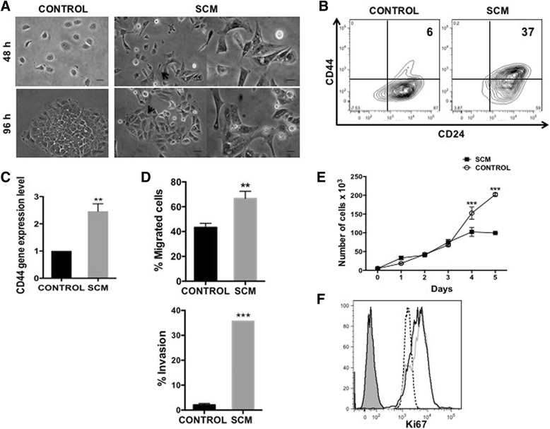
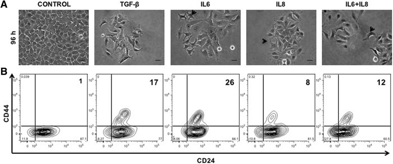
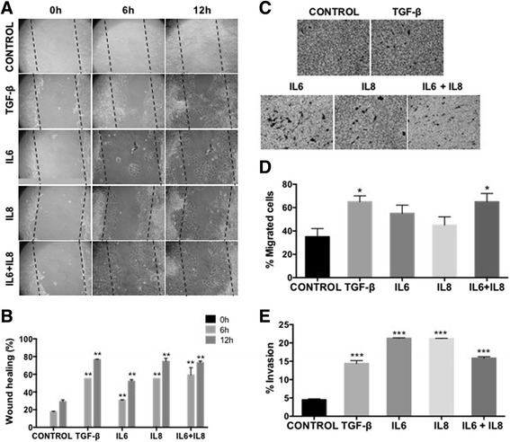
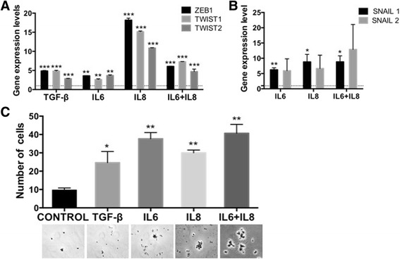
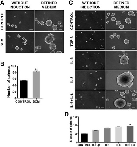
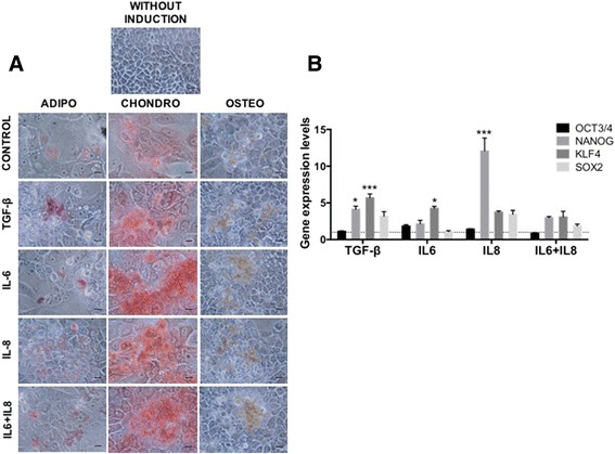
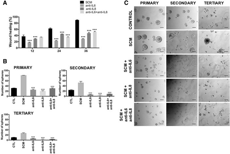
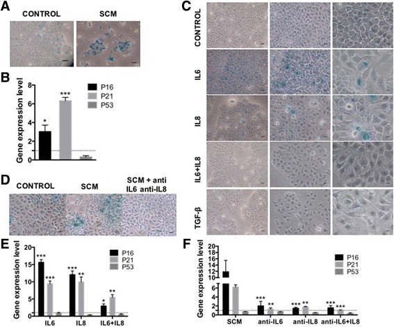
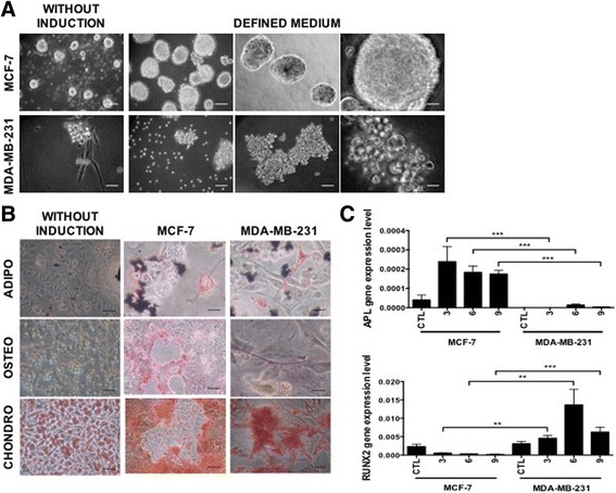
BACKGROUND: There is compelling evidence associating senescent cells with the malignant progression of tumours. Of all senescence-related mechanisms, the so-called senescence-associated secretory phenotype (SASP) has attracted much attention. Since the pro-inflammatory cytokines IL-6 and IL-8 are consistently present in the SASP, and secreted by highly aggressive breast cancer cell lines, we aimed at elucidating their role on the less aggressive breast cancer cell line MCF-7, which does not secret these cytokines. METHODS: The MCF-7 cell line was treated with either senescence-conditioned medium (SCM), IL-6 or IL-8 and then evaluated for phenotypic (CD44 and CD24 by FACS) and functional changes associated with an EMT program (migration/invasion) and for the acquisition of stem cell properties: mammosphere-forming capacity, expression of reprogramming factors (by qRT-PCR) and multilineage differentiation potential. We also evaluated the role of IL6 and IL8 in the cytokine-secreting, highly tumorigenic cell line MDA-MB-231. RESULTS: Our results show that treatment of MCF-7 cells with IL6 and IL8, alone or together, induced the appearance of cells with fibroblastoid morphology, increased CD44 expression and migration, self-renewal and multilineage differentiation capacity, all characteristics compatible with an EMT program and stemness. These changes closely resembled those induced by a SCM. Interestingly, SCM treatments further increased IL6 and IL8 secretion by MCF-7 cells, thus suggesting the participation of an autocrine loop. Indeed, neutralizing antibodies against IL6 and IL8 reversed the effects of SCM on MCF-7, pinpointing these cytokines as major mediators of EMT and stemness-related effects associated with the senescent microenvironment. Additionally, prolonged exposure of MCF cells to IL6 or IL8 induced the appearance of senescent cells, suggesting a mechanism by which senescence and inflammation are reinforced favouring the acquisition of EMT and stem-like features at the population level, thus increasing tumour aggressiveness. Strikingly, our results also show that both IL6 and IL8 are important to maintain aggressive traits in MDA-MB-231 cells, a highly tumorigenic cell line, which appears to be devoid of stemness-related features. CONCLUSIONS: This study demonstrates that, similar to what is observed with a senescent microenvironment, purified IL6 and IL8 induce a self- and cross-reinforced senescence/inflammatory milieu responsible for the emergence of epithelial plasticity and stemness features, thus conferring more aggressive phenotypes to a luminal breast cancer cell line. On the other hand, the basal-like MDA-MB-231 cells, whose aggressiveness-related features depend on IL6 and IL8 secretion, almost completely lack mammosphere formation and differentiation capacities, suggesting that tumour aggressiveness is not always related to stemness.
Overview
The claims and statistics of this paper haven’t been checked yet.
Run the check — free, no account needed. It reads the open-access text and checks the sources it cites.
Integrity signals
Registry
Cites
Statistics
Figures
Verification
Claims the paper makes
The claims and statistics of this paper haven’t been checked yet.
Run the check — free, no account needed. It reads the open-access text and checks the sources it cites.
Claims other papers make about this one
Each is checked against this paper’s own text: does it say what the citing paper says it does?
The source backs part of the claim, not all of it — or backs it for a different population, condition or comparison.
In the citing paper
“IL-6 and IL-8, two well- characterized and abundant SASP factors, are known drivers of cancer proliferation 163,164 .”
From Senescence and cancer — role and therapeutic opportunities (2022) · cites this paper as [164]
Experimental — accuracy not yet measuredThe source backs part of the claim, not all of it — or backs it for a different population, condition or comparison.
In the citing paper
“but also enhance SASP autocrine loop, as found in multiple previous studies,124,125,126,127”
From Single-cell senescence identification reveals senescence heterogeneity, trajectory, and modulators (2024) · cites this paper as [126]
Experimental — accuracy not yet measured
Self-check
Percentages, sums and reported differences re-computed from the paper’s own numbers.
The self-check needs the paper’s full text.
Citations
Cites
Analyzing real-time PCR data by the comparative CT method
Schmittgen, T. D., & Livak, K. J. (2008) · Nature Protocols · doi:10.1038/nprot.2008.73 · Cited by 26,190
The Epithelial-Mesenchymal Transition Generates Cells with Properties of Stem Cells
Mani, S. A., Guo, W., Liao, M.-J., Eaton, E. N., Ayyanan, A., Zhou, A. Y., Brooks, M. W., Reinhard, F., Zhang, C. C., Shipitsin, M. V., et al. (2008) · Cell · doi:10.1016/j.cell.2008.03.027 · Cited by 8,839
p53 Mutations in Human Cancers
Hollstein, M., Sidransky, D., Vogelstein, B., & Harris, C. C. (1991) · Science · doi:10.1126/science.1905840 · Cited by 8,018
Specific Association of Human Telomerase Activity with Immortal Cells and Cancer
Kim, N. W., Piatyszek, M. A., Prowse, K. R., Harley, C. B., West, M. D., Ho, P. L. C., Coviello, G. M., Wright, W. E., Weinrich, S. L., & Shay, J. W. (1994) · Science · doi:10.1126/science.7605428 · Cited by 7,091
The senescence-associated secretory phenotype: the dark side of tumor suppression
Coppé, J.-P., Desprez, P.-Y., Krtolica, A., & Campisi, J. (2010) · Annual Review of Pathology: Mechanisms of Disease · doi:10.1146/annurev-pathol-121808-102144 · Cited by 5,206
TGF-β-induced epithelial to mesenchymal transition
Xu, J., Lamouille, S., & Derynck, R. (2009) · Cell Research · doi:10.1038/cr.2009.5 · Cited by 2,804
A survey of telomerase activity in human cancer
Shay, J. W., & Bacchetti, S. (1997) · European Journal of Cancer · doi:10.1016/s0959-8049(97)00062-2 · Cited by 2,794
Ben‐Porath, I., Thomson, M., Carey, V. J., Ge, R., Bell, G. W., Regev, A., & Weinberg, R. A. (2008) · Nature Genetics · doi:10.1038/ng.127 · Cited by 2,571
Senescence and tumour clearance is triggered by p53 restoration in murine liver carcinomas
Xue, W., Zender, L., Miething, C., Dickins, R. A., Hernando, E., Krizhanovsky, V., Cordon-Cardo, C., & Lowe, S. W. (2007) · Nature · doi:10.1038/nature05529 · Cited by 2,473
A complex secretory program orchestrated by the inflammasome controls paracrine senescence
Acosta, J. C., Banito, A., Wuestefeld, T., Georgilis, A., Janich, P., Morton, J. P., Athineos, D., Kang, T.-W., Lasitschka, F., Andrulis, M., et al. (2013) · Nature Cell Biology · doi:10.1038/ncb2784 · Cited by 2,278 · cited 1×
“This paracrineinduced senescence has also been shown recently for TGF-β, VEGF and CCL2 suggesting that this is a general mechanism for senescence propagation involving not only tumour cells but also surrounding stromal cells [81].”
BackgroundPersistent DNA damage signalling triggers senescence-associated inflammatory cytokine secretion
Rodier, F., Coppé, J.-P., Patil, C. K., Hoeijmakers, W. A. M., Muñoz, D. P., Raza, S. R., Freund, A., Campeau, E., Davalos, A. R., & Campisi, J. (2009) · Nature Cell Biology · doi:10.1038/ncb1909 · Cited by 2,232
The Interleukin-8 Pathway in Cancer
Waugh, D. J. J., & Wilson, C. (2008) · Clinical Cancer Research · doi:10.1158/1078-0432.ccr-07-4843 · Cited by 2,103
Oncogene-Induced Senescence Relayed by an Interleukin-Dependent Inflammatory Network
Kuilman, T., Michaloglou, C., Vredeveld, L. C. W., Douma, S., van Doorn, R., Desmet, C. J., Aarden, L. A., Mooi, W. J., & Peeper, D. S. (2008) · Cell · doi:10.1016/j.cell.2008.03.039 · Cited by 2,086 · cited 1×
“Additionally, both cytokines play an essential role in mediating oncogenic-induced senescence acting in an autocrine and cell-autonomous fashion [36, 37].”
BackgroundOncogene-induced senescence is part of the tumorigenesis barrier imposed by DNA damage checkpoints
Bartkova, J., Rezaei, N., Liontos, M., Karakaidos, P., Kletsas, D., Issaeva, N., Vassiliou, L.-V. F., Kolettas, E., Niforou, K., Zoumpourlis, V. C., et al. (2006) · Nature · doi:10.1038/nature05268 · Cited by 1,986
Transcriptional control of adipocyte formation
Farmer, S. R. (2006) · Cell Metabolism · doi:10.1016/j.cmet.2006.07.001 · Cited by 1,836
Chemokine signaling via the CXCR2 receptor reinforces senescence
Acosta, J. C., O'Loghlen, A., Banito, A., Guijarro, M. V., Augert, A., Raguz, S., Fumagalli, M., Da Costa, M., Brown, C., Popov, N., et al. (2008) · Cell · doi:10.1016/j.cell.2008.03.038 · Cited by 1,801 · cited 1×
“Additionally, both cytokines play an essential role in mediating oncogenic-induced senescence acting in an autocrine and cell-autonomous fashion [36, 37].”
BackgroundSmoldering and polarized inflammation in the initiation and promotion of malignant disease
Balkwill, F., Charles, K. A., & Mantovani, A. (2005) · Cancer Cell · doi:10.1016/j.ccr.2005.02.013 · Cited by 1,769
Krtolica, A., Parrinello, S., Lockett, S., Desprez, P.-Y., & Campisi, J. (2001) · Proceedings of the National Academy of Sciences · doi:10.1073/pnas.211053698 · Cited by 1,730
Generation of Breast Cancer Stem Cells through Epithelial-Mesenchymal Transition
Morel, A., Lièvre, M., Thomas, C., Hinkal, G. W., Ansieau, S., & Puisieux, A. (2008) · PLoS ONE · doi:10.1371/journal.pone.0002888 · Cited by 1,725
Cellular senescence in cancer and aging.
Collado, M., Blasco, M. A., & Serrano, M. (2007) · Cell · doi:10.1016/j.cell.2007.07.003 · Cited by 1,708
Choosing the right cell line for breast cancer research
Holliday, D. L., & Speirs, V. (2011) · Breast Cancer Research · doi:10.1186/bcr2889 · Cited by 1,600
Rothwell, P. M., Fowkes, F. G. R., Belch, J. F., Ogawa, H., Warlow, C. P., & Meade, T. W. (2011) · The Lancet · doi:10.1016/s0140-6736(10)62110-1 · Cited by 1,497
Charafe-Jauffret, E., Ginestier, C., Iovino, F., Wicinski, J., Cervera, N., Finetti, P., Hur, M., Diebel, M. E., Monville, F., Dutcher, J., et al. (2009) · Cancer Research · doi:10.1158/0008-5472.can-08-2741 · Cited by 1,210
Sheridan, C. A., Kishimoto, H., Fuchs, R. K., Mehrotra, S., Bhat‐Nakshatri, P., Turner, C. H., Goulet, R. J., Badve, S., & Nakshatri, H. (2006) · Breast Cancer Research · doi:10.1186/bcr1610 · Cited by 1,013
Regulation of bone development and extracellular matrix protein genes by RUNX2
Komori, T. (2009) · Cell and Tissue Research · doi:10.1007/s00441-009-0832-8 · Cited by 806
TGF-β signaling and epithelial–mesenchymal transition in cancer progression
Katsuno, Y., Lamouille, S., & Derynck, R. (2012) · Current Opinion in Oncology · doi:10.1097/cco.0b013e32835b6371 · Cited by 806
Sansone, P., Storci, G., Tavolari, S., Guarnieri, T., Giovannini, C., Taffurelli, M., Ceccarelli, C., Santini, D., Paterini, P., Marcu, K. B., et al. (2007) · Journal of Clinical Investigation · doi:10.1172/jci32533 · Cited by 779
A senescent cell bystander effect: senescence‐induced senescence
Nelson, G., Wordsworth, J., Wang, C., Jurk, D., Lawless, C., Martin‐Ruiz, C., & von Zglinicki, T. (2012) · Aging Cell · doi:10.1111/j.1474-9726.2012.00795.x · Cited by 755
Interleukin-6 signaling pathway in targeted therapy for cancer
Guo, Y., Xu, F., Lu, T., Duan, Z., & Zhang, Z. (2012) · Cancer Treatment Reviews · doi:10.1016/j.ctrv.2012.04.007 · Cited by 693
Interleukin-8 and human cancer biology
Xie, K. (2001) · Cytokine & Growth Factor Reviews · doi:10.1016/s1359-6101(01)00016-8 · Cited by 690
Iliopoulos, D., Hirsch, H. A., Wang, G., & Struhl, K. (2011) · Proceedings of the National Academy of Sciences · doi:10.1073/pnas.1018898108 · Cited by 645
Senescent cells as a source of inflammatory factors for tumor progression
Davalos, A. R., Coppe, J.-P., Campisi, J., & Desprez, P.-Y. (2010) · Cancer and Metastasis Reviews · doi:10.1007/s10555-010-9220-9 · Cited by 636
Beyond tumorigenesis: cancer stem cells in metastasis
Li, F., Tiede, B., Massagué, J., & Kang, Y. (2006) · Cell Research · doi:10.1038/sj.cr.7310118 · Cited by 630
Körkaya, H., Kim, G.-I., Davis, A., Malik, F. A., Henry, N. L., Ithimakin, S., Quraishi, A. A., Tawakkol, N., D'Angelo, R. C., Paulson, A. K., et al. (2012) · Molecular Cell · doi:10.1016/j.molcel.2012.06.014 · Cited by 546
Yadav, A., Kumar, B., Datta, J., Teknos, T. N., & Kumar, P. (2011) · Molecular Cancer Research · doi:10.1158/1541-7786.mcr-11-0271 · Cited by 503
Liu, D., & Hornsby, P. J. (2007) · Cancer Research · doi:10.1158/0008-5472.can-06-3452 · Cited by 499
Regulation of Osteoblast Formation and Function
Aubin, J. E. (2001) · Reviews in Endocrine and Metabolic Disorders · doi:10.1023/a:1010011209064 · Cited by 468
Bavik, C., Coleman, I., Dean, J. P., Knudsen, B., Plymate, S., & Nelson, P. S. (2006) · Cancer Research · doi:10.1158/0008-5472.can-05-1716 · Cited by 453
Cellular senescence: a link between cancer and age-related degenerative disease?
Campisi, J., Andersen, J. K., Kapahi, P., & Melov, S. (2011) · Seminars in Cancer Biology · doi:10.1016/j.semcancer.2011.09.001 · Cited by 421
Hartman, Z. C., Poage, G. M., den Hollander, P., Tsimelzon, A. I., Hill, J. L., Panupinthu, N., Zhang, Y., Mazumdar, A., Hilsenbeck, S. G., Mills, G. B., et al. (2013) · Cancer Research · doi:10.1158/0008-5472.can-12-4524-t · Cited by 416
Significance of interleukin-6 (IL-6) in breast cancer (review)
Knüpfer, H., & Preiß, R. (2006) · Breast Cancer Research and Treatment · doi:10.1007/s10549-006-9328-3 · Cited by 410
Senescence-associated inflammatory responses: aging and cancer perspectives
Lasry, A., & Ben-Neriah, Y. (2015) · Trends in Immunology · doi:10.1016/j.it.2015.02.009 · Cited by 408
Oestrogen receptor negative breast cancers exhibit high cytokine content
Chavey, C., Bibeau, F., Gourgou-Bourgade, S., Burlinchon, S., Boissière, F., Laune, D., Roques, S., & Lazennec, G. (2007) · Breast Cancer Research · doi:10.1186/bcr1648 · Cited by 357
Benoy, I. H., Salgado, R., van Dam, P. A., Geboers, K., Marck, E. V., Scharpé, S., Vermeulen, P. B., & Dirix, L. Y. (2004) · Clinical Cancer Research · doi:10.1158/1078-0432.ccr-04-0812 · Cited by 354
Miyazono, K. (2009) · Proceedings of the Japan Academy Series B · doi:10.2183/pjab.85.314 · Cited by 338
May, C. D., Sphyris, N., Evans, K. W., Werden, S. J., Guo, W., & Mani, S. A. (2011) · Breast Cancer Research · doi:10.1186/bcr2789 · Cited by 338
The transcriptional basis of adipocyte development.
Rosen, E. (2005) · Prostaglandins, Leukotrienes and Essential Fatty Acids · doi:10.1016/j.plefa.2005.04.004 · Cited by 320
Bachelot, T., Ray-Coquard, I., Ménétrier‐Caux, C., Rastkha, M., Duc, A., & Blay, J. (2003) · British Journal of Cancer · doi:10.1038/sj.bjc.6600956 · Cited by 317
Epithelial mesenchymal transition traits in human breast cancer cell lines
Blick, T., Widodo, E., Hugo, H. J., Waltham, M. C., Lenburg, M. E., Neve, R. M., & Thompson, E. W. (2008) · Clinical & Experimental Metastasis · doi:10.1007/s10585-008-9170-6 · Cited by 315
Kim, S. Y., Kang, J. W., Song, X., Kim, B. K., Yoo, Y. D., Kwon, Y. T., & Lee, Y. J. (2013) · Cellular Signalling · doi:10.1016/j.cellsig.2013.01.007 · Cited by 306
Coppé, J.-P., Patil, C. K., Rodier, F., Sun, Y., Muñoz, D. P., Goldstein, J., Nelson, P. S., Desprez, P.-Y., & Campisi, J. (2008) · PLoS Biology · doi:10.1371/journal.pbio.0060301 · Cited by 302 · cited 15×
“Since the pro-inflammatory cytokines IL-6 and IL-8 are consistently present in the SASP, and secreted by highly aggressive breast cancer cell lines, we aimed at elucidating their role on the less aggressive breast cancer cell line MCF-7, which does not secret these cytokines.”
BackgroundMethodInfluentialHwang, W., Yang, M., Tsai, M., Lan, H., Su, S., Chang, S., Teng, H., Yang, S., Lan, Y., Chiou, S., et al. (2011) · Gastroenterology · doi:10.1053/j.gastro.2011.04.008 · Cited by 300
Recent advances reveal IL-8 signaling as a potential key to targeting breast cancer stem cells
Singh, J. K., Simões, B. M., Howell, S. J., Farnie, G., & Clarke, R. B. (2013) · Breast Cancer Research · doi:10.1186/bcr3436 · Cited by 255
The role of interleukin-8 in cancer cells and microenvironment interaction
Yuan, A. (2005) · Frontiers in bioscience · doi:10.2741/1579 · Cited by 253
Mammary epithelial cell interactions with fibronectin stimulate epithelial-mesenchymal transition
Park, J., & Schwarzbauer, J. E. (2013) · Oncogene · doi:10.1038/onc.2013.118 · Cited by 243
Inactivation of p16 in Human Mammary Epithelial Cells by CpG Island Methylation
Foster, S. A., Wong, D. J., Barrett, M. T., & Galloway, D. A. (1998) · Molecular and Cellular Biology · doi:10.1128/mcb.18.4.1793 · Cited by 228
Freund, A., Chauveau, C., Brouillet, J., Lucas, A., Lacroix, M., Licznar, A., Vignon, F., & Lazennec, G. (2003) · Oncogene · doi:10.1038/sj.onc.1206113 · Cited by 225
Interleukin‐6 is a potent growth factor for ER‐α‐positive human breast cancer
Sasser, A. K., Sullivan, N. J., Studebaker, A. W., Hendey, L. F., Axel, A. E., & Hall, B. M. (2007) · The FASEB Journal · doi:10.1096/fj.07-8832com · Cited by 222
Morel, A., Hinkal, G. W., Thomas, C., Fauvet, F., Courtois-Cox, S., Wierinckx, A., Devouassoux‐Shisheboran, M., Treilleux, I., Tissier, A., Gras, B., et al. (2012) · PLoS Genetics · doi:10.1371/journal.pgen.1002723 · Cited by 207
Lin, Y., Huang, R., Chen, L., Li, S., Shi, Q., Jordan, C., & Huang, R. (2004) · International Journal of Cancer · doi:10.1002/ijc.11724 · Cited by 205
Dontu, G., & Wicha, M. S. (2005) · Journal of Mammary Gland Biology and Neoplasia · doi:10.1007/s10911-005-2542-5 · Cited by 191
Sommers, C. L., Thompson, E. W., Torri, J. A., Kemler, R., Gelmann, E. P., & Byers, S. W. (1991) · PubMed · Cited by 180
Jarrard, D. F., Sarkar, S., Shi, Y., Yeager, T. R., Magrane, G., Kinoshita, H., Nassif, N., Meisner, L. F., Newton, M. A., Waldman, F. M., et al. (1999) · PubMed · Cited by 156
Sarrió, D., Franklin, C. K., Mackay, A. G., Reis-Filho, J. S., & Isacke, C. M. (2011) · Stem Cells · doi:10.1002/stem.791 · Cited by 153
Liu, H., Ren, G., Wang, T., Chen, Y., Gong, C., Bai, Y., Wang, B., Qi, H., Shen, J., Zhu, L., et al. (2015) · Carcinogenesis · doi:10.1093/carcin/bgv017 · Cited by 150
Glucocorticoid-induced osteogenesis is negatively regulated by Runx2/Cbfa1 serine phosphorylation
Phillips‐Cremins, J. E., Gersbach, C. A., Wojtowicz, A. M., & Garcı́a, A. J. (2006) · Journal of Cell Science · doi:10.1242/jcs.02758 · Cited by 141
In vitro stage‐specific chondrogenesis of mesenchymal stem cells committed to chondrocytes
Chen, W., Lai, M., Wu, A. T. H., Wu, C., Gelovani, J. G., Lin, C., Hung, S., Chiu, W., & Deng, W. (2009) · Arthritis & Rheumatism · doi:10.1002/art.24265 · Cited by 119
Bartholomew, J. N., Volonté, D., & Galbiati, F. (2009) · Cancer Research · doi:10.1158/0008-5472.can-08-2857 · Cited by 106
Miao, J., Liú, L., & Huang, J. (2014) · International Journal of Oncology · doi:10.3892/ijo.2014.2422 · Cited by 105
Mendoza-Villanueva, D., Zeef, L. A. H., & Shore, P. (2011) · Breast Cancer Research · doi:10.1186/bcr3048 · Cited by 103
Aspirin use and survival after the diagnosis of breast cancer: a population-based cohort study
Fraser, D. M., Sullivan, F., Thompson, A. M., & McCowan, C. (2014) · British Journal of Cancer · doi:10.1038/bjc.2014.264 · Cited by 103
Multi-lineage potential of human mesenchymal stem cells following clonal expansion.
Halleux, C. M., Sottile, V., Gasser, J. A., & Seuwen, K. (2001) · PubMed · Cited by 83
Cancer stem cell heterogeneity in hereditary breast cancer
Wicha, M. S. (2008) · Breast Cancer Research · doi:10.1186/bcr1990 · Cited by 81
Case–Control Study of Aspirin Use and Risk of Pancreatic Cancer
Streicher, S. A., Yu, H., Lu, L., Kidd, M. S., & Risch, H. A. (2014) · Cancer Epidemiology Biomarkers & Prevention · doi:10.1158/1055-9965.epi-13-1284 · Cited by 72
Zhang, B., Shi, L., Lu, S., Sun, X., Liu, Y., Li, H., Wang, X., Zhao, C., Zhang, H., & Wang, Y. (2015) · Cancer Biology & Therapy · doi:10.1080/15384047.2015.1028702 · Cited by 70
Interleukin-6 and pro inflammatory status in the breast tumor microenvironment
Sanguinetti, A., Santini, D., Bonafè, M., Taffurelli, M., & Avenia, N. (2015) · World Journal of Surgical Oncology · doi:10.1186/s12957-015-0529-2 · Cited by 57
Jiang, X. P., Yang, D. C., Elliott, R. L., & Head, J. F. (2000) · Cytokine · doi:10.1006/cyto.1999.0591 · Cited by 55
Bhatia, B., Multani, A. S., Patrawala, L., Chen, X., Calhoun‐Davis, T., Zhou, J., Schroeder, L., Schneider‐Broussard, R., Shen, J., Pathak, S., et al. (2008) · International Journal of Cancer · doi:10.1002/ijc.23222 · Cited by 47
Effect of Cellular Senescence on the Growth of HER2-Positive Breast Cancers
Fluck, M. F. Z., Morancho, B., Vicario, R., Luque-García, A., Escorihuela, M., Villanueva, J., Rubio, I. T., & Arribas, J. (2015) · JNCI Journal of the National Cancer Institute · doi:10.1093/jnci/djv020 · Cited by 45
The senescent microenvironment promotes the emergence of heterogeneous cancer stem-like cells
Castro‐Vega, L. J., Jouravleva, K., Ortíz-Montero, P., Liu-Bordes, W.-Y., Galeano, J. L., Romero, M. L., Popova, T. G., Bacchetti, S., Vernot, J., & Londoño‐Vallejo, A. (2015) · Carcinogenesis · doi:10.1093/carcin/bgv101 · Cited by 43
Self-citationLuo, F., Liu, T., Wang, J., Li, J. J., Ma, P., Ding, H., Feng, G., Lin, D., Xu, Y., & Yang, K. (2016) · Oncotarget · doi:10.18632/oncotarget.9045 · Cited by 42
Yang, Z., Schmitt, J. F., & Lee, E. H. (2011) · Methods in molecular biology · doi:10.1007/978-1-60761-999-4_26 · Cited by 37
Zhang, R.-L., Peng, L.-X., Yang, J.-P., Zheng, L.-S., XIE, P., Wang, M.-Y., Huang, B., ZHAO, H.-R., Bao, Y.-X., & Qian, C.-N. (2015) · International Journal of Oncology · doi:10.3892/ijo.2015.3226 · Cited by 28
Jung, K., Gupta, N., Wang, P., Lewis, J. T., Gopal, K., Wu, F., Ye, X., Alshareef, A., Abdulkarim, B., Douglas, D. N., et al. (2015) · Oncotarget · doi:10.18632/oncotarget.3590 · Cited by 24
Role of interleukin-8 in the progression of estrogen receptor-negative breast cancer
Yao, C., Lin, Y., Ye, C., Bi, J., Zhu, Y., & Wang, S. (2007) · Chinese Medical Journal · doi:10.1097/00029330-200710020-00007 · Cited by 22
The Ability to Generate Senescent Progeny as a Mechanism Underlying Breast Cancer Cell Heterogeneity
Mumcuoğlu, M., Baǧışlar, S., Yuzugullu, H., Alotaibi, H., Şentürk, Ş., Telkoparan‐Akillilar, P., Dedeoğlu, B. G., Cingoz, B., Bozkurt, B., Tazebay, U. H., et al. (2010) · PLoS ONE · doi:10.1371/journal.pone.0011288 · Cited by 20
The potential role of liver stem cells in initiation of primary liver cancer
Zhi, X., Xiong, J., Zi, X., & Hu, Y. (2016) · Hepatology International · doi:10.1007/s12072-016-9730-9 · Cited by 12
Cited by
Sorted by citations
Aging and aging-related diseases: from molecular mechanisms to interventions and treatments
Guo, J., Huang, X., Dou, L., Yan, M., Shen, T., Tang, W., & Li, J. (2022) · Signal Transduction and Targeted Therapy · doi:10.1038/s41392-022-01251-0 · Cited by 1,976
Cancer stem cells—Origins and biomarkers: Perspectives for targeted personalized therapies
Walcher, L., Kistenmacher, A.-K., Suo, H., Kitte, R., Dluczek, S., Strauß, A., Blaudszun, A.-R., Yevsa, T., Fricke, S., & Kossatz-Boehlert, U. (2020) · Frontiers in Immunology · doi:10.3389/fimmu.2020.01280 · Cited by 1,007
Unmasking senescence: context-dependent effects of SASP in cancer
Faget, D. V., Ren, Q., & Stewart, S. A. (2019) · Nature Reviews Cancer · doi:10.1038/s41568-019-0156-2 · Cited by 863
The senescence-associated secretory phenotype and its physiological and pathological implications
Wang, B., Han, J., Elisseeff, J. H., & Demaria, M. (2024) · Nature Reviews Molecular Cell Biology · doi:10.1038/s41580-024-00727-x · Cited by 948
Senescence and cancer — role and therapeutic opportunities
Schmitt, C. A., Wang, B., & Demaria, M. (2022) · Nature Reviews Clinical Oncology · doi:10.1038/s41571-022-00668-4 · Cited by 794
Metadata
- Authors
- Ortiz-Montero, P., Londoño-Vallejo, A., & Vernot, J.-P.
- Affiliations
- Universidad Nacional de Colombia (CO)
- Corresponding author
- Vernot, Jean-Paul
- ORCID
- Vernot, Jean-Paul
- Venue
- Cell Communication and Signaling
- Published
- 2017-05-04
- Volume
- 15
- Issue
- 1
- Pages
- 17-17
- ISSN
- 1478-811X (ISSN-L)
- PMC
- PMC5418812
- OpenAlex
- W2611479744
- Cited by
- 428
- References
- 88
- Citations by year
- 2026: 58 · 2025: 87 · 2024: 57 · 2023: 35 · 2022: 47 · 2021: 63 · 2020: 37 · 2019: 20 · 2018: 21 · 2017: 3
- FWCI
- 16.40
- Type
- journal article
- Language
- en
- Open access
- gold
- doi.org
- Licence
- cc-by
- Locations
- Cell Communication and Signaling (publishedVersion, cc-by)
- PubMed Central (publishedVersion, cc-by)
- PubMed (publishedVersion)
- HAL (submittedVersion, cc-by)
- DOAJ (DOAJ: Directory of Open Access Journals) (submittedVersion, cc-by-sa)
- Funders
- Patrimonio Autónomo Fondo Nacional de Financiamiento para la Ciencia, la Tecnología y la Innovación, Francisco José de Caldas, Colciencias (Contract No RC-692–2013)
- DIEB and Faculty of Medicine, Universidad Nacional de Colombia. (30174)
- Topics
- Life Sciences › Biochemistry, Genetics and Molecular Biology › Cell Biology › Telomeres, Telomerase, and Senescence
- Health Sciences › Medicine › Oncology › Cancer Cells and Metastasis
- Life Sciences › Biochemistry, Genetics and Molecular Biology › Molecular Biology › TGF-β signaling in diseases
- Keywords
- senescence-associated secretory phenotype, interleukin-6, interleukin-8, senescence, breast cancer, MCF-7 cells, MDA-MB-231 cells, epithelial-mesenchymal transition, cancer stemness, tumorigenicity, autocrine loop, inflammation, multilineage differentiation, CD44, CD24
- MeSH terms
- Breast Neoplasms, Humans, Inflammation, Phenotype, Interleukin-6, Interleukin-8, Cellular Senescence, Culture Media, Conditioned, MCF-7 Cells, Carcinogenesis
About these checks
Registry, retraction, Cites and statistics results are computed, not judged, with one exception: a printed reference that no index lists is read once by a model before it counts as a citation error. Claims are extracted by a model and kept only when their quote appears verbatim in the text; each cited source is then checked, with a model’s help, for whether it says what the claim relies on. Those verdicts can be wrong — read the claim’s quote and the cited source before relying on one.
Statistics and arithmetic are recomputed, not judged.
Confidence bands are shown only for fields calibrated on our annotated test set. None is calibrated yet, so no band is shown.
What counts as a claim ›How we measure confidence ›How we check this ›
Are you an author, or do you see a problem with this page? Request a review
