

The DOI matches its Crossref record; 48 of the 48 references it cites were found in a registry or an index.
Asiatic acid attenuates methamphetamine-induced neuroinflammation and neurotoxicity through blocking of NF-kB/STAT3/ERK and mitochondria-mediated apoptosis pathway
Open accessAbstract
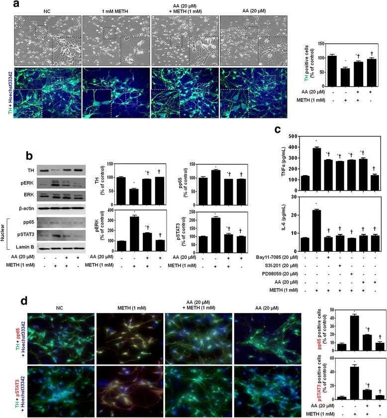
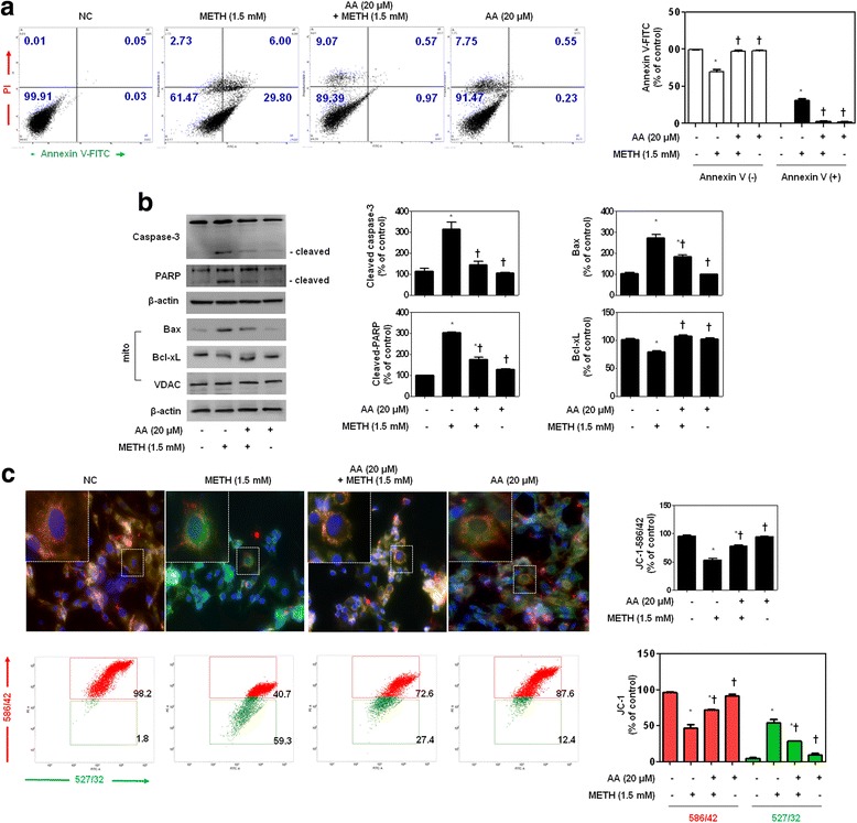
BACKGROUND: Methamphetamine (METH) is a commonly abused drug that may result in neurotoxic effects. Recent studies have suggested that involvement of neuroinflammatory processes in brain dysfunction is induced by misuse of this drug. However, the mechanism underlying METH-induced inflammation and neurotoxicity in neurons is still unclear. In this study, we investigated whether asiatic acid (AA) effected METH-mediated neuroinflammation and neurotoxicity in dopaminergic neuronal cells. And we further determined whether the effect involved in the nuclear factor kappa-light-chain-enhancer of activated B cells (NF-κB) and signal transducer and activator of transcription (STAT)3 and extracellular signal-regulated kinase (ERK) pathway. METHODS: We used the human dopaminergic neuroblastoma SH-SY5Y cell line, murine microglial BV2 cell line, and primary culture of rat embryo mesencephalic neurons. Pro-inflammatory cytokine production was monitored by ELISA and RT/real-time PCR. The cell cycle distribution and mitochondrial membrane integrity was analyzed by flow cytometry. We used immunoblotting, DNA-binding activity, and immunofluorescence staining to analyze the effect of AA on activation of the NF-κB, STAT3, MAPK-ERK, and apoptosis signaling pathways. RESULTS: METH induced TNF receptor (TNFR) expression and led to morphological changes of cells. Additionally, this drug increased pro-inflammatory cytokine (TNFα and IL-6) expression. AA significantly suppressed METH-induced TNFR expression in concentration dependent. Increased secretion of TNFα and IL-6 was inhibited in METH-stimulated neuronal cells by AA administration. AA showed significant protection against METH-induced translocation of NF-κB/STAT3 and ERK phosphorylation. AA inhibited METH-induced proteolytic fragmentation of caspase-3 and PARP. The pro-apoptotic protein Bax was significantly decreased, while the anti-apoptotic protein Bcl-xL was increased by AA treatment in METH-stimulated cells. A similar protective effect of AA on mitochondrial membrane integrity was also confirmed by flow cytometry and immunofluorescence staining. CONCLUSIONS: Based on the literatures and our findings, AA is a promising candidate for an anti-neurotoxic agent, and it can potentially be used for the prevention and treatment of various neurological disorders.
Overview
The claims and statistics of this paper haven’t been checked yet.
Run the check — free, no account needed. It reads the open-access text and checks the sources it cites.
Integrity signals
Registry
Cites
Statistics
Figures
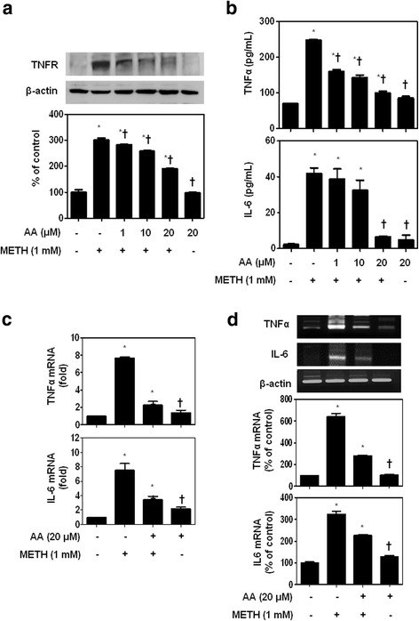
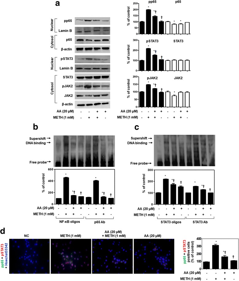
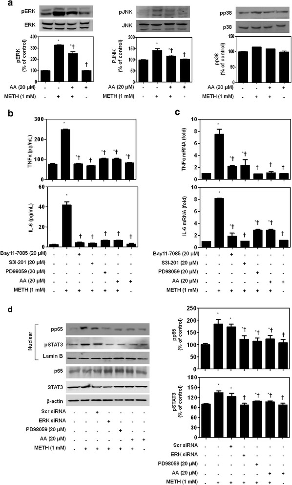
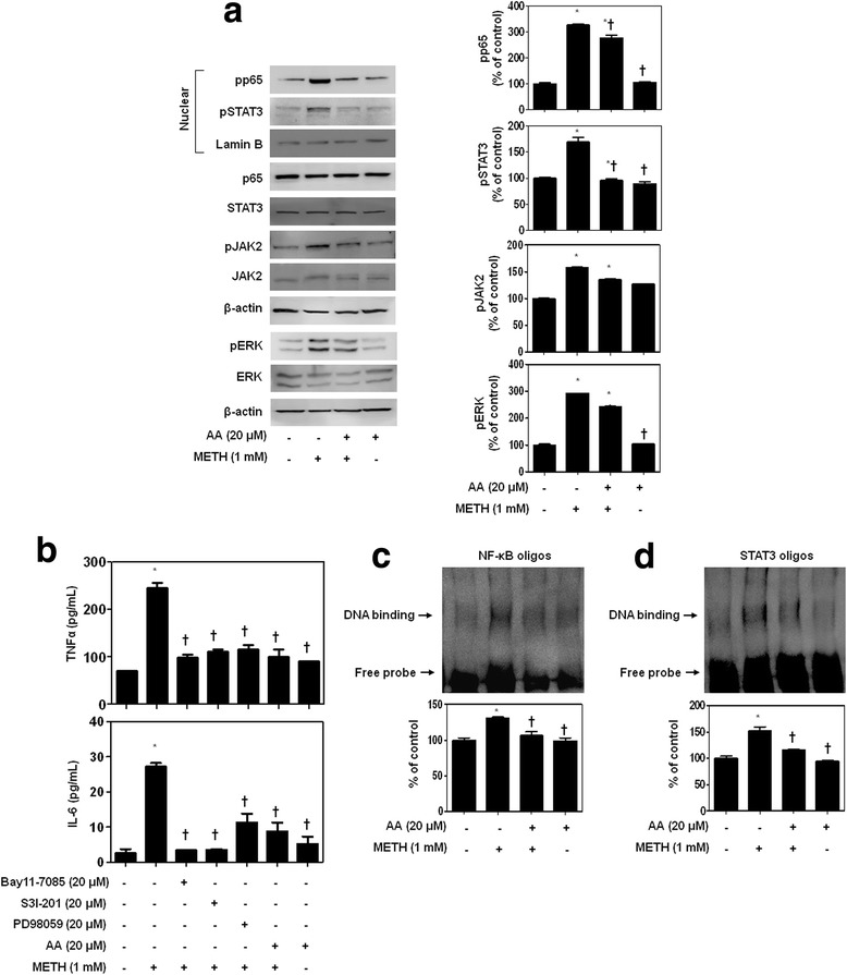
Verification
Claims the paper makes
The claims and statistics of this paper haven’t been checked yet.
Run the check — free, no account needed. It reads the open-access text and checks the sources it cites.
Claims other papers make about this one
Each is checked against this paper’s own text: does it say what the citing paper says it does?
In the citing paper
“Previously, in vitro studies have shown that both CAW whole extract and isolated compounds (detailed in a review [32]) facilitate neurite outgrowth [6,28,106], and elicit neuroprotection [107,108,109,110,111].”
From Centella Asiatica Improves Memory and Promotes Antioxidative Signaling in 5XFAD Mice (2019) · cites this paper as [108]
Self-check
Percentages, sums and reported differences re-computed from the paper’s own numbers.
The self-check needs the paper’s full text.
Citations
Cites
1 retracted
Tsao, S., & Yin, M.-C. (2015) · Journal of Agricultural and Food Chemistry · doi:10.1021/acs.jafc.5b00102 · Cited by 56
RetractedThe pro- and anti-inflammatory properties of the cytokine interleukin-6
Scheller, J., Chalaris, A., Schmidt-Arras, D., & Rose-John, S. (2011) · Biochimica et Biophysica Acta (BBA) - Molecular Cell Research · doi:10.1016/j.bbamcr.2011.01.034 · Cited by 3,212
Molecular basis of long-term plasticity underlying addiction
Nestler, E. J. (2001) · Nature reviews. Neuroscience · doi:10.1038/35053570 · Cited by 1,893
TNF and MAP kinase signalling pathways
Sabio, G., & Davis, R. J. (2014) · Seminars in Immunology · doi:10.1016/j.smim.2014.02.009 · Cited by 738
Neuropeptides and Microglial Activation in Inflammation, Pain, and Neurodegenerative Diseases
Carniglia, L., Ramírez, D., Durand, D., Saba, J., Turati, J., Caruso, C. M., Scimonelli, T. N., & Lasaga, M. (2017) · Mediators of Inflammation · doi:10.1155/2017/5048616 · Cited by 239
Annexin A1: A Central Player in the Anti-Inflammatory and Neuroprotective Role of Microglia
McArthur, S., Cristante, E., Paterno, M., Christian, H., Roncaroli, F., Gillies, G. E., & Solito, E. (2010) · The Journal of Immunology · doi:10.4049/jimmunol.1001095 · Cited by 213
Venkatesan, R., Ji, E., & Kim, S. Y. (2015) · BioMed Research International · doi:10.1155/2015/814068 · Cited by 212
Xu, M.-F., Xiong, Y.-Y., Liu, J.-K., Qian, J.-J., Zhu, L., & Gao, J. (2012) · Acta Pharmacologica Sinica · doi:10.1038/aps.2012.3 · Cited by 156
Krishnamurthy, R. G., Senut, M., Zemke, D., Min, J., Frenkel, M. B., Greenberg, E. J., Yu, S., Ahn, N., Goudreau, J., Kassab, M., et al. (2009) · Journal of Neuroscience Research · doi:10.1002/jnr.22071 · Cited by 155
Gonçalves, J., Baptista, S., Martins, T., Milhazes, N., Borges, F., Ribeiro, C. F., Malva, J. O., & Silva, A. P. (2010) · European Journal of Neuroscience · doi:10.1111/j.1460-9568.2009.07059.x · Cited by 152
Cited by
Sorted by citations
Neurotoxicity of methamphetamine: Main effects and mechanisms
Jayanthi, S., Daiwile, A. P., & Cadet, J. L. (2021) · Experimental Neurology · doi:10.1016/j.expneurol.2021.113795 · Cited by 238
Tian, Z., Ji, X., & Liu, J. (2022) · International Journal of Molecular Sciences · doi:10.3390/ijms23116224 · Cited by 223
Meeran, M. F. N., Goyal, S. N., Suchal, K., Sharma, C., Patil, C. R., & Ojha, S. K. (2018) · Frontiers in Pharmacology · doi:10.3389/fphar.2018.00892 · Cited by 201
Tian, Y., Liu, B., Li, Y., Zhang, Y., Shao, J., Wu, P., Xu, C., Chen, G., & Shi, H. (2022) · Frontiers in Immunology · doi:10.3389/fimmu.2022.839796 · Cited by 143
Paeoniflorin: A neuroprotective monoterpenoid glycoside with promising anti-depressive properties
Wang, X., Feng, S., Wang, Y., Chen, N., Wang, Z., & Zhang, Y. (2021) · Phytomedicine · doi:10.1016/j.phymed.2021.153669 · Cited by 120
Basheer, A. S., Abas, F., Othman, I., & Naidu, R. (2021) · Cancers · doi:10.3390/cancers13164226 · Cited by 120
Methamphetamine-Induced Neuronal Damage: Neurotoxicity and Neuroinflammation
Kim, B., Yun, J., & Park, B. (2020) · Biomolecules & Therapeutics · doi:10.4062/biomolther.2020.044 · Cited by 116
Self-citationMethamphetamine and its immune-modulating effects
Papageorgiou, M., Raza, A., Fraser, S. J., Nurgali, K., & Apostolopoulos, V. (2018) · Maturitas · doi:10.1016/j.maturitas.2018.12.003 · Cited by 90
Centella Asiatica Improves Memory and Promotes Antioxidative Signaling in 5XFAD Mice
Matthews, D. G., Caruso, M., Murchison, C. F., Zhu, J. Y., Wright, K. M., Harris, C. J., Gray, N. E., Quinn, J. F., & Soumyanath, A. (2019) · Antioxidants · doi:10.3390/antiox8120630 · Cited by 86
Tan, S. C., Bhattamisra, S. K., Chellappan, D. K., & Candasamy, M. (2021) · Applied Sciences · doi:10.3390/app11188475 · Cited by 72
Qian, X., Zhang, X., Sun, L., Xing, W.-F., Wang, Y., Sun, S., Ma, M., Cheng, Z., Wu, Z.-D., Chen, X., et al. (2021) · Phytomedicine · doi:10.1016/j.phymed.2021.153696 · Cited by 65
Recent Advances Regarding the Molecular Mechanisms of Triterpenic Acids: A Review (Part I)
Mioc, M., Milan, A., Malita, D. C., Mioc, A., Prodea, A., Racoviceanu, R., Ghiulai, R. M., Cristea, A. M., Căruntu, F. R., & Şoica, C. (2022) · International Journal of Molecular Sciences · doi:10.3390/ijms23147740 · Cited by 66
Fan, J., Chen, Q., Wei, L., Zhou, X., Wang, R., & Zhang, H. (2018) · Drug Design Development and Therapy · doi:10.2147/dddt.s179876 · Cited by 60
Tan, X.-H., Zhang, K.-K., Xu, J., Qu, D., Chen, L., Li, J.-H., Wang, Q., Wang, H., & Xie, X. (2020) · Food and Chemical Toxicology · doi:10.1016/j.fct.2020.111179 · Cited by 60
Cheng, W., Chen, W., Wang, P., & Chu, J. (2018) · Life Sciences · doi:10.1016/j.lfs.2018.07.016 · Cited by 57
Gao, H., Yu, X., Feng, Y., Yu, Y., Hu, H.-B., Zhao, Y., Zhang, J., Liu, K., Zhang, Y., Fu, L., et al. (2023) · Nutrients · doi:10.3390/nu15030502 · Cited by 55
Zhao, Y., Zhao, W., Liu, M., Liu, L., & Wang, Y. (2020) · Oxidative Medicine and Cellular Longevity · doi:10.1155/2020/8874304 · Cited by 53
Meng, X., Zhang, C., Guo, Y., Han, Y., Wang, C., Chu, H., Kong, L., & Ma, H. (2020) · Oxidative Medicine and Cellular Longevity · doi:10.1155/2020/8787156 · Cited by 49
The Crosstalk Between Neurons and Glia in Methamphetamine-Induced Neuroinflammation
Shi, S., Chen, T., & Zhao, M. (2022) · Neurochemical Research · doi:10.1007/s11064-021-03513-9 · Cited by 53
Shen, S., Zhao, J., Dai, Y., Chen, F., Zhang, Z., Yu, J., & Wang, K. (2019) · Toxicology Letters · doi:10.1016/j.toxlet.2019.12.020 · Cited by 46
Meng, Z., Li, H., Si, C., Liu, Y., & Teng, S. (2019) · International Immunopharmacology · doi:10.1016/j.intimp.2019.105712 · Cited by 46
Hanapi, N. A., Arshad, A. S. M., Abdullah, J. M., Muhammad, T. S. T., & Yusof, S. R. (2020) · Journal of Pharmaceutical Sciences · doi:10.1016/j.xphs.2020.09.015 · Cited by 45
Ham, H. J., Lee, Y. S., Yun, J., Son, D. J., Lee, H. P., Han, S., & Hong, J. T. (2020) · Journal of Neuroinflammation · doi:10.1186/s12974-020-02022-w · Cited by 42
The Role of Oxidative Stress and Inflammation in the Pathogenesis and Treatment of Vascular Dementia
Altahrawi, A. Y., James, A. W., & Shah, Z. A. (2025) · Cells · doi:10.3390/cells14080609 · Cited by 41
Targeting STATs in neuroinflammation: The road less traveled!
Nabavi, S. M., Ahmed, T., Nawaz, M., Devi, K. P., Balan, D. J., Pittalà, V., Argüelles, S., Testai, L., Khan, H. Z., Sureda, A., et al. (2018) · Pharmacological Research · doi:10.1016/j.phrs.2018.12.004 · Cited by 43
Ferlazzo, N., Cirmi, S., Maugeri, A., Russo, C., Lombardo, G. E., Gangemi, S., Calapai, G., Mollace, V., & Navarra, M. (2020) · Pharmaceutics · doi:10.3390/pharmaceutics12040326 · Cited by 40
Research Progress on Anti-Inflammatory Effects and Related Mechanisms of Astragalin
Ruan, J., Shi, Z., Cao, X., Dang, Z., Zhang, Q., Zhang, W., Wu, L., Zhang, Y., & Wang, T. (2024) · International Journal of Molecular Sciences · doi:10.3390/ijms25084476 · Cited by 39
Hong, G., Zhou, L., Han, X., Sun, P., Chen, Z., He, W., Tickner, J. C., Chen, L., Shi, X., & Xu, J. (2020) · Frontiers in Pharmacology · doi:10.3389/fphar.2020.00331 · Cited by 36
Islamie, R., Myint, S. L. L., Rojanaratha, T., Ritthidej, G. C., Wanakhachornkrai, O., Wattanathamsan, O., & Rodsiri, R. (2023) · BMC Complementary Medicine and Therapies · doi:10.1186/s12906-023-04125-2 · Cited by 35
Zhang, K.-K., Wang, H., Qu, D., Chen, L., Wang, L., Li, J.-H., Liu, J.-L., Xu, L., Yoshida, J. S., Xu, J., et al. (2021) · Frontiers in Pharmacology · doi:10.3389/fphar.2021.641917 · Cited by 35
Park, J., Jang, K. M., & Park, K. (2020) · International Journal of Molecular Sciences · doi:10.3390/ijms21124319 · Cited by 32
Self-citationCao, S., Zhu, C. X., Feng, J., Zhu, L., Yin, J., Xu, Y., Yang, H., Huang, Y., & Zhang, Q. (2019) · Helicobacter · doi:10.1111/hel.12677 · Cited by 30
Zhang, K.-K., Chen, L., Li, J.-H., Liu, J.-L., Wang, L., Xu, L., Yang, J.-Z., Li, X., Xie, X., & Wang, Q. (2022) · Frontiers in Microbiology · doi:10.3389/fmicb.2022.755189 · Cited by 30
Mushtaq, Z., Imran, M., Hussain, M., Saeed, F., Imran, A., Umar, M., Abdelgawad, M. A., El-Ghorab, A. H., Ahmed, A., Alsagaby, S. A., et al. (2023) · International Journal of Food Properties · doi:10.1080/10942912.2023.2209702 · Cited by 33
Jiang, L., Zhu, R., Bu, Q., Li, Y., Shao, X., Gu, H., Kong, J., Luo, L., Long, H., Guo, W., et al. (2018) · Neurotherapeutics · doi:10.1007/s13311-018-0613-8 · Cited by 29
Methamphetamine-induced dopaminergic neurotoxicity as a model of Parkinson’s disease
Shin, E., Jeong, J. H., Hwang, Y., Sharma, N., Dang, D., Nguyen, B.-T., Nah, S.-Y., Jang, C., Bing, G., Nabeshima, T., et al. (2021) · Archives of Pharmacal Research · doi:10.1007/s12272-021-01341-7 · Cited by 31
Shi, R., Tan, W., Jin, H., Chan, S. I., Li, W., Lei, S. S., Cui, G., Wang, Y., Yang, D., & Zhong, Z. (2025) · Advanced Science · doi:10.1002/advs.202505921 · Cited by 30
Asiatic acid and its derivatives: Pharmacological insights and applications
Liang, C., Ma, Y., Ding, M., Gao, F., Yu, K., Wang, S., Qu, Y., Hu, X., & Li, D. (2025) · European Journal of Medicinal Chemistry · doi:10.1016/j.ejmech.2025.117429 · Cited by 30
Jones, B., Balasubramaniam, M., Lebowitz, J. J., Taylor, A., Villalta, F., Khoshbouei, H., Grueter, C. A., Grueter, B. A., Dash, C., & Pandhare, J. (2021) · Scientific Reports · doi:10.1038/s41598-020-80917-7 · Cited by 27
Park, J., Jang, K. M., & Park, K. (2022) · International Journal of Molecular Sciences · doi:10.3390/ijms232315255 · Cited by 27
Self-citationLee, H., Kim, E., & Jeong, G. (2020) · International Journal of Molecular Sciences · doi:10.3390/ijms21051617 · Cited by 26
Elevated serum levels of TNF‐α, IL‐6, and IL‐18 in chronic methamphetamine users
Luo, Y., He, H., Ou, Y., Zhou, Y., & Fan, N. (2021) · Human Psychopharmacology Clinical and Experimental · doi:10.1002/hup.2810 · Cited by 28
Zhang, M., Wáng, Y., Wong, R. M. S., Yung, K. K. L., & Li, R. (2021) · NeuroToxicology · doi:10.1016/j.neuro.2021.11.012 · Cited by 26
Xie, X., Zhou, W., Zhang, K.-K., Chen, L., & Wang, Q. (2018) · Frontiers in Neuroscience · doi:10.3389/fnins.2018.00802 · Cited by 26
Wu, L., Wang, L., Du, Y., Zhang, Y., & Ren, J. T. (2024) · Acta Pharmacologica Sinica · doi:10.1038/s41401-024-01367-9 · Cited by 25
Tooth Loss Induces Memory Impairment and Gliosis in App Knock-In Mouse Models of Alzheimer’s Disease
Taslima, F., Jung, C., Zhou, C., Abdelhamid, M., Abdullah, M., Goto, T., Saito, T., Saido, T. C., & Michikawa, M. (2021) · Journal of Alzheimer s Disease · doi:10.3233/jad-201055 · Cited by 26
Du, X., Liu, M., Tai, W., Yu, H., Hao, X., Loor, J. J., Jiang, Q., Fang, Z., Gao, X., Fan, M., et al. (2022) · Journal of Dairy Science · doi:10.3168/jds.2022-22009 · Cited by 27
Yu, G., Song, Y., Xie, C.-W., Tao, L., Wan, F., Jiang, L., Wang, J., & Tang, J. (2019) · Toxicology and Applied Pharmacology · doi:10.1016/j.taap.2019.03.019 · Cited by 23
Role of Microglia in Psychostimulant Addiction
da Silva, M. C. M., Iglesias, L. P., Candelario‐Jalil, E., Khoshbouei, H., Moreira, F. A., & de Oliveira, A. C. P. (2022) · Current Neuropharmacology · doi:10.2174/1570159x21666221208142151 · Cited by 26
Huang, J., Tu, T., Wang, W., Zhou, G., Zhang, W., Wu, X., & Liu, W. (2019) · Photochemistry and Photobiology · doi:10.1111/php.13160 · Cited by 23
Pentacyclic triterpenoids: diversity, distribution and their propitious pharmacological potential
Malik, J., & Mandal, S. C. (2024) · Phytochemistry Reviews · doi:10.1007/s11101-024-10056-8 · Cited by 23
Gou, H., Sun, D., Hao, L., An, M., Xie, B., Cong, B., Ma, C., & Wen, D. (2020) · NeuroToxicology · doi:10.1016/j.neuro.2020.09.001 · Cited by 22
Pantia, S., Kangsamaksin, T., Janvilisri, T., & Komyod, W. (2023) · Pharmaceuticals · doi:10.3390/ph16060902 · Cited by 23
Lü, C.-W., Lin, T., Wang, S., & Huang, S.-K. (2019) · European Journal of Pharmacology · doi:10.1016/j.ejphar.2019.172781 · Cited by 22
Re, G.-F., Li, H., Yang, J.-Q., Li, Y., Zhang, Z., Wu, X., Zhou, R., Kong, D., Luo, H., Kuang, Y., et al. (2022) · Frontiers in Molecular Neuroscience · doi:10.3389/fnmol.2022.955799 · Cited by 22
Qing, X., Dou, R., Wang, P., Zhou, M., Cao, C., Zhang, H., Qiu, G., Yang, Z., Zhang, J., Liu, H., et al. (2023) · Journal of Nanobiotechnology · doi:10.1186/s12951-023-02230-5 · Cited by 21
Wu, Z.-C., Song, Y., Wang, Y., Zhou, H., Chen, L., Zhan, Y., Li, T., Xie, G., & Wu, H. (2024) · Cell Biochemistry and Function · doi:10.1002/cbf.4056 · Cited by 21
Fan, R., Shen, Y., Li, X., Luo, H., Zhang, P., Liu, Y., Si, Z., Zhou, W., & Liu, Y. (2022) · Drug and Alcohol Dependence · doi:10.1016/j.drugalcdep.2022.109537 · Cited by 20
RetractedLiang, M., Zhu, L., Wang, R., Su, H., Ma, D., Wang, H., & Chen, T. (2022) · Frontiers in Cellular Neuroscience · doi:10.3389/fncel.2022.892757 · Cited by 18
Hwang, J. S., Cha, E.-H., Ha, E., Park, B., & Seo, J. H. (2020) · Life Sciences · doi:10.1016/j.lfs.2020.117917 · Cited by 18
Self-citationZhu, Y., Lu, Y., Yuan, L., Ling, W., Jiang, X., Chen, S., & Hu, B. (2021) · International Journal of Medical Microbiology · doi:10.1016/j.ijmm.2021.151515 · Cited by 17
Natural Products in Modulating Methamphetamine-Induced Neuronal Apoptosis
Zeng, Y., Chen, Y., Zhang, S., Ren, H., Xia, J., Liu, M., Shan, B., & Ren, Y. (2022) · Frontiers in Pharmacology · doi:10.3389/fphar.2021.805991 · Cited by 17
Apigenin ameliorates imiquimod-induced psoriasis in C57BL/6J mice by inactivating STAT3 and NF-κB
Meng, X., Zheng, S., Yin, Z., Wang, X., Yang, D., Zou, T., Li, H., Chen, Y., Liao, C., Xie, Z., et al. (2023) · Food Science and Human Wellness · doi:10.26599/fshw.2022.9250018 · Cited by 17
Wang, T., Xie, Y.-Q., Miao, G., Cui, H., Liu, K., Li, Y., Li, Y., & Zhao, J. (2020) · Life Sciences · doi:10.1016/j.lfs.2020.117421 · Cited by 17
Wei, J., Mou, C., Bao, Y., Xie, Y., Jin, H., Shen, H., Zhou, W., Zhang, J., He, S., Chen, B., et al. (2021) · Journal of Functional Foods · doi:10.1016/j.jff.2021.104713 · Cited by 18
Yan, D., Gao, L., Lang, J., Gao, X., & Ma, H. (2021) · Environmental Toxicology · doi:10.1002/tox.23294 · Cited by 16
Sharma, N., Shin, E., Pham, D. T., Sharma, G., Dang, D.-K., Duong, C. X., Kang, S. W., Nah, S.-Y., Jang, C., Lei, X. G., et al. (2021) · Food and Chemical Toxicology · doi:10.1016/j.fct.2021.112313 · Cited by 16
MicroRNAs in Methamphetamine-Induced Neurotoxicity and Addiction
Bi, D., Zhang, Z., Zhou, H., Zhang, X., Niu, S., Yan, X., & Yan, J. (2022) · Frontiers in Pharmacology · doi:10.3389/fphar.2022.875666 · Cited by 16
Hippocampal blood–brain barrier of methamphetamine self‐administering HIV‐1 transgenic rats
Ohene‐Nyako, M., Persons, A. L., & Napier, T. C. (2020) · European Journal of Neuroscience · doi:10.1111/ejn.14925 · Cited by 16
Bai, Y., Guo, N., Xu, Z., Chen, Y., Zhang, W., Chen, Q., & Bi, Z. (2022) · Molecular Medicine Reports · doi:10.3892/mmr.2022.12917 · Cited by 15
Yang, J., Sun, P., Xu, X., Liu, X., Lan, L., Yi, M., Xiao, C., Ni, R., & Fan, Y. (2023) · Aging and Disease · doi:10.14336/ad.2023.0219 · Cited by 15
Neuroprotective prospectives of triterpenoids
Bankar, A. A., Nagulwar, V. P., Kotagale, N. R., & Inamdar, N. (2024) · Exploration of neuroscience · doi:10.37349/en.2024.00047 · Cited by 17
Yuan, Z., Yang, M., Liang, Z., Yang, C., Kong, X., Wu, Y., Wang, S., Fan, H. J., Ning, C., Xiao, W., et al. (2022) · Journal of Ethnopharmacology · doi:10.1016/j.jep.2022.116028 · Cited by 15
Translocator Protein 18 kDa (TSPO) as a Novel Therapeutic Target for Chronic Pain
Liu, J., Huang, J., Zhang, Z., Zhang, R., Zhang, Z., Liu, Y., & Ma, B. (2022) · Neural Plasticity · doi:10.1155/2022/8057854 · Cited by 16
Lokman, M. S., Kassab, R. B., Salem, F. A. M., Elshopakey, G. E., Hussein, A., Aldarmahi, A. A., Theyab, A., Alzahrani, K. J., Hassan, K. E., Alsharif, K. F., et al. (2024) · Bioscience Reports · doi:10.1042/bsr20232004 · Cited by 14
Oladapo, A., Kannan, M., Deshetty, U. M., Singh, S., Buch, S. J., & Periyasamy, P. (2025) · Redox Biology · doi:10.1016/j.redox.2025.103517 · Cited by 15
Wang, Y., Zhang, H., Wang, J., Yu, M., Zhang, Q., Yan, S., You, D., Shi, L., Zhang, L., Wang, L., et al. (2021) · Pharmaceutical Biology · doi:10.1080/13880209.2021.1900879 · Cited by 12
Zhang, T., Li, S., Li, Y., Cao, J., Xue, H. Y., Wang, C., & Jin, C. (2022) · Molecules · doi:10.3390/molecules27092946 · Cited by 13
Baba, K., Hiramatsu, R., Suradej, B., Tanigaki, R., Koeda, S., Waku, T., & Kataoka, T. (2018) · Biological and Pharmaceutical Bulletin · doi:10.1248/bpb.b18-00276 · Cited by 14
Shi, S., Sun, Y., Zan, G., & Zhao, M. (2025) · Journal of Neuroinflammation · doi:10.1186/s12974-025-03372-z · Cited by 19
Ouyang, W., Zhu, C., Li, Y., Huang, H., Li, F., & Li, L. (2025) · Ecotoxicology and Environmental Safety · doi:10.1016/j.ecoenv.2025.118225 · Cited by 12
Lee, H., & Jeong, G. (2021) · The American Journal of Chinese Medicine · doi:10.1142/s0192415x21500051 · Cited by 12
Asiatic acid alleviates Ang-II induced cardiac hypertrophy and fibrosis via miR-126/PIK3R2 signaling
Li, H., Tian, X., Ruan, Y., Xing, J., & Meng, Z. (2021) · Nutrition & Metabolism · doi:10.1186/s12986-021-00596-7 · Cited by 14
Varada, S., Chamberlin, S. R., Bui, L., Brandes, M. S., Gladen-Kolarsky, N., Harris, C., Hack, W. R., Neff, C. J., Brumbach, B. H., Soumyanath, A., et al. (2025) · Nutrients · doi:10.3390/nu17040729 · Cited by 15
Jiang, T., Xu, J., Yang, L., Chen, X., & Li, Y. (2022) · Mediators of Inflammation · doi:10.1155/2022/1708030 · Cited by 11
EGFR mediated the renal cell apoptosis in rhabdomyolysis-induced model via upregulation of autophagy
Sun, T., Wu, D., Deng, Y., & Zhang, D. (2022) · Life Sciences · doi:10.1016/j.lfs.2022.121050 · Cited by 11
Ham, H. J., Lee, Y.-S., Yun, J., Han, S., Son, D.-J., & Hong, J. (2020) · Behavioural Brain Research · doi:10.1016/j.bbr.2020.112518 · Cited by 12
Atractylodin: An Alkyne Compound with Anticancer Potential
Zhong, W., & Zhang, Q. (2024) · The American Journal of Chinese Medicine · doi:10.1142/s0192415x24500551 · Cited by 10
Oladapo, A., Deshetty, U. M., Callen, S. E., Buch, S. J., & Periyasamy, P. (2025) · International Journal of Molecular Sciences · doi:10.3390/ijms26020649 · Cited by 11
Gynura bicoloraqueous extract attenuated H2O2induced injury in PC12 cells
Yang, Y., Wu, W., Mong, M., & Wang, Z. (2019) · Biomedicine · doi:10.1051/bmdcn/2019090212 · Cited by 10
Asiatic acid inhibits osteosarcoma cell migration and invasion via the AKT / Sp1 / MMP1 axis
Law, Y., Lee, H., Lin, C., Chen, P., Wang, P., Hsieh, Y., & Chen, C. (2024) · Environmental Toxicology · doi:10.1002/tox.24246 · Cited by 10
Yang, L., Guo, Y., Huang, M., Wu, X., Li, X., Chen, G., Li, Y., & Bai, J. (2018) · Frontiers in Neurology · doi:10.3389/fneur.2018.00049 · Cited by 10
Ding, J., Wang, Y., Huang, J. A., Lian, Y., Meng, Y., Li, C., He, Y., & Qiu, P. (2020) · Neuroreport · doi:10.1097/wnr.0000000000001495 · Cited by 10
Methamphetamine: Mechanism of Action and Chinese Herbal Medicine Treatment for Its Addiction
Zeng, R., Pu, H., Zhang, X., Yao, M., & Sun, Q. (2023) · Chinese Journal of Integrative Medicine · doi:10.1007/s11655-023-3635-y · Cited by 10
5-HTP decreases goat mammary epithelial cells apoptosis through MAPK/ERK/Bcl-3 pathway
Zhao, H., Chen, S., Hu, K., Zhang, Z., Yan, X., Gao, H., Du, W., & Zheng, H. (2020) · Gene · doi:10.1016/j.gene.2020.145240 · Cited by 10
Qiu, W., Jiang, W., Su, Y., Huang, H., Ye, J., Wang, R., Tang, Z., & Su, R. (2024) · Journal of Animal Physiology and Animal Nutrition · doi:10.1111/jpn.14070 · Cited by 9
Dong, Y., Cao, K., Xiang, J., Qi, X.-L., Xiao, Y., Yu, W., He, Y., Hong, W., & Guan, Z.-Z. (2023) · Neuroscience · doi:10.1016/j.neuroscience.2023.04.023 · Cited by 10
Moon, H.-R., & Yun, J. (2023) · Journal of Medicinal Food · doi:10.1089/jmf.2023.k.0022 · Cited by 9
Asiatic acid prevents glucocorticoid-induced femoral head osteonecrosis via PI3K/AKT pathway
Lin, C., Jiang, H., Lou, C., Wang, W., Cai, T., Lin, Z., Jiang, L., Lin, S., Xue, X., & Pan, X. (2024) · International Immunopharmacology · doi:10.1016/j.intimp.2024.111758 · Cited by 9
Liu, Z., Yoon, C., Lee, H., Yim, J. H., Yim, J. H., Kim, T. K., Oh, H., & Lee, D. (2024) · Biochemical Pharmacology · doi:10.1016/j.bcp.2024.116654 · Cited by 9
Metadata
- Authors
- Park, J.-H., Seo, Y. H., Jang, J.-H., Jeong, C.-H., Lee, S., & Park, B.
- Affiliations
- Keimyung University (KR)
- Corresponding author
- Park, Byoungduck
- Venue
- Journal of Neuroinflammation
- Published
- 2017-12
- Volume
- 14
- Issue
- 1
- Pages
- 240-240
- ISSN
- 1742-2094 (ISSN-L)
- PMC
- PMC5725763
- OpenAlex
- W2771852048
- Cited by
- 175
- References
- 48
- Citations by year
- 2026: 20 · 2025: 30 · 2024: 25 · 2023: 17 · 2022: 23 · 2021: 19 · 2020: 22 · 2019: 9 · 2018: 10
- FWCI
- 5.59
- Type
- journal article
- Language
- en
- Open access
- gold
- doi.org
- Licence
- cc-by
- Locations
- Journal of Neuroinflammation (publishedVersion, cc-by)
- Europe PMC (PubMed Central) (publishedVersion, cc-by)
- PubMed Central (publishedVersion, cc-by)
- PubMed (publishedVersion)
- DOAJ (DOAJ: Directory of Open Access Journals) (submittedVersion, cc-by-sa)
- Funders
- Korea Brain Research Institute
- National Research Foundation (NRF-2016R1A6A1A03011325)
- Korea University
- National Research Foundation of Korea (NRF‐2016R1A6A1A03011325, - 2016R1A6A1A03011325)
- Topics
- Life Sciences › Neuroscience › Neurology › Neuroinflammation and Neurodegeneration Mechanisms
- Life Sciences › Biochemistry, Genetics and Molecular Biology › Cancer Research › NF-κB Signaling Pathways
- Life Sciences › Neuroscience › Biological Psychiatry › Tryptophan and brain disorders
- Keywords
- asiatic acid, methamphetamine, neuroinflammation, neurotoxicity, dopaminergic neurons, NF-κB, STAT3, MAPK/ERK pathway, apoptosis, mitochondrial membrane integrity, TNF receptor, inflammatory cytokines, caspase-3, neuroprotection
- MeSH terms
- Central Nervous System Stimulants, Animals, Cell Line, Humans, Inflammation, Methamphetamine, Mitochondria, Neurons, Signal Transduction, NF-kappa B, Apoptosis, Neuroprotective Agents, Extracellular Signal-Regulated MAP Kinases, STAT3 Transcription Factor, Mice, Rats, Pentacyclic Triterpenes
About these checks
Registry, retraction, Cites and statistics results are computed, not judged, with one exception: a printed reference that no index lists is read once by a model before it counts as a citation error. Claims are extracted by a model and kept only when their quote appears verbatim in the text; each cited source is then checked, with a model’s help, for whether it says what the claim relies on. Those verdicts can be wrong — read the claim’s quote and the cited source before relying on one.
Statistics and arithmetic are recomputed, not judged.
Confidence bands are shown only for fields calibrated on our annotated test set. None is calibrated yet, so no band is shown.
What counts as a claim ›How we measure confidence ›How we check this ›
Are you an author, or do you see a problem with this page? Request a review
