

The DOI matches its Crossref record; 43 of the 43 references it cites were found in a registry or an index.
Porphyromonas gingivalis lipopolysaccharide induces cognitive dysfunction, mediated by neuronal inflammation via activation of the TLR4 signaling pathway in C57BL/6 mice
Open accessAbstract
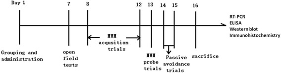
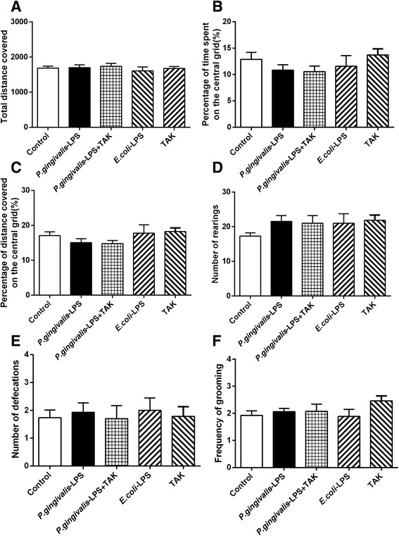
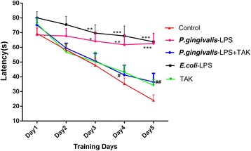
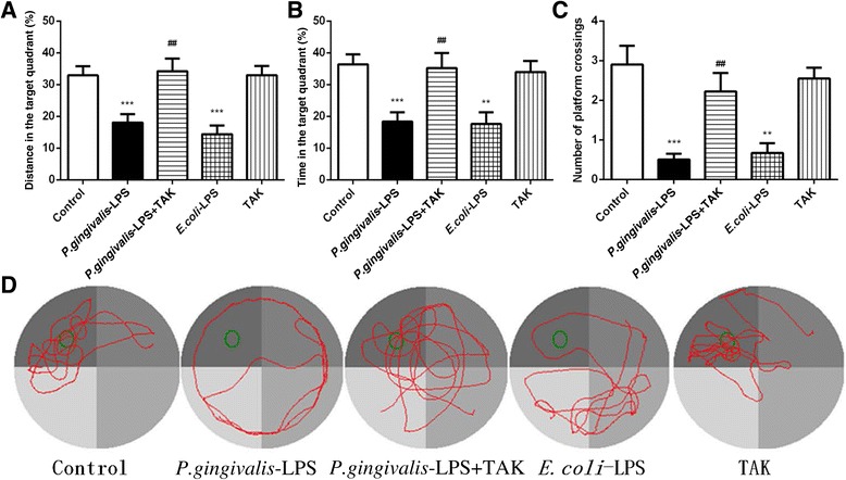
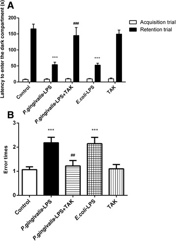
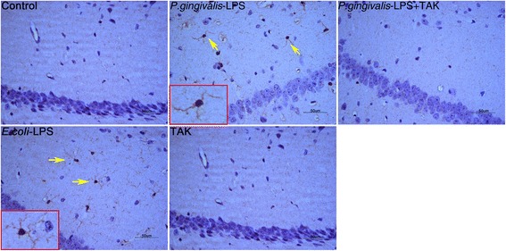
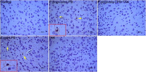
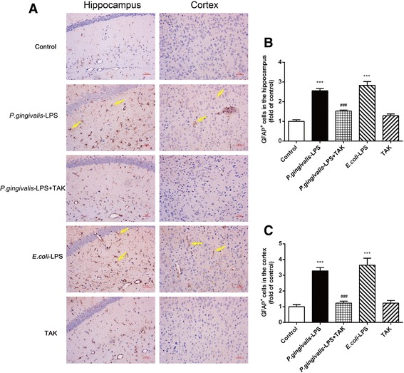
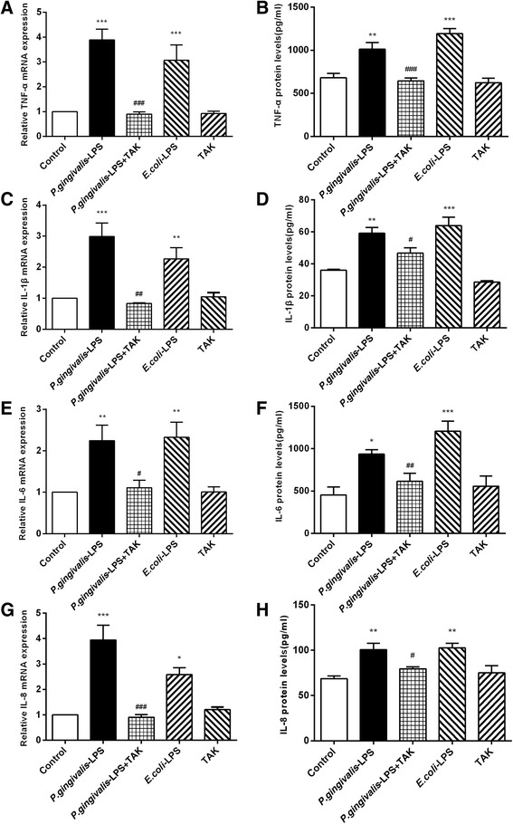
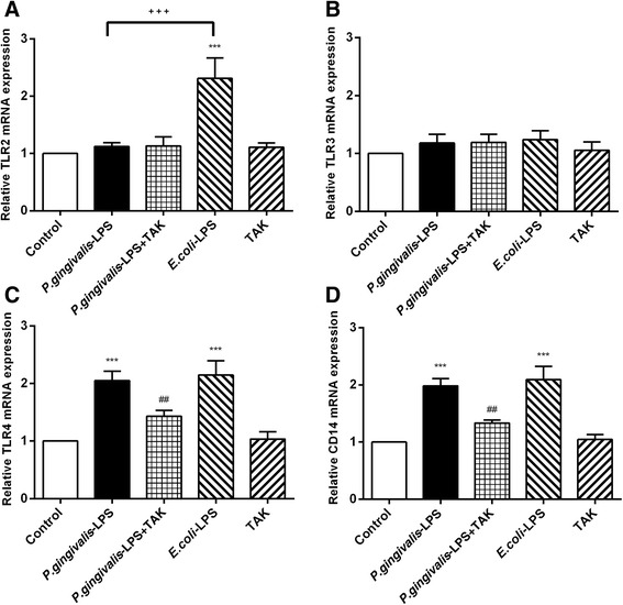
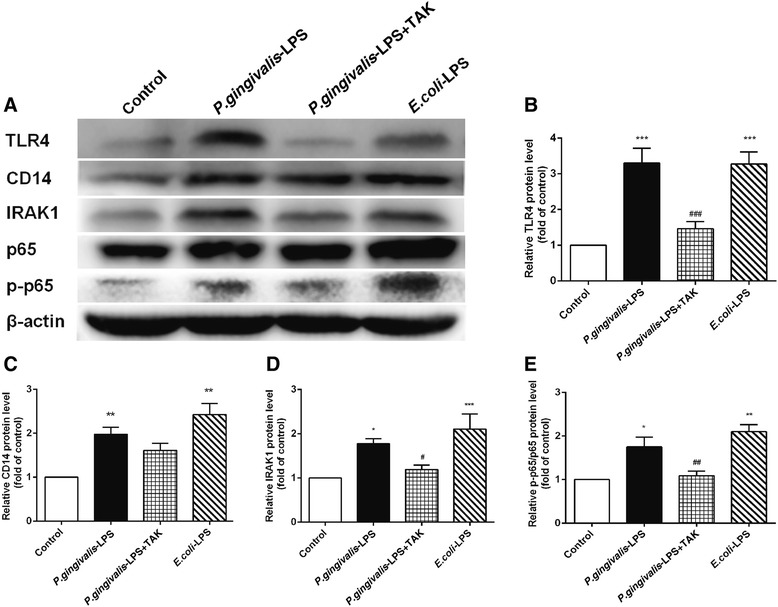
BACKGROUND: Porphyromonas gingivalis lipopolysaccharide (P. gingivalis-LPS) is one of the major pathogenic factors of chronic periodontitis (CP). Few reports on the correlation between P. gingivalis-LPS and cognitive function exist. Thus, the present study aimed to investigate the effects of P. gingivalis-LPS on cognitive function and the associated underlying mechanism in C57BL/6 mice. METHODS: ). After 7 days, behavioral alterations were assessed with the open field test (OFT), Morris water maze (MWM) test, and passive avoidance test (PAT). The activation of astrocytes and microglia in the cerebral cortex and hippocampus of mice was observed by immunohistochemistry. The expression of inflammatory cytokines (TNF-α, IL-1β, IL-6, and IL-8), TLRs (TLR2, TLR3, and TLR4), and CD14 and the activation of the NF-κB signaling pathway (IRAK1, p65, and p-p65) in the cerebral cortex of the mice were evaluated by RT-PCR, ELISA, and western blot. RESULTS: The OFT showed that P. gingivalis-LPS did not affect the initiative and activity of mice. Administration of P. gingivalis-LPS significantly impaired spatial learning and memory during the MWM test and attenuated the ability of passive avoidance learning during the PAT. Both astrocytes and microglia were activated in the cortex and hippocampus. The messenger RNA (mRNA) and protein expression of inflammatory cytokines (TNF-α, IL-1β, IL-6, and IL-8) was upregulated by P. gingivalis-LPS in the cortex. In addition, the TLR4/NF-κB signaling pathway was activated (TLR4, CD14, IRAK1, and p-p65). These effects were effectively alleviated by TAK-242. CONCLUSIONS: Administration of P. gingivalis-LPS can lead to learning and memory impairment in C57BL/6 mice. This impairment is mediated by activation of the TLR4 signaling pathway. Our study suggests that P. gingivalis-LPS-induced neuroinflammation plays an important role in cognitive impairment. It also reveals that endotoxins of periodontal pathogens could represent a risk factor for cognitive disorders.
Overview
The claims and statistics of this paper haven’t been checked yet.
Run the check — free, no account needed. It reads the open-access text and checks the sources it cites.
Integrity signals
Registry
Cites
Statistics
Figures
Verification
Claims the paper makes
The claims and statistics of this paper haven’t been checked yet.
Run the check — free, no account needed. It reads the open-access text and checks the sources it cites.
Claims other papers make about this one
Each is checked against this paper’s own text: does it say what the citing paper says it does?
The source backs part of the claim, not all of it — or backs it for a different population, condition or comparison.
In the citing paper
“Chronic oral application of P. gingivalis or injection of P. gingivalis LPS results in brain inflammation and neurodegeneration in wild-type mice [70, 89].”
From The endotoxin hypothesis of neurodegeneration (2019) · cites this paper as [89]
Experimental — accuracy not yet measured
Self-check
Percentages, sums and reported differences re-computed from the paper’s own numbers.
The self-check needs the paper’s full text.
Citations
Cites
Developments of a water-maze procedure for studying spatial learning in the rat
Morris, R. (1984) · Journal of Neuroscience Methods · doi:10.1016/0165-0270(84)90007-4 · Cited by 7,180
Neuroinflammation in Alzheimer's disease
Heneka, M. T., Carson, M. J., Khoury, J. E., Landreth, G. E., Brosseron, F., Feinstein, D. L., Jacobs, A. H., Wyss-Coray, T., Vitorica, J., Ransohoff, R. M., et al. (2015) · The Lancet Neurology · doi:10.1016/s1474-4422(15)70016-5 · Cited by 6,226
Toll-like Receptor-4 Mediates Lipopolysaccharide-induced Signal Transduction
Chow, J. C., Young, D. W., Golenbock, D. T., Christ, W. J., & Gusovsky, F. (1999) · Journal of Biological Chemistry · doi:10.1074/jbc.274.16.10689 · Cited by 1,924
Integrated Systems Approach Identifies Genetic Nodes and Networks in Late-Onset Alzheimer’s Disease
Zhang, B., Gaiteri, C., Bodea, L.-G., Wang, Z., McElwee, J., Podtelezhnikov, A. A., Zhang, C., Xie, T., Tran, L., Dobrin, R., et al. (2013) · Cell · doi:10.1016/j.cell.2013.03.030 · Cited by 1,657
Diabetes mellitus and periodontitis: a tale of two common interrelated diseases
Lalla, E., & Papapanou, P. N. (2011) · Nature Reviews Endocrinology · doi:10.1038/nrendo.2011.106 · Cited by 1,063
Neuroinflammation: The role and consequences
Lyman, M., Lloyd, D. G., Ji, X., Vizcaychipi, M. P., & Ma, D. (2013) · Neuroscience Research · doi:10.1016/j.neures.2013.10.004 · Cited by 776
Hirschfeld, M. J., Weis, J. J., Toshchakov, V. Y., Salkowski, C. A., Cody, M. J., Ward, D. C., Qureshi, N., Michalek, S. M., & Vogel, S. N. (2001) · Infection and Immunity · doi:10.1128/iai.69.3.1477-1482.2001 · Cited by 655
Poole, S., Singhrao, S. K., Kesavalu, L. N., Curtis, M. A., & Crean, S. (2013) · Journal of Alzheimer s Disease · doi:10.3233/jad-121918 · Cited by 610
Michelucci, A., Heurtaux, T., Grandbarbe, L., Morga, E., & Heuschling, P. (2009) · Journal of Neuroimmunology · doi:10.1016/j.jneuroim.2009.02.003 · Cited by 411
Microglia: activation and their significance in the central nervous system
Nakajima, K., & Kohsaka, S. (2001) · Journal of Biochemistry · doi:10.1093/oxfordjournals.jbchem.a002969 · Cited by 371
Cited by
Sorted by citations
Items 201–298 of 298
Yu, W., Lu, L., Ji, X., Qian, Q., Lin, X., & Wang, H. (2021) · Journal of Alzheimer s Disease · doi:10.3233/jad-215143 · Cited by 7
[Research progress of correlation between periodontal pathogens and systemic diseases].
Du, Q., & Ma, X. (2020) · PubMed Central · doi:10.12122/j.issn.1673-4254.2020.05.24 · Cited by 7
Mechanism and effects of artesunate on the liver function of rats with type 1 diabetic periodontitis
Bai, S., Zhou, J., Nong, X., Shi, R., Yuan, Ž., Ma, C., & Li, J. (2022) · Canadian Journal of Physiology and Pharmacology · doi:10.1139/cjpp-2021-0665 · Cited by 7
Dumitru, C. N., Dumitru, A. O., Popa, G. V., Marcu, T., Ursu, M., Nechita, A., & Matei, N. M. (2026) · Dentistry Journal · doi:10.3390/dj14050289 · Cited by 6
“…toward the pro-inflammatory M1 phenotype: circulating LPS engaging microglial TLR4 [80]; pro-inflammatory cytokines traversing a disrupted BBB [77,81]; gingipains and other bacterial virulence factors potentially reaching the brain parenchyma [32,45]; and loss of butyrate-mediated HDAC…”
Vo, T. T. T., Tsai, M.-H., Cheng, C.-Y., Wang, Y.-L., Lee, W.-J., & Lee, I. (2025) · Biochemical Pharmacology · doi:10.1016/j.bcp.2025.117362 · Cited by 7
Ameliorative Effect of Medicarpin on Scopolamine-Induced Cognitive Impairment in Mice
Oh, J., Park, J., Mun, S., Yee, S., & Kim, H. (2023) · Processes · doi:10.3390/pr11020385 · Cited by 6
SorCS2 is involved in promoting periodontitis‐induced depression‐like behaviour in mice
Li, Y., Yang, Y., Guan, X., Liu, Z., Pan, L., Wang, Y., Jia, X., Yang, J., & Hou, T. (2024) · Oral Diseases · doi:10.1111/odi.14944 · Cited by 6
Peri-implantitis with a potential axis to brain inflammation: an inferential review
Tessarin, G. W. L., Toro, L. F., Pereira, R. F., dos Santos, R. M., & Azevedo, R. G. (2024) · Odontology · doi:10.1007/s10266-024-00936-y · Cited by 7
Kordi, R., Andrews, T. J., & Hicar, M. D. (2025) · Virology · doi:10.1016/j.virol.2025.110523 · Cited by 6
Ishida, E., Furusho, H., Renn, T., Shiba, F., Chang, H., Oue, H., Terayama, R., Ago, Y., Tsuga, K., & Miyauchi, M. (2023) · Frontiers in Pediatrics · doi:10.3389/fped.2023.1203894 · Cited by 6
Liccardo, D., Valletta, A., Spagnuolo, G., Vinciguerra, C., Lauria, M. R., Perrotta, A., Giudice, C. D., Luca, F. D., Rengo, G., Rengo, S., et al. (2024) · Journal of Biotechnology · doi:10.1016/j.jbiotec.2024.07.009 · Cited by 6
Karakaya, E., Abdul, Y., Chowdhury, N., Wellslager, B., Jamil, S., Albayram, Ö., Yilmaz, Ö., & Ergul, A. (2022) · Canadian Journal of Physiology and Pharmacology · doi:10.1139/cjpp-2022-0035 · Cited by 6
Li, Q., Zhou, D.-N., Tu, Y.-Q., Wu, X., Pei, D., & Xiong, Y. (2022) · Experimental and Therapeutic Medicine · doi:10.3892/etm.2022.11405 · Cited by 6
PORPHYROMONAS GINGIVALIS DAN PATOGENESIS DISFUNGSI KOGNITIF: ANALISIS PERAN SITOKIN NEUROINFLAMASI
Putri, C. F., & Bachtiar, E. W. (2020) · Cakradonya Dental Journal · doi:10.24815/cdj.v12i1.17068 · Cited by 6
Oue, K., Yamawaki, Y., Ouhara, K., Imado, E., Tamura, T., Doi, M., Shimizu, Y., Yoshida, M., Mizuno, N., Morioka, N., et al. (2024) · Journal of Oral Microbiology · doi:10.1080/20002297.2024.2419155 · Cited by 5
Sung, W., Yeh, T.-M., & Shih, W. (2024) · BMC Complementary Medicine and Therapies · doi:10.1186/s12906-024-04682-0 · Cited by 5
Sun, Q., & Li, M. (2025) · Clinical Epidemiology and Global Health · doi:10.1016/j.cegh.2025.102020 · Cited by 4
Effects of Experimental Periodontitis and Periodontal Treatment on Alzheimer‐Like Pathology in Rats
Yavuz, S., Elibol, B., Demir, E., Kinsiz, B., Toluk, Ö., & Günpınar, S. (2025) · Oral Diseases · doi:10.1111/odi.70001 · Cited by 5
The Immune System Response to Porphyromonas gingivalis in Neurological Diseases
Franciotti, R., Pignatelli, P., D’Antonio, D. L., Mancinelli, R., Fulle, S., Rosa, M. A. D., Puca, V., Piattelli, A., Thomas, A. M., ONOFRJ, M. C., et al. (2023) · Microorganisms · doi:10.3390/microorganisms11102555 · Cited by 5
Druggable targets for the immunopathy of Alzheimer's disease
Weaver, D. F. (2023) · RSC Medicinal Chemistry · doi:10.1039/d3md00096f · Cited by 5
Matsuo, I., Ohnuki, Y., Suita, K., Ishikawa, M., Mototani, Y., Ito, A., Hayakawa, Y., Nariyama, M., Morii, A., Kiyomoto, K., et al. (2021) · Journal of Oral Biosciences · doi:10.1016/j.job.2021.10.001 · Cited by 6
Porphyromonas Gingivalis in the Pathogenesis of Alzheimer’s Disease and Its Therapeutic Target
Seymour, T., & Zhang, J. (2021) · Journal of Exploratory Research in Pharmacology · doi:10.14218/jerp.2021.00030 · Cited by 5
Self-citationLin, J., Li, M., Wang, L., Lu, X., Xu, Q., Chen, H., & Dai, D. (2025) · Frontiers in Cell and Developmental Biology · doi:10.3389/fcell.2025.1512238 · Cited by 4
Self-citationJin, S., Wu, X., Zhang, Y., Xu, F., Zhen, W., Sun, Y., Pan, Y., Lu, Y., Yang, Y., Sun, W., et al. (2026) · hLife · doi:10.1016/j.hlife.2026.03.001 · Cited by 6
Zhao, Y., Ji, H., Feng, H., Xu, Q.-F., Zhang, H., Liu, D., Wei, J., Xu, D., Jiang, L., Du, J.-K., et al. (2024) · Journal of Pharmaceutical Analysis · doi:10.1016/j.jpha.2024.101153 · Cited by 4
Bradley, A., Mashburn‐Warren, L., Blalock, L. C., Scarpetti, F., & Lauber, C. L. (2025) · PLoS ONE · doi:10.1371/journal.pone.0310482 · Cited by 7
Matar, S., Gomaa, R. A., Wakil, A. E., & Essawy, A. E. (2025) · Metabolic Brain Disease · doi:10.1007/s11011-025-01646-2 · Cited by 4
RetractedWutzke, M. L. S., Zazula, M. F., Peretti, A. L., Marin, E., Gomes, J. C., de Carvalho, A. R., Beu, C. C. L., Brancalhão, R. M. C., Ribeiro, L. D. F. C., & Bertolini, G. R. F. (2024) · Physiology International · doi:10.1556/2060.2024.00404 · Cited by 4
da Silva, A. F., Gomes, A., Gonçalves, L., Fernandes, A. M. A., & Almeida, A. J. (2025) · Pharmaceutics · doi:10.3390/pharmaceutics17020141 · Cited by 4
Momeni, M., Ebadi, A. G., & Sadegh, M. (2021) · Archives of Oral Biology · doi:10.1016/j.archoralbio.2021.105076 · Cited by 4
Is there a role of epigenetics in gut microbiome and neurological disorders?
Muhammad, J. S., Siddiqui, R., & Khan, N. A. (2022) · Apmis · doi:10.1111/apm.13257 · Cited by 4
Yang, X., Shi, Z., Wang, X., Wang, X., Sun, D., Zhu, B., Song, F., Zhu, B., Ding, S., Zou, Y., et al. (2023) · International Immunopharmacology · doi:10.1016/j.intimp.2023.110774 · Cited by 5
Oral and Dental Infections: Bacteria
Fu, Y., Rubio, A. H., Gscheider, C., Espina, M. D. T., Flores-Vallejo, R. D. C., van Dijl, J. M., & Gabarrini, G. (2021) · Elsevier eBooks · doi:10.1016/b978-0-12-818731-9.00207-x · Cited by 4
Vinciguerra, C., Bellia, L., Corbi, G., Rengo, S., & Cannavò, A. (2024) · Journal of Traditional and Complementary Medicine · doi:10.1016/j.jtcme.2024.11.017 · Cited by 3
Neuroinflammation-mediated relationship between oral microbiome and neurodegenerative diseases
Kong, Z., Hu, S., Lei, X., Yang, H., Liu, Y., Wang, Y., & Deng, S. (2025) · Critical Reviews in Microbiology · doi:10.1080/1040841x.2025.2479533 · Cited by 3
Oral microbiota: Roles and treatment in radiation injury (Review)
Song, J., & Lei, X. (2025) · Oncology Letters · doi:10.3892/ol.2025.15218 · Cited by 3
Periodontitis and neuropathic diseases: A literature review
Cabanillas, J., Risco, R., Munive-Degregori, A., Guerrero, M. E., Mauricio, F., & Mayta‐Tovalino, F. (2024) · Journal of International Society of Preventive and Community Dentistry · doi:10.4103/jispcd.jispcd_68_22 · Cited by 3
[Relationship between Oral Microbiota and Alzheimer's Disease].
Li, Y.-C., Liu, Y., Chen, F., Chu, M., & Chen, X.-C. (2022) · 四川大学学报:医学版 · doi:10.12182/20220360304 · Cited by 6
Huang, S., Hu, K., Lin, M., Tseng, C., Wu, B., Dai, Z., Hsu, J., & Yeh, J. (2025) · Journal of Periodontal Research · doi:10.1111/jre.13393 · Cited by 3
Phospho-tau 181 is enhanced in saliva and plasma of edentulous patients: a first sign of dementia?
Zürcher, C., Defrancesco, M., & Humpel, C. (2025) · Frontiers in Oral Health · doi:10.3389/froh.2025.1627681 · Cited by 3
Jia, G.-D., & Ren, Z. (2023) · Critical Reviews in Eukaryotic Gene Expression · doi:10.1615/critreveukaryotgeneexpr.2023048819 · Cited by 2
Chen, M., Li, J., Ren, P., Yang, S., Zhong, F., Zhu, Y., Fan, Y., Chen, J., Xu, M., & Wu, W. (2025) · Chinese Medicine · doi:10.1186/s13020-025-01235-6 · Cited by 2
Zheng, X., Li, L., Ma, H., Li, X., & Feng, X. (2025) · Journal of Neuroinflammation · doi:10.1186/s12974-025-03576-3 · Cited by 3
Huang, Y.-Y., Li, C.-Y., Li, Y., Fang, H., & Ke, X.-Y. (2026) · World Journal of Psychiatry · doi:10.5498/wjp.v16.i2.111012 · Cited by 2
Yuan, Y., Zhu, B., Su, X., & Chen, X. (2021) · Genetic Testing and Molecular Biomarkers · doi:10.1089/gtmb.2021.0090 · Cited by 2
THERAPEUTICS APPROACHES OF INVERTEBRATE ANIMAL TOXINS: A REVIEW
Sharma, S., & Upadhyay, R. K. (2022) · International Journal of Pharmacy and Pharmaceutical Sciences · doi:10.22159/ijpps.2022v14i9.45520 · Cited by 2
Fang, Y., Shen, P. F., Xu, L., Shi, Y., Wang, L., & Yang, M. (2024) · Brain Injury · doi:10.1080/02699052.2024.2361623 · Cited by 2
Aono, Y., Saito, K., Kawashima, H., Watanabe, A., Kôno, T., Hashizume‐Takizawa, T., OKADA, H. M., Kosuge, Y., Senpuku, H., Waddington, J. L., et al. (2024) · PLoS ONE · doi:10.1371/journal.pone.0292830 · Cited by 2
Tang, Q., Xu, W., Zhang, F., Yuan, G., & Zhou, D. (2025) · Frontiers in Oral Health · doi:10.3389/froh.2025.1588737 · Cited by 4
Asiaticoside alleviates migraine-induced cognitive impairment via TLR4-Mediated apoptosis regulation
Jiang, W., Feng, X., Yu, P., Li-xi, Z., Cai, M.-T., Qu, K., Yang, Y. T., & Dong, M. (2025) · European Journal of Pharmacology · doi:10.1016/j.ejphar.2025.178204 · Cited by 4
Zhou, Y., Li, J., Wang, D., Peng, L., & Chen, K. (2025) · Neuropharmacology · doi:10.1016/j.neuropharm.2025.110760 · Cited by 2
Zhong, Q., Li, Q., Jia, X., Hu, L., Zhang, Y., Zu, J., He, Y., Li, X., Wang, Y., Feng, H., et al. (2025) · bioRxiv (Cold Spring Harbor Laboratory) · doi:10.1101/2025.02.26.640260 · Cited by 2
Singhrao, S. K., & Consoli, C. (2025) · Journal of Alzheimer s Disease Reports · doi:10.1177/25424823251332874 · Cited by 1
Deng, Y., Du, S.-B., Cao, G.-Q., Li, X.-R., Wang, J.-Y., Ling-Wu, Lin, G.-Y., Wu, Z., & Li, H. (2025) · Journal of Neuroimmunology · doi:10.1016/j.jneuroim.2025.578720 · Cited by 1
Breivik, T. J., Gjermo, P. E., Opstad, P. K., Murison, R. C. C., von Hörsten, S., & Fristad, I. (2025) · Life · doi:10.3390/life15101572 · Cited by 2
Patil, S., Chaudhary, H. R., & Shah, U. A. (2026) · Learning and Motivation · doi:10.1016/j.lmot.2026.102242 · Cited by 1
Neuroimmune Crosstalk and Gut Dysbiosis in Neurodegenerative Diseases
Kaur, J., Thakran, A., & Naqvi, S. (2026) · Springer eBooks · doi:10.1007/978-981-95-5088-3_1 · Cited by 1
Type 2 neuroimmune interaction-mediated functional homeostasis and tissue repair
Chang, X., Zhang, J., Li, J. N., Liu, L., & Wang, F. (2026) · Oral Science and Homeostatic Medicine · doi:10.26599/oshm.2026.9610057 · Cited by 1
Self-citationBachtiar, E. W., Putri, C. F., Soejoedono, R. D., & Bachtiar, B. M. (2021) · F1000Research · doi:10.12688/f1000research.26749.3 · Cited by 1
Transcriptome analysis on pulmonary inflammation between periodontitis and COPD
Wang, K., Gao, X., Yang, H., Tian, H., Zhang, Z., & Wang, Z. (2024) · Heliyon · doi:10.1016/j.heliyon.2024.e28828 · Cited by 1
Bachtiar, E. W., & Septiwidyati, T. R. (2021) · Journal of International Society of Preventive and Community Dentistry · doi:10.4103/jispcd.jispcd_108_21 · Cited by 1
Sreelatha, I., Choi, G.-Y., Lee, I.-S., Omkaram, I., Lee, H., Park, Y.-N., Lee, C.-W., Maeng, S., & Park, J. H. (2024) · Preprints.org · doi:10.20944/preprints202408.1470.v1 · Cited by 1
Bradley, A., Mashburn‐Warren, L., Blalock, L. C., Scarpetti, F., & Lauber, C. L. (2024) · bioRxiv (Cold Spring Harbor Laboratory) · doi:10.1101/2024.09.03.611094 · Cited by 1
Gut microbiota and brain function and pathophysiology
Duttaroy, A. K. (2021) · Elsevier eBooks · doi:10.1016/b978-0-12-822405-2.00014-1
Мінухін, В. В., Knysh, O. V., Ждамарова, Л. А., Bolshakova, H., & Voyda, Y. V. (2022) · Zaporozhye Medical Journal · doi:10.14739/2310-1210.2022.1.236932
Bachtiar, E. W., Putri, C. F., Soejoedono, R. D., & Bachtiar, B. M. (2020) · F1000Research · doi:10.12688/f1000research.26749.1
Bachtiar, E. W., Putri, C. F., Soejoedono, R. D., & Bachtiar, B. M. (2021) · F1000Research · doi:10.12688/f1000research.26749.2
Zhang, Z., Guo, L., Yang, F., Peng, S., Wang, D., Lai, X., Su, B., & Xie, H. (2022) · Research Square · doi:10.21203/rs.3.rs-2117207/v1
Aggregatibacter actinomycetemcomitans
Fu, Y. (2023) · doi:10.33612/diss.621543190
Ran, X., Li, Y., & Liu, Z. (2023) · Research Square · doi:10.21203/rs.3.rs-2757157/v1
Straka, M., Džupa, P., & Šupler, M. (2023) · Stomatológ. · doi:10.69658/sto.2023.011
Neuroinflammation and Cognitive Function
Sharma, K., & Veena, N. (2025) · Advances in medical diagnosis, treatment, and care (AMDTC) book series · doi:10.4018/979-8-3693-7432-0.ch009
da Silva, I. F., Felipe, C. D., Alves, A. D., Peixoto, P., & Rodrigues, L. C. D. M. (2025) · Phytotherapy Research · doi:10.1002/ptr.8500
Siddiqui, H. P., Kuijpers‐Jagtman, A. M., Sennimalai, K., Meazzini, M. C., Selvaraj, M., & Machawal, J. (2025) · Orthodontics and Craniofacial Research · doi:10.1111/ocr.70014
Hu, Y., Zhang, X., Zhang, J., Xia, X., Li, H., Qiu, C., Liao, Y., Chen, H. W., Zhiyan, H., Song, Z., et al. (2021) · Figshare · doi:10.6084/m9.figshare.14274541
Self-citationChen, H., Liao, Y., Zhang, X., Shen, H., Shang, D., Zhiyan, H., Zhou, W., & Song, Z. (2023) · Figshare · doi:10.6084/m9.figshare.23590509
Self-citationZhao, C., Kuraji, R., Ye, C., Gao, L., Radaic, A., Kamarajan, P., Taketani, Y., & Kapila, Y. L. (2024) · Figshare · doi:10.6084/m9.figshare.26623867
Chen, H., Liao, Y., Zhang, X., Shen, H., Shang, D., Zhiyan, H., Zhou, W., & Song, Z. (2023) · Figshare · doi:10.6084/m9.figshare.23590506
Self-citationWang, S.-Y., Cai, Y., Hu, X. Y., Li, F., Qian, X.-H., Xia, L.-Y., Gao, B., Wu, L., Xie, W.-Z., Gu, J.-M., et al. (2024) · Figshare · doi:10.6084/m9.figshare.25857559
Chen, H., Liao, Y., Zhang, X., Shen, H., Shang, D., Zhiyan, H., Zhou, W., & Song, Z. (2023) · Figshare · doi:10.6084/m9.figshare.23590521
Self-citationChen, H., Liao, Y., Zhang, X., Shen, H., Shang, D., Zhiyan, H., Zhou, W., & Song, Z. (2023) · Figshare · doi:10.6084/m9.figshare.23590515
Self-citationZhao, C., Kuraji, R., Ye, C., Gao, L., Radaic, A., Kamarajan, P., Taketani, Y., & Kapila, Y. L. (2024) · Figshare · doi:10.6084/m9.figshare.26623864
Cheng, X., Chi, L., Lin, T., Liang, F., Pei, Z., Sun, J., & Teng, W. (2023) · Figshare · doi:10.6084/m9.figshare.22611200
Chenghan, M., Zhong, Q., Xiaojie, L., Yao, H., Zhang, T., Zhao, B., Jiangshang, L., & Zhihui, Z. (2026) · bioRxiv (Cold Spring Harbor Laboratory) · doi:10.64898/2026.02.17.706258
Wang, H., Wang, M., Shen, J., Zhang, H., & Duan, C. (2026) · Frontiers in Neuroscience · doi:10.3389/fnins.2026.1790970
Cai, Y., Han, S., Zhao, M., Xiang, X., Zhang, Y., Zhang, L., Lai, Q., Wang, J., & Liao, L. (2026) · Journal of Oral Microbiology · doi:10.1080/20002297.2026.2652170
Li, J. N., Liu, L., Chang, X., Jiang, Y. N., Li, M., Zhang, J., Yang, Y., Zhang, A., & Wang, F. (2026) · Research Square · doi:10.21203/rs.3.rs-9421608/v1
Self-citationSpantidakis, N., Zisis, V., Charisi, C., Fytros, F., Poulopoulos, K., Chontos, T., Yiannouras, A., Papadopoulos, P., Katsagkolis, A., Arsoudi, V., et al. (2026) · Cureus · doi:10.7759/cureus.109402
Dentists’ awareness of hypertension
Ибрагимовна, С. А., Karshina, O. O., uulu, J. E., Asylbekov, I. A., Barataliev, C. D., & Sabirov, I. S. (2026) · Kazan medical journal · doi:10.17816/kmj698610
Oral microbiota dysbiosis and the oral-X axis in chronic diseases
Li, E., Huang, J., Yao, J., Lei, M., & Wu, M. (2026) · Genes & Diseases · doi:10.1016/j.gendis.2026.102274
A mechanistic framework linking the oral microbiome to Alzheimer's disease through neuroinflammation
Evers, M. J. A. P., Krom, B. P., & de Jongh, C. A. (2026) · Journal of Alzheimer s Disease · doi:10.1177/13872877261456324
Sabouri, E., Shahmoradi, T., Keshvari, N. Z., khodaee, P., Saleki, K., & Rezaei, N. (2026) · Reviews in the Neurosciences · doi:10.1515/revneuro-2026-0064
ВЛИЯНИЕ ХРОНИЧЕСКОГО ПЕРИОДОНТИТА НА РАЗВИТИЕ НЕЙРОДЕГЕНЕРАТИВНЫХ ПРОЦЕССОВ (ОБЗОР ЛИТЕРАТУРЫ)
Попова, О. А., Скоробогатый, Р. В., Чичкан, А. С., & Гончаренко, А. Н. (2026) · Military and tactical medicine Emergency medicine · doi:10.55359/2782-3296.2026.58.24.007
Gao, X., Wang, R., Chen, H., Chen, X., & Wang, X. (2026) · Frontiers in Pharmacology · doi:10.3389/fphar.2026.1817366
Self-citationNakahara, M., Kataoka, K., Maruyama, T., Nurhamim, M., Zhang, Y., Fukuhara, D., Uchida‐Fukuhara, Y., Islam, M. M., Morita, M., Saito, T., et al. (2026) · International Journal of Molecular Sciences · doi:10.3390/ijms27177542
Liu, A., Li, Q., Zhong, X., Xing, D., Liu, F., Wang, L., & Zou, Y. (2026) · Journal of Agriculture and Food Research · doi:10.1016/j.jafr.2026.103261
Hu, W., Wang, J., Tong, J., Xu, Z., Chen, W., He, R., Wei, W., Xu, S., Sun, X., & Tu, J. (2026) · Frontiers in Immunology · doi:10.3389/fimmu.2026.1863200
The Neural Symphony in the Periodontium: Health, Inflammation, and Regeneration
Zhan, C., Huang, M., Dai, Q., Jin, L., & Lin, Y. (2026) · Journal of Dental Research · doi:10.1177/00220345261479223
Metadata
- Authors
- Zhang, J., Yu, C., Zhang, X., Chen, H., Dong, J., Lu, W., Song, Z., & Zhou, W.
- Affiliations
- Shanghai Jiao Tong University (CN)
- Corresponding author
- Song, Zhongchen
- Zhou, Wei
- Venue
- Journal of Neuroinflammation
- Published
- 2018-02-09
- Volume
- 15
- Issue
- 1
- Pages
- 37-37
- ISSN
- 1742-2094 (ISSN-L)
- PMC
- PMC5810193
- OpenAlex
- W2802436906
- Cited by
- 297
- References
- 43
- Citations by year
- 2026: 22 · 2025: 41 · 2024: 53 · 2023: 49 · 2022: 44 · 2021: 45 · 2020: 25 · 2019: 13 · 2018: 5
- FWCI
- 9.90
- Type
- journal article
- Language
- en
- Open access
- gold
- doi.org
- Licence
- cc-by
- Locations
- Journal of Neuroinflammation (publishedVersion, cc-by)
- Europe PMC (PubMed Central) (publishedVersion, cc-by)
- PubMed Central (publishedVersion, cc-by)
- PubMed (publishedVersion)
- DOAJ (DOAJ: Directory of Open Access Journals) (submittedVersion, cc-by-sa)
- Funders
- National Natural Science Foundation of China (81670992, 81273519)
- Shanghai Summit & Plateau Disciplines (20152523)
- Chinese Stomatological Association (CSAY2015-01)
- Interdisciplinary Program of Shanghai JiaoTong University (YG2015MS07, YG2015MS04)
- Topics
- Life Sciences › Neuroscience › Neurology › Neuroinflammation and Neurodegeneration Mechanisms
- Health Sciences › Dentistry › Periodontics › Oral microbiology and periodontitis research
- Life Sciences › Neuroscience › Neurology › Neurological Disease Mechanisms and Treatments
- Keywords
- Porphyromonas gingivalis, lipopolysaccharide, cognitive impairment, neuroinflammation, TLR4 signaling, NF-κB signaling, chronic periodontitis, spatial learning and memory, passive avoidance learning, astrocyte activation, microglial activation, inflammatory cytokines, TAK-242, periodontal pathogens, endotoxin
- MeSH terms
- Animals, Inflammation, Lipopolysaccharides, Male, Mice, Inbred C57BL, Random Allocation, Sulfonamides, Signal Transduction, Porphyromonas gingivalis, Maze Learning, Toll-Like Receptor 4, Mice, Cognitive Dysfunction
About these checks
Registry, retraction, Cites and statistics results are computed, not judged, with one exception: a printed reference that no index lists is read once by a model before it counts as a citation error. Claims are extracted by a model and kept only when their quote appears verbatim in the text; each cited source is then checked, with a model’s help, for whether it says what the claim relies on. Those verdicts can be wrong — read the claim’s quote and the cited source before relying on one.
Statistics and arithmetic are recomputed, not judged.
Confidence bands are shown only for fields calibrated on our annotated test set. None is calibrated yet, so no band is shown.
What counts as a claim ›How we measure confidence ›How we check this ›
Are you an author, or do you see a problem with this page? Request a review
