

The DOI matches its Crossref record; 54 of the 54 references it cites were found in a registry or an index.
Activation of retinoid X receptor by bexarotene attenuates neuroinflammation via PPARγ/SIRT6/FoxO3a pathway after subarachnoid hemorrhage in rats
Abstract
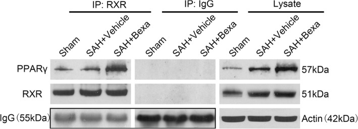
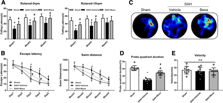
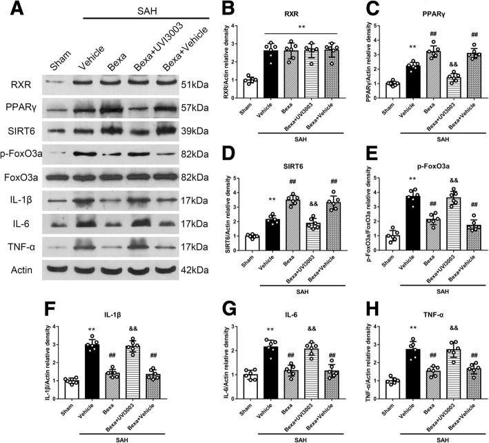
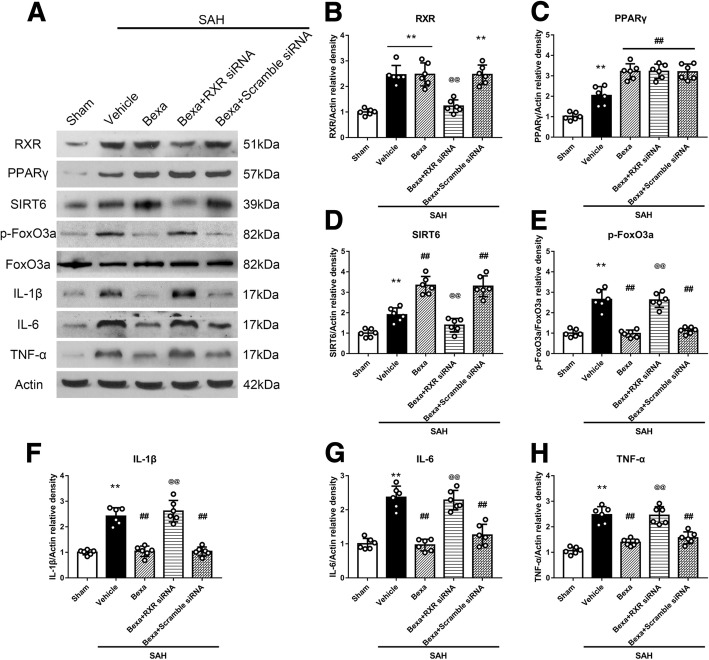
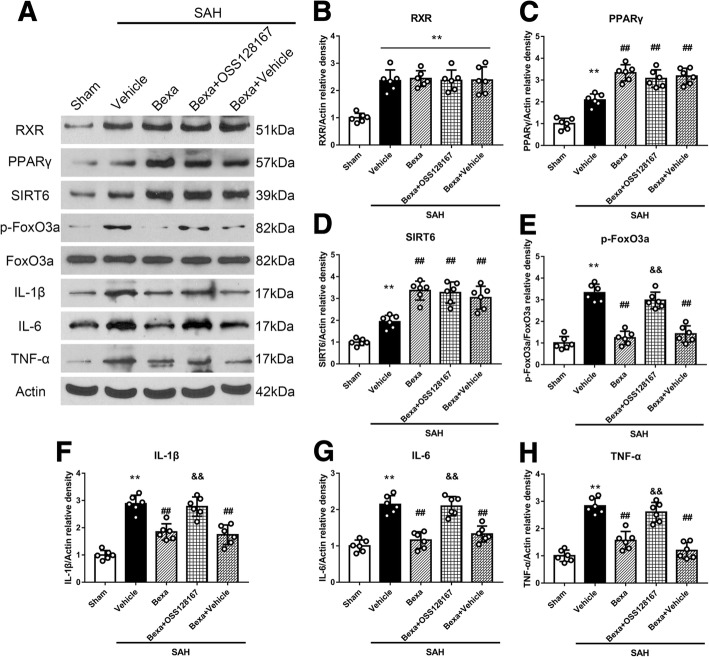
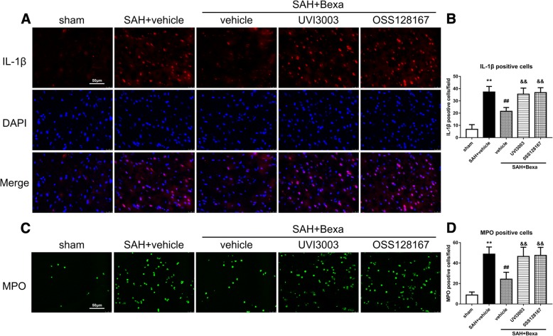
BACKGROUND: Subarachnoid hemorrhage (SAH) is a life-threatening subtype of stroke with high mortality and disabilities. Retinoid X receptor (RXR) has been shown to be neuroprotective against ischemia/reperfusion injury. This study aimed to investigate the effects of the selective RXR agonist bexarotene on neuroinflammation in a rat model of SAH. METHODS: Two hundred male Sprague-Dawley rats were used. The endovascular perforation induced SAH. Bexarotene was administered intraperitoneally at 1 h after SAH induction. To investigate the underlying mechanism, the selective RXR antagonist UVI3003 and RXR siRNA or SIRT6 inhibitor OSS128167 was administered via intracerebroventricular 1 h before SAH induction. Post-SAH assessments including SAH grade, neurological score, brain water content, Western blot, and immunofluorescence were performed. RESULTS: The endogenous RXR and sirtuin 6 (SIRT6) protein levels were increased after SAH. Bexarotene treatment significantly reduced brain edema and improved the short-/long-term neurological deficit after SAH. Mechanistically, bexarotene increased the levels of PPARγ and SIRT6; decreased the expression of phosphorylated FoxO3a (p-FoxO3a), IL-6, IL-1β, and TNF-a; and inhibited the microglia activation and neutrophils infiltration at 24 h after SAH. Either UVI3003, OSS128167, or RXR siRNA abolished the neuroprotective effects of bexarotene and its regulation on protein levels of PPARγ/SIRT6/p-FoxO3a after SAH. CONCLUSIONS: The activation of RXR by bexarotene attenuated neuroinflammation and improved neurological deficits after SAH. The anti-neuroinflammatory effect was at least partially through regulating PPARγ/SIRT6/FoxO3a pathway. Bexarotene may be a promising therapeutic strategy in the management of SAH patients.
Overview
The claims and statistics of this paper haven’t been checked yet.
Run the check — free, no account needed. It reads the open-access text and checks the sources it cites.
Integrity signals
Registry
Cites
Statistics
Figures
Verification
Claims the paper makes
The claims and statistics of this paper haven’t been checked yet.
Run the check — free, no account needed. It reads the open-access text and checks the sources it cites.
Claims other papers make about this one
Each is checked against this paper’s own text: does it say what the citing paper says it does?
The cited source says what the claim attributes to it.
In the citing paper
“After SAH, a large number of microglia are activated and release inflammatory factors, which would lead to inflammatory responses and aggravate the neurological deficit [7–9].”
Self-check
Percentages, sums and reported differences re-computed from the paper’s own numbers.
The self-check needs the paper’s full text.
Citations
Cites
1 retracted
Liu, F., Chen, Y., Hu, Q., Li, B., Tang, J., He, Y., Guo, Z., Feng, H., Tang, J., & Zhang, J. H. (2015) · Experimental Neurology · doi:10.1016/j.expneurol.2015.04.016 · Cited by 51
Self-citationRetractedvan Gijn, J., Kerr, R. S. C., & Rinkel, G. J. E. (2007) · The Lancet · doi:10.1016/s0140-6736(07)60153-6 · Cited by 1,725
ApoE-Directed Therapeutics Rapidly Clear β-Amyloid and Reverse Deficits in AD Mouse Models
Cramer, P. E., Cirrito, J. R., Wesson, D. W., Lee, C. Y. D., Karlo, J. C., Zinn, A. E., Casali, B. T., Restivo, J. L., Goebel, W. D., James, M. J., et al. (2012) · Science · doi:10.1126/science.1217697 · Cited by 1,050
Sugawara, T., Ayer, R., Jadhav, V., & Zhang, J. H. (2008) · Journal of Neuroscience Methods · doi:10.1016/j.jneumeth.2007.08.004 · Cited by 569
Self-citationRAR and RXR modulation in cancer and metabolic disease
Altucci, L., Leibowitz, M. D., Ogilvie, K. M., de Lera, A. R., & Gronemeyer, H. (2007) · Nature Reviews Drug Discovery · doi:10.1038/nrd2397 · Cited by 517
Salman, R. A., M, W. P. A. N. E. T. H. H. H. A., Davenport, R. J., Lindsay Kenneth, W., & 周立新 (2007) · doi:10.3760/cma.j.issn.1007-9742.2007.02.122 · Cited by 474
Retinoid signalling in the development of the central nervous system
Maden, M. (2002) · Nature Reviews Neuroscience · doi:10.1038/nrn963 · Cited by 428
Primary cutaneous T-cell lymphoma (mycosis fungoides and Sézary syndrome)
Jawed, S. I., Myskowski, P. L., Horwitz, S., Moskowitz, A., & Querfeld, C. (2014) · Journal of the American Academy of Dermatology · doi:10.1016/j.jaad.2013.08.033 · Cited by 347
Natrajan, M. S., de la Fuente, A. G., Crawford, A. H., Linehan, E., Núñez, V., Johnson, K. R., Wu, T., Fitzgerald, D. C., Ricote, M., Bielekova, B., et al. (2015) · Brain · doi:10.1093/brain/awv289 · Cited by 227
Mo, J., Enkhjargal, B., Travis, Z. D., Zhou, K., Wu, P., Zhang, G., Zhu, Q., Zhang, T., Peng, J., Xu, W., et al. (2018) · Redox Biology · doi:10.1016/j.redox.2018.09.022 · Cited by 170
Self-citationWang, X., Wang, X.-L., Tong, M.-M., Gan, L., Chen, H., Wu, S.-S., Chen, J., Li, R.-L., Wu, Y., Zhang, H.-Y., et al. (2016) · Basic Research in Cardiology · doi:10.1007/s00395-016-0531-z · Cited by 163
Zhu, Q., Enkhjargal, B., Huang, L., Zhang, T., Sun, C., Xie, Z., Wu, P., Mo, J., Tang, J., Xie, Z., et al. (2018) · Journal of Neuroinflammation · doi:10.1186/s12974-018-1211-8 · Cited by 161
Self-citationDrew, P. D., Johnson, J. W., Douglas, J. C., Phelan, K. D., & Kane, C. J. M. (2015) · Alcoholism Clinical and Experimental Research · doi:10.1111/acer.12639 · Cited by 154
Nuclear Receptors and Inflammation Control: Molecular Mechanisms and Pathophysiological Relevance
Huang, W. J. M., & Glass, C. K. (2010) · Arteriosclerosis Thrombosis and Vascular Biology · doi:10.1161/atvbaha.109.191189 · Cited by 143
Natural products as modulators of the nuclear receptors and metabolic sensors LXR, FXR and RXR
Hiebl, V., Ladurner, A., Latkolik, S. L., & Dirsch, V. M. (2018) · Biotechnology Advances · doi:10.1016/j.biotechadv.2018.03.003 · Cited by 142
Aragon, M. J., Topper, L. A., Steadman, C. R., Sanchez, B., Zychowski, K. E., Young, T. L., Herbert, G., Hall, P. R., Erdely, A., Eye, T., et al. (2017) · Proceedings of the National Academy of Sciences · doi:10.1073/pnas.1616070114 · Cited by 140
Retinoid X receptors in macrophage biology
Rőszer, T., Menéndez-Gutiérrez, M. P., Cedenilla, M., & Ricote, M. (2013) · Trends in Endocrinology & Metabolism · doi:10.1016/j.tem.2013.04.004 · Cited by 137
Sirtuin 6 protects the brain from cerebral ischemia/reperfusion injury through NRF2 activation
Zhang, W., Wei, R., Zhang, L., Tan, Y., & Qian, C. (2017) · Neuroscience · doi:10.1016/j.neuroscience.2017.09.035 · Cited by 135
Zhao, X., Gonzales, N. R., & Aronowski, J. A. (2014) · CNS Neuroscience & Therapeutics · doi:10.1111/cns.12350 · Cited by 123
Jin, J., Albertz, J., Guo, Z., Peng, Q., Rudow, G. L., Troncoso, J. C., Ross, C. A., & Duan, W. (2013) · Journal of Neurochemistry · doi:10.1111/jnc.12190 · Cited by 122
The High Cost of Stroke and Stroke Cytoprotection Research
Lapchak, P. A., & Zhang, J. H. (2016) · Translational Stroke Research · doi:10.1007/s12975-016-0518-y · Cited by 122
Self-citationChen, S., Zhao, L., Sherchan, P., Ding, Y., Yu, J., Nowrangi, D., Tang, J., Xia, Y., & Zhang, J. H. (2018) · Journal of Neuroinflammation · doi:10.1186/s12974-018-1140-6 · Cited by 122
Self-citationRetinoic acid: A key player in immunity
Pino‐Lagos, K., Guo, Y., & Noelle, R. J. (2010) · BioFactors · doi:10.1002/biof.117 · Cited by 109
Bonet-Costa, V., Herranz‐Pérez, V., Blanco-Gandía, M. C., Mas‐Bargues, C., Inglés, M., García-Tárraga, P., Rodríguez‐Arias, M., Miñarro, J., Borrás, C., García‐Verdugo, J. M., et al. (2016) · Journal of Alzheimer s Disease · doi:10.3233/jad-151020 · Cited by 107
Suzuki, H. (2014) · Translational Stroke Research · doi:10.1007/s12975-014-0380-8 · Cited by 100
Xie, Z., Enkhjargal, B., Wu, L., Zhou, K., Sun, C., Hu, X., Gospodarev, V., Tang, J., You, C., & Zhang, J. H. (2017) · Neuropharmacology · doi:10.1016/j.neuropharm.2017.09.040 · Cited by 98
Self-citationZhao, Y., Guan, Y., Zhou, X., Li, G., Li, Z., Zhou, C., Wang, P., & Miao, C. (2015) · Stroke · doi:10.1161/strokeaha.115.009216 · Cited by 91
Review: PPARs as new therapeutic targets for the treatment of cerebral ischemia/reperfusion injury
Collino, M., Patel, N. S. A., & Thiemermann, C. (2008) · Therapeutic Advances in Cardiovascular Disease · doi:10.1177/1753944708090924 · Cited by 87
Iniaghe, L. O., Krafft, P. R., Klebe, D. W., Omogbai, E. K. I., Zhang, J. H., & Tang, J. (2015) · Neurobiology of Disease · doi:10.1016/j.nbd.2015.07.001 · Cited by 78
Self-citationYang, S. J., Choi, J. M., Chae, S. W., Kim, W. J., Park, S. E., Rhee, E., Lee, W., Oh, K. W., Park, S. W., Kim, S. W., et al. (2011) · PLoS ONE · doi:10.1371/journal.pone.0017057 · Cited by 77
Xie, Z., Huang, L., Enkhjargal, B., Reis, C., Wan, W., Tang, J., Cheng, Y. Z., & Zhang, J. H. (2017) · Brain Behavior and Immunity · doi:10.1016/j.bbi.2017.11.012 · Cited by 77
Self-citationCambray, S., Arber, C., Little, G. E., Dougalis, A., Paola, V. D., Ungless, M. A., Li, M. C., & Rodríguez, T. A. (2012) · Nature Communications · doi:10.1038/ncomms1817 · Cited by 71
Activation of RXR/PPARγ underlies neuroprotection by bexarotene in ischemic stroke
Certo, M., Endo, Y., Ohta, K., Sakurada, S., Bagetta, G., & Amantea, D. (2015) · Pharmacological Research · doi:10.1016/j.phrs.2015.10.009 · Cited by 66
Bexarotene protects against traumatic brain injury in mice partially through apolipoprotein E
Zhong, J., Cheng, C., Liu, H., Huang, Z., Wu, Y., Teng, Z., He, J., Zhang, H., Wu, J., Cao, F., et al. (2016) · Neuroscience · doi:10.1016/j.neuroscience.2016.05.033 · Cited by 65
Liu, L., Fujimoto, M., Nakano, F., Nishikawa, H., Okada, T., Kawakita, F., Imanaka‐Yoshida, K., Yoshida, T., & Suzuki, H. (2018) · Molecular Neurobiology · doi:10.1007/s12035-018-1006-z · Cited by 65
Bexarotene-Activated Retinoid X Receptors Regulate Neuronal Differentiation and Dendritic Complexity
Mounier, A., Georgiev, D., Nam, K. N., Fitz, N. F., Castranio, E. L., Wolfe, C. M., Cronican, A. A., Schug, J., Lefterov, I., & Koldamova, R. (2015) · Journal of Neuroscience · doi:10.1523/jneurosci.1001-15.2015 · Cited by 64
Isoflurane on brain inflammation
Altay, O., Suzuki, H., Hasegawa, Y., Ostrowski, R. P., Tang, J., & Zhang, J. H. (2013) · Neurobiology of Disease · doi:10.1016/j.nbd.2013.09.016 · Cited by 63
Self-citationDheer, Y., Chitranshi, N., Gupta, V., Abbasi, M., Mirzaei, M., You, Y., Chung, R., Graham, S. L., & Gupta, V. (2018) · Molecular Neurobiology · doi:10.1007/s12035-018-1041-9 · Cited by 63
Siler, D. A., Gonzalez, J. A., Wang, R. K., Cetas, J. S., & Alkayed, N. J. (2014) · Translational Stroke Research · doi:10.1007/s12975-014-0329-y · Cited by 59
Hu, Y., Li, R., Yang, H., Luo, H., & Chen, Z. (2014) · Journal of Stroke and Cerebrovascular Diseases · doi:10.1016/j.jstrokecerebrovasdis.2014.10.006 · Cited by 53
Muroi, C., Fujioka, M., Mishima, K., Irie, K., Fujimura, Y., Nakano, T., Fandino, J., Keller, E., Iwasaki, K., & Fujiwara, M. (2014) · Journal of Thrombosis and Haemostasis · doi:10.1111/jth.12511 · Cited by 50
Drug repurposing for immune modulation in acute ischemic stroke
Amantea, D., & Bagetta, G. (2015) · Current Opinion in Pharmacology · doi:10.1016/j.coph.2015.11.006 · Cited by 47
Retinoid receptors trigger neuritogenesis in retinal degenerations
Lin, Y., Jones, B. W., Liu, A., Tucker, J. F., Rapp, K. D., Luo, L., Baehr, W., Bernstein, P. S., Watt, C. B., Yang, J., et al. (2011) · The FASEB Journal · doi:10.1096/fj.11-192914 · Cited by 41
Veldeman, M., Coburn, M., Rossaint, R., Clusmann, H., Nolte, K. W., Kremer, B., & Höllig, A. (2017) · Frontiers in Neurology · doi:10.3389/fneur.2017.00511 · Cited by 41
Xiao, Y.-C., Li, G., Chen, Y., Zuo, Y., Rashid, K., He, T., Feng, H., Zhang, J. H., & Liu, F. (2018) · Frontiers in Neurology · doi:10.3389/fneur.2018.00096 · Cited by 39
Self-citation9‐Cis‐retinoic acid reduces ischemic brain injury in rodents via bone morphogenetic protein
Shen, H., Luo, Y., Kuo, C., Deng, X., Chang, C., Harvey, B. K., Hoffer, B. J., & Wang, Y. (2008) · Journal of Neuroscience Research · doi:10.1002/jnr.21865 · Cited by 35
Huuskonen, M. T., Loppi, S. H., Dhungana, H., Keksa-Goldsteine, V., Lemarchant, S., Korhonen, P., Wojciechowski, S., Pollari, E., Valonen, P. K., Koponen, J., et al. (2016) · Scientific Reports · doi:10.1038/srep33176 · Cited by 34
Casali, B. T., Reed-Geaghan, E. G., & Landreth, G. E. (2018) · Journal of Neuroinflammation · doi:10.1186/s12974-018-1091-y · Cited by 30
Jiang, Y., Wang, J., Rozewski, D. M., Kolli, S., Wu, C.-H., Chen, C., Yang, X., Hofmeister, C. C., Byrd, J. C., Johnson, A. J., et al. (2013) · Journal of Pharmaceutical and Biomedical Analysis · doi:10.1016/j.jpba.2013.08.036 · Cited by 24
9-cis retinoic acid induces neurorepair in stroke brain
Yu, S., Airavaara, M., Wu, K., Harvey, B. K., Liu, H. S., Yang, Y., Zacharek, A., Chen, J., & Wang, Y. (2017) · Scientific Reports · doi:10.1038/s41598-017-04048-2 · Cited by 20
Rosiglitazone attenuates early brain injury after experimental subarachnoid hemorrhage in rats
Gu, C., Wang, Y., Li, J., Chen, J., Yan, F., Wu, C., & Chen, G. (2015) · Brain Research · doi:10.1016/j.brainres.2015.07.025 · Cited by 19
Tu, L., Yang, X., Zhang, Q., Wang, Q., Tian, T., Liu, D., Qu, X., & Tian, J. (2018) · Neurological Research · doi:10.1080/01616412.2018.1463900 · Cited by 16
Relaunching an old drug: the potential role of bexarotene in neurodegenerative diseases
Riancho, J., Berciano, M. T., Berciano, J., & Lafarga, M. (2016) · Journal of Neurology · doi:10.1007/s00415-015-8004-0 · Cited by 13
Effects of retinoic acid signaling on extraocular muscle myogenic precursor cells in vitro
Hebert, S. L., Fitzpatrick, K. R., McConnell, S. A., Cucak, A., Yuan, C., & McLoon, L. K. (2017) · Experimental Cell Research · doi:10.1016/j.yexcr.2017.10.007 · Cited by 8
Cited by
Chen, X., Jiang, M., Li, H., Wang, Y., Shen, H., Li, X., Zhang, Y., Wu, J., Yu, Z., & Chen, G. (2020) · Journal of Neuroinflammation · doi:10.1186/s12974-020-01882-6 · Cited by 85
Metadata
- Authors
- Zuo, Y., Huang, L., Enkhjargal, B., Xu, W., Umut, O., Travis, Z. D., Zhang, G., Tang, J., Liu, F., & Zhang, J. H.
- ORCID
- Zhang, John H.
- Venue
- Journal of Neuroinflammation
- Published
- 2019-02-21
- Volume
- 16
- Issue
- 1
- ISSN
- 1742-2094
- PMC
- PMC6385420
- Cited by
- 88
- References
- 54
- Language
- en
- Licence
- creativecommons.org/licenses/by/4.0/ (tdm)
About these checks
Registry, retraction, Cites and statistics results are computed, not judged, with one exception: a printed reference that no index lists is read once by a model before it counts as a citation error. Claims are extracted by a model and kept only when their quote appears verbatim in the text; each cited source is then checked, with a model’s help, for whether it says what the claim relies on. Those verdicts can be wrong — read the claim’s quote and the cited source before relying on one.
Statistics and arithmetic are recomputed, not judged.
Confidence bands are shown only for fields calibrated on our annotated test set. None is calibrated yet, so no band is shown.
What counts as a claim ›How we measure confidence ›How we check this ›
Are you an author, or do you see a problem with this page? Request a review
Data sources
Bibliographic data: Semantic Scholar, OpenAlex, Crossref.
Contains information from the Semantic Scholar Academic Graph Datasets, made available under the ODC Attribution License (ODC-BY 1.0).
Semantic Scholar’s data is described in: Kinney, R., et al. (2023). The Semantic Scholar Open Data Platform. arXiv:2301.10140.
