

The DOI matches its Crossref record; 4 of the 4 references it cites were found in a registry or an index.
Astrocyte-derived exosomes enriched with miR-873a-5p inhibit neuroinflammation via microglia phenotype modulation after traumatic brain injury
Published version of a preprint
Abstract
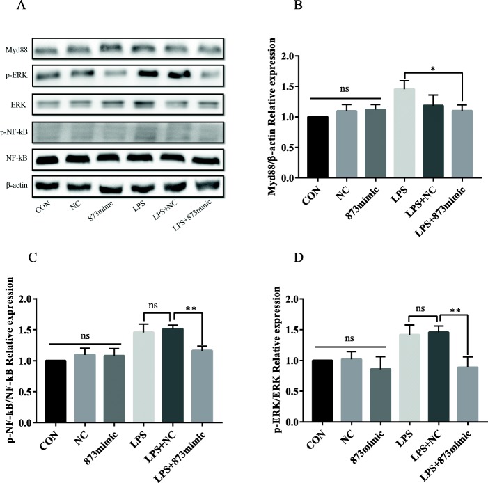
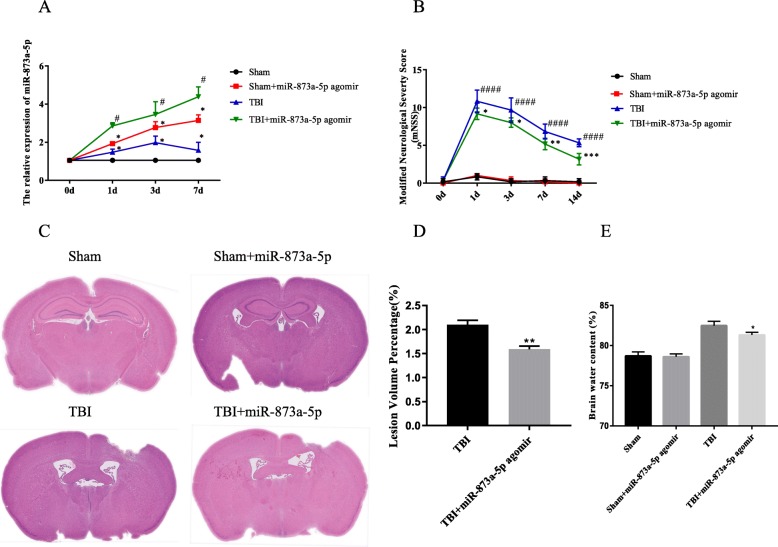
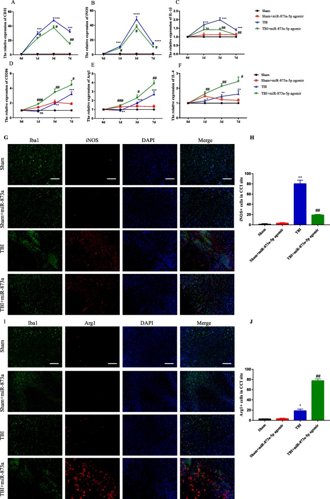
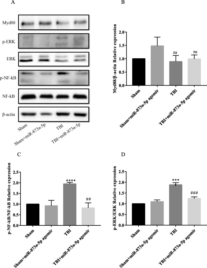
Background: The interaction between astrocytes and microglia plays a vital role in the damage and repair of brain lesions due to traumatic brain injury (TBI). Recent studies have shown that exosomes act as potent mediators involved in intercellular communication. Methods: In the current study, the expression of inflammatory factors and miR-873a-5p in the lesion area and oedema area was evaluated in 15 patients with traumatic brain injury. Exosomes secreted by astrocytes were detected by immunofluorescence, Western blot and electron microscopy. A mouse model of TBI and an in vitro model of LPS-induced primary microglia were established to study the protective mechanism of exosomes from miR-873a-5p overexpressing in TBI-induced nerve injury. Results: We discovered that exosomes derived from activated astrocytes promote microglial M2 phenotype transformation following TBI. More than 100 miRNAs were detected in these astrocyte-derived exosomes. miR-873a-5p is a major component that was highly expressed in human traumatic brain tissue. Moreover, miR-873a-5p significantly inhibited LPS-induced microglial M1 phenotype transformation and the subsequent inflammation through decreased phosphorylation of ERK and NF-κB p65. This effect also greatly improved the modified neurological severity score (mNSS) and attenuated brain injury in a strictly controlled cortical impact mouse model. Conclusions: Taken together, our research indicates that miRNAs in the exosomes derived from activated astrocytes play a key role in the astrocyte-microglia interaction. miR-873a-5p, as one of the main components of these astrocyte-derived exosomes, attenuated microglia-mediated neuroinflammation and improved neurological deficits following TBI by inhibiting the NF-κB signalling pathway. These findings suggest a potential role for miR-873a-5p in treating traumatic brain injury.
Overview
The claims and statistics of this paper haven’t been checked yet.
Run the check — free, no account needed. It reads the open-access text and checks the sources it cites.
Integrity signals
Registry
Cites
Statistics
Figures
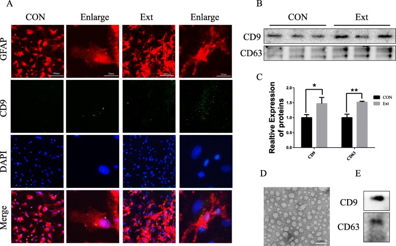
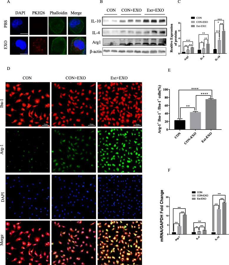
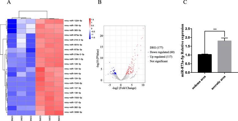
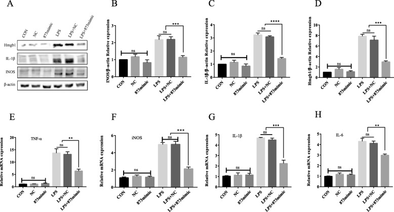
Verification
Claims the paper makes
The claims and statistics of this paper haven’t been checked yet.
Run the check — free, no account needed. It reads the open-access text and checks the sources it cites.
Claims other papers make about this one
Each is checked against this paper’s own text: does it say what the citing paper says it does?
The source backs part of the claim, not all of it — or backs it for a different population, condition or comparison.
In the citing paper
“Exosomal-transported miR-124 prevents microglial activation by inhibiting myosin heavy chain 9 expression in microglia; thus inhibiting the NF-κB signaling pathway and consequently preventing the shift to M1 polarization (Long et al. 2020).”
From MiR-124 and MiR-155 as Therapeutic Targets in Microglia-Mediated Inflammation in Multiple Sclerosis (2025) · cites this paper as Long et al. 2020
Experimental — accuracy not yet measured
Self-check
Percentages, sums and reported differences re-computed from the paper’s own numbers.
The self-check needs the paper’s full text.
Citations
Cites
Neurotoxic reactive astrocytes are induced by activated microglia
Liddelow, S. A., Guttenplan, K. A., Clarke, L. E., Bennett, F. C., Bohlen, C. J., Schirmer, L., Bennett, M. L., Münch, A. E., Chung, W.-S., Peterson, T. C., et al. (2017) · Nature · doi:10.1038/nature21029 · Cited by 7,971
Astrocyte barriers to neurotoxic inflammation
Sofroniew, M. V. (2015) · Nature Reviews Neuroscience · doi:10.1038/nrn3898 · Cited by 1,202
SIRT1 Protects against Microglia-dependent Amyloid-β Toxicity through Inhibiting NF-κB Signaling
Chen, J., Zhou, Y., Mueller-Steiner, S., Chen, L.-F., Kwon, H., Yi, S., Mucke, L., & Gan, L. (2005) · Journal of Biological Chemistry · doi:10.1074/jbc.m509329200 · Cited by 740
Dickens, A. M., Tovar-y-Romo, L. B., Yoo, S.-W., Trout, A. L., Bae, M., Kanmogne, M., Megra, B., Williams, D. W., Witwer, K. W., Gacias, M., et al. (2017) · Science Signaling · doi:10.1126/scisignal.aai7696 · Cited by 285
Research works printed but not linked: 26
- Andelic N. The epidemiology of traumatic brain injury. Lancet Neurol. 2013;12:28–9. · DOI
- Faden AI, Wu J, Stoica BA, Loane DJ. Progressive inflammation-mediated neurodegeneration after traumatic brain or spinal cord injury. Br J Pharmacol. 2016;173:681–91. · DOI
- Loane DJ, Kumar A. Microglia in the TBI brain: the good, the bad, and the dysregulated. Exp Neurol. 2016;275(Pt 3):316–27. · DOI
- Xia CY, Zhang S, Gao Y, Wang ZZ, Chen NH. Selective modulation of microglia polarization to M2 phenotype for stroke treatment. Int Immunopharmacol. 2015;25:377–82. · DOI
- Chen Y, Swanson RA. Astrocytes and brain injury. J Cereb Blood Flow Metab. 2003;23:137–49. · DOI
- Burda JE, Bernstein AM, Sofroniew MV. Astrocyte roles in traumatic brain injury. Exp Neurol. 2016;275(Pt 3):305–15. · DOI
- Farina C, Aloisi F, Meinl E. Astrocytes are active players in cerebral innate immunity. Trends Immunol. 2007;28:138–45. · DOI · Paper page
- Min KJ, Yang MS, Kim SU, Jou I, Joe EH. Astrocytes induce hemeoxygenase-1 expression in microglia: a feasible mechanism for preventing excessive brain inflammation. J Neurosci. 2006;26:1880–7. · DOI
- Quek C, Hill AF. The role of extracellular vesicles in neurodegenerative diseases. Biochem Biophys Res Commun. 2017;483:1178–86. · DOI · Paper page
- Chivet M, Hemming F, Pernet-Gallay K, Fraboulet S, Sadoul R. Emerging role of neuronal exosomes in the central nervous system. Front Physiol. 2012;3:145. · DOI
- Lafourcade C, Ramirez JP, Luarte A, Fernandez A, Wyneken U. MiRNAs in astrocyte-derived exosomes as possible mediators of neuronal plasticity. J Exp Neurosci. 2016;10:1–9.
- Yao X, Liu S, Ding W, Yue P, Jiang Q, Zhao M, Hu F, Zhang H. TLR4 signal ablation attenuated neurological deficits by regulating microglial M1/M2 phenotype after traumatic brain injury in mice. J Neuroimmunol. 2017;310:38–45. · DOI
- Chen X, Li Y, Wang L, Katakowski M, Zhang L, Chen J, Xu Y, Gautam SC, Chopp M. Ischemic rat brain extracts induce human marrow stromal cell growth factor production. Neuropathology. 2002;22:275–9. · DOI
- Xin H, Li Y, Buller B, Katakowski M, Zhang Y, Wang X, Shang X, Zhang ZG, Chopp M. Exosome-mediated transfer of miR-133b from multipotent mesenchymal stromal cells to neural cells contributes to neurite outgrowth. Stem Cells. 2012;30:1556–64. · DOI
- Wei W, Wang H, Wu Y, Ding K, Li T, Cong Z, Xu J, Zhou M, Huang L, Ding H, Wu H. Alpha lipoic acid inhibits neural apoptosis via a mitochondrial pathway in rats following traumatic brain injury. Neurochem Int. 2015;87:85–91. · DOI
- Jang E, Kim JH, Lee S, Kim JH, Seo JW, Jin M, Lee MG, Jang IS, Lee WH, Suk K. Phenotypic polarization of activated astrocytes: the critical role of lipocalin-2 in the classical inflammatory activation of astrocytes. J Immunol. 2013;191:5204–19. · DOI
- Hu G, Liao K, Niu F, Yang L, Dallon BW, Callen S, Tian C, Shu J, Cui J, Sun Z, et al. Astrocyte EV-induced lincRNA-Cox2 regulates microglial phagocytosis: implications for morphine-mediated neurodegeneration. Mol Ther Nucleic Acids. 2018;13:450–63. · DOI
- Sobue A, Ito N, Nagai T, Shan W, Hada K, Nakajima A, Murakami Y, Mouri A, Yamamoto Y, Nabeshima T, et al. Astroglial major histocompatibility complex class I following immune activation leads to behavioral and neuropathological changes. Glia. 2018;66:1034–52. · DOI
- Xu L, Cao H, Xie Y, Zhang Y, Du M, Xu X, Ye R, Liu X. Exosome-shuttled miR-92b-3p from ischemic preconditioned astrocytes protects neurons against oxygen and glucose deprivation. Brain Res. 1717;2019:66–73.
- Hu G, Niu F, Liao K, Periyasamy P, Sil S, Liu J, Dravid SM, Buch S. HIV-1 tat-induced astrocytic extracellular vesicle miR-7 impairs synaptic architecture. J NeuroImmune Pharmacol. 2019:1–16. https://doi.org/10.1007/s11481-019-09869-8. · DOI
- Wang L, Jiang F, Ma F, Zhang B. MiR-873-5p suppresses cell proliferation and epithelial-mesenchymal transition via directly targeting Jumonji domain-containing protein 8 through the NF-kappaB pathway in colorectal cancer. J Cell Commun Signal. 2019;13:549–60. · DOI
- Wang Q, Zhu W. MicroRNA-873 inhibits the proliferation and invasion of endometrial cancer cells by directly targeting hepatoma-derived growth factor. Exp Ther Med. 2019;18:1291–8.
- Zhang Y, Zhang C, Zhao Q, Wei W, Dong Z, Shao L, Li J, Wu W, Zhang H, Huang H, et al. The miR-873/NDFIP1 axis promotes hepatocellular carcinoma growth and metastasis through the AKT/mTOR-mediated Warburg effect. Am J Cancer Res. 2019;9:927–44.
- Cui J, Yang Y, Li H, Leng Y, Qian K, Huang Q, Zhang C, Lu Z, Chen J, Sun T, et al. MiR-873 regulates ERalpha transcriptional activity and tamoxifen resistance via targeting CDK3 in breast cancer cells. Oncogene. 2015;34:3895–907. · DOI
- Gao Y, Xue Q, Wang D, Du M, Zhang Y, Gao S. miR-873 induces lung adenocarcinoma cell proliferation and migration by targeting SRCIN1. Am J Transl Res. 2015;7:2519–26.
- Liu X, He F, Pang R, Zhao D, Qiu W, Shan K, Zhang J, Lu Y, Li Y, Wang Y. Interleukin-17 (IL-17)-induced microRNA 873 (miR-873) contributes to the pathogenesis of experimental autoimmune encephalomyelitis by targeting A20 ubiquitin-editing enzyme. J Biol Chem. 2014;289:28971–86. · DOI
Cited by
Sorted by citations
Emerging role of non-coding RNAs in neuroinflammation mediated by microglia and astrocytes
Yang, R., Yang, B., Liu, W., Tan, C., Chen, H., & Wang, X. (2023) · Journal of Neuroinflammation · doi:10.1186/s12974-023-02856-0 · Cited by 91
The link between neuroinflammation and the neurovascular unit in synucleinopathies
Wang, Q., Zheng, J., Pettersson, S., Reynolds, R., & Tan, E.-K. (2023) · Science Advances · doi:10.1126/sciadv.abq1141 · Cited by 81
Pistono, C., Bister, N., Stanová, I., & Malm, T. (2021) · Frontiers in Cell and Developmental Biology · doi:10.3389/fcell.2020.623771 · Cited by 75
Chivero, E. T., Liao, K., Niu, F., Tripathi, A., Tian, C., Buch, S., & Hu, G. (2020) · Frontiers in Cell and Developmental Biology · doi:10.3389/fcell.2020.00573 · Cited by 69
Li, Q., Fu, X., Kou, Y., & Han, N. (2023) · Theranostics · doi:10.7150/thno.84971 · Cited by 63
Extracellular vesicles (exosomes and ectosomes) play key roles in the pathology of brain diseases
Meldolesi, J. (2021) · Molecular Biomedicine · doi:10.1186/s43556-021-00040-5 · Cited by 47
Role of Glia-Derived Extracellular Vesicles in Neurodegenerative Diseases
Li, T., Tan, X., Li, S., Al-Nusaif, M., & Le, W. (2021) · Frontiers in Aging Neuroscience · doi:10.3389/fnagi.2021.765395 · Cited by 33
Li, Y., Zhang, Y., Wang, Q., Wu, C., Du, G., & Yang, L. (2023) · Pharmaceuticals · doi:10.3390/ph16040621 · Cited by 24
MiR-124 and MiR-155 as Therapeutic Targets in Microglia-Mediated Inflammation in Multiple Sclerosis
Asl, E. R., Hosseini, S. E., Tahmasebi, F., Bolandi, N., & Barati, S. (2025) · Cellular and Molecular Neurobiology · doi:10.1007/s10571-025-01578-6 · Cited by 19
Clinical Theragnostic Signature of Extracellular Vesicles in Traumatic Brain Injury (TBI)
Dey, A., Ghosh, S., Bhuniya, T., Koley, M., Bera, A., Guha, S., Chakraborty, K., Muthu, S., Gorai, S., Vorn, R., et al. (2023) · ACS Chemical Neuroscience · doi:10.1021/acschemneuro.3c00386 · Cited by 16
Metadata
- Authors
- Long, X., Yao, X., Jiang, Q., Yang, Y., He, X., Tian, W., Zhao, K., & Zhang, H.
- Venue
- Journal of Neuroinflammation
- Published
- 2020-03-19
- Volume
- 17
- Issue
- 1
- ISSN
- 1742-2094
- PMC
- PMC7082961
- Cited by
- 294
- References
- 4
- Language
- en
- Funders
- National Natural Science Foundation of China (81371381, 81602204)
About these checks
Registry, retraction, Cites and statistics results are computed, not judged, with one exception: a printed reference that no index lists is read once by a model before it counts as a citation error. Claims are extracted by a model and kept only when their quote appears verbatim in the text; each cited source is then checked, with a model’s help, for whether it says what the claim relies on. Those verdicts can be wrong — read the claim’s quote and the cited source before relying on one.
Statistics and arithmetic are recomputed, not judged.
Confidence bands are shown only for fields calibrated on our annotated test set. None is calibrated yet, so no band is shown.
What counts as a claim ›How we measure confidence ›How we check this ›
Are you an author, or do you see a problem with this page? Request a review
