

The DOI matches its Crossref record; 15 of the 15 references it cites were found in a registry or an index.
Cerebrovascular amyloid Angiopathy in bioengineered vessels is reduced by high-density lipoprotein particles enriched in Apolipoprotein E
Abstract
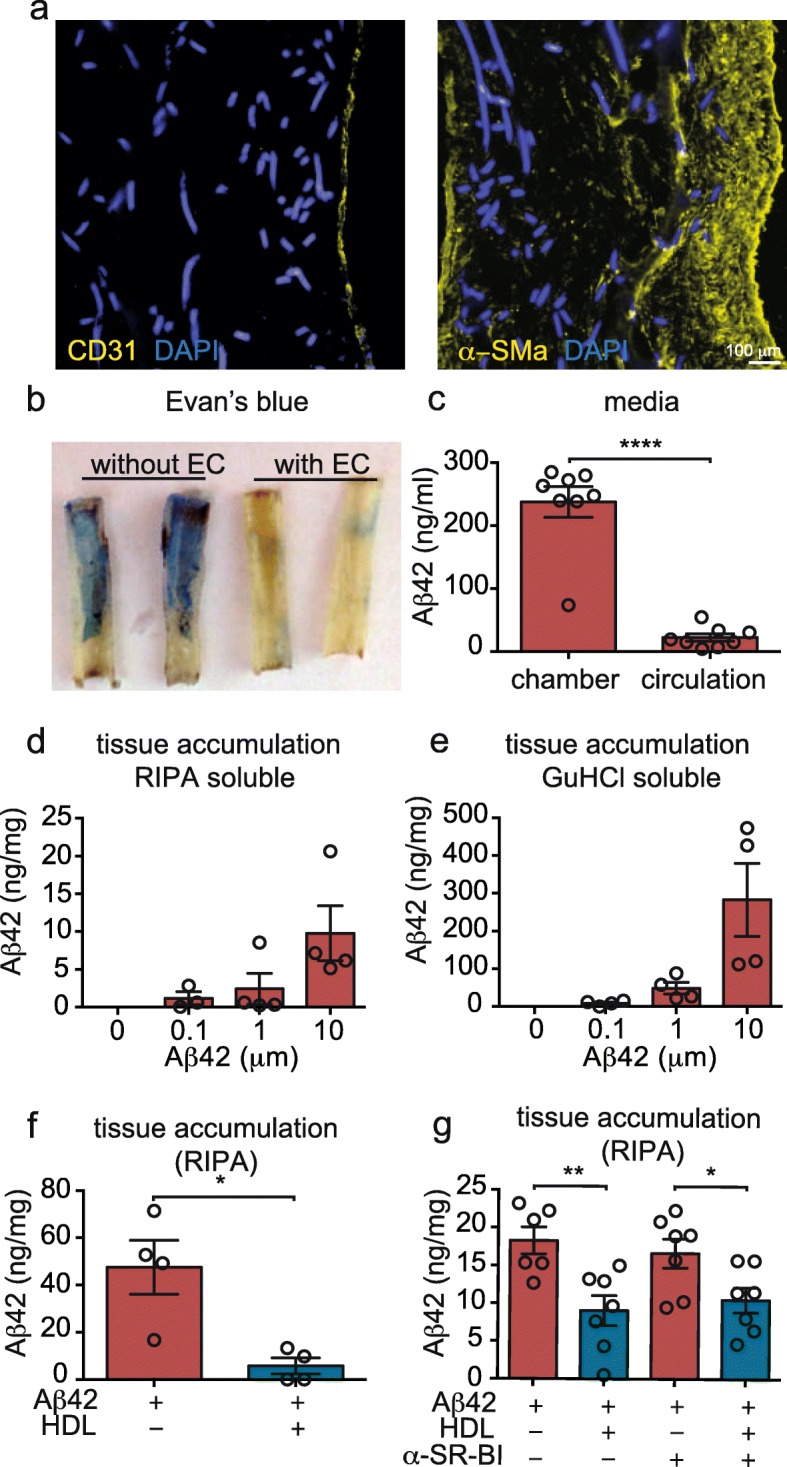
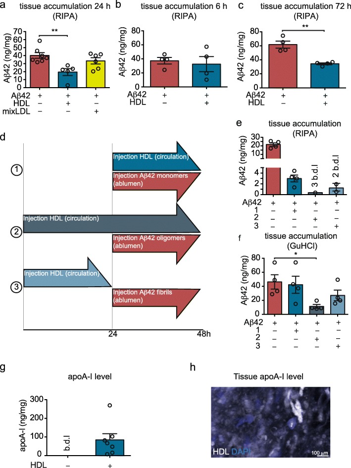
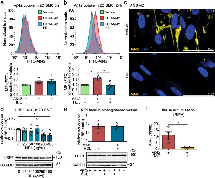
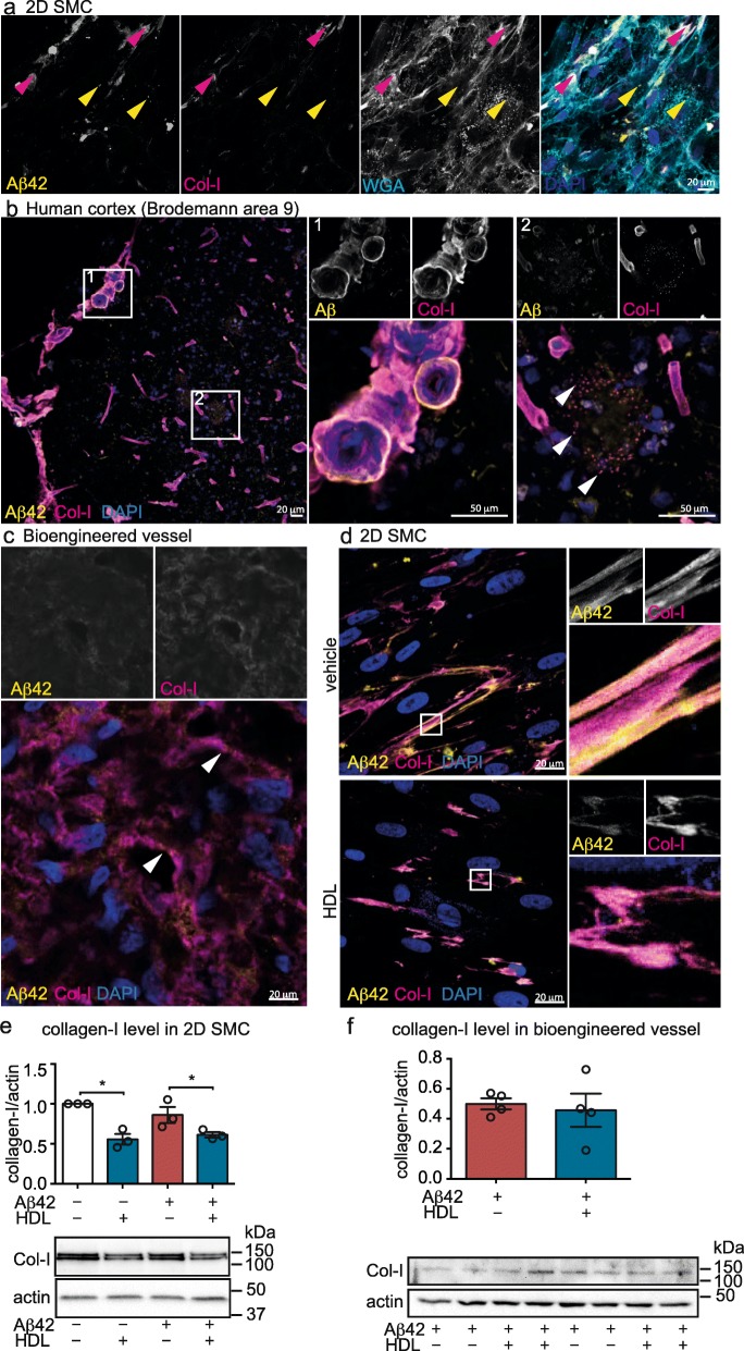
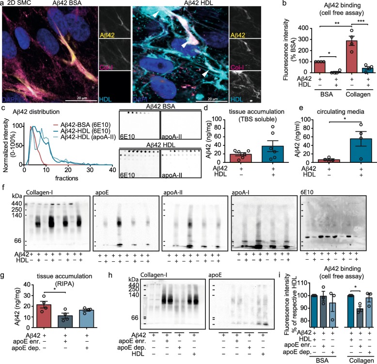
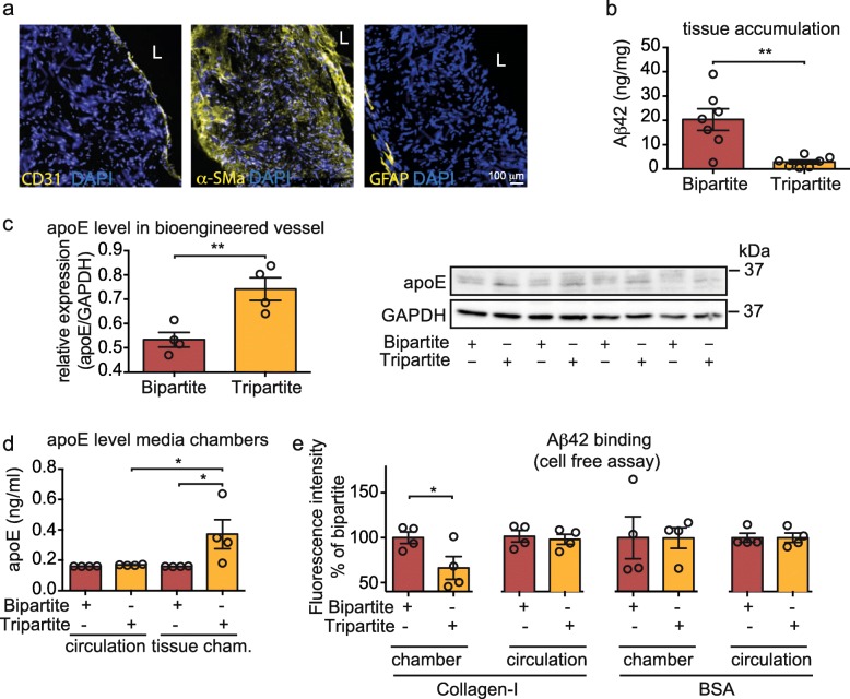
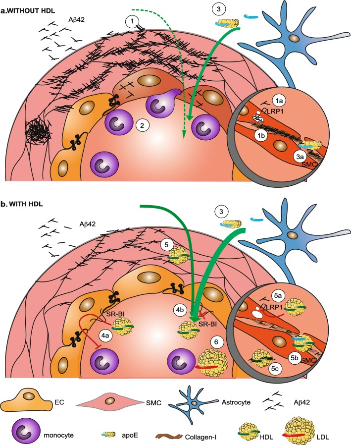
Background: Several lines of evidence suggest that high-density lipoprotein (HDL) reduces Alzheimer’s disease (AD) risk by decreasing vascular beta-amyloid (Aβ) deposition and inflammation, however, the mechanisms by which HDL improve cerebrovascular functions relevant to AD remain poorly understood. Methods: Here we use a human bioengineered model of cerebral amyloid angiopathy (CAA) to define several mechanisms by which HDL reduces Aβ deposition within the vasculature and attenuates endothelial inflammation as measured by monocyte binding. Results: We demonstrate that HDL reduces vascular Aβ accumulation independently of its principal binding protein, scavenger receptor (SR)-BI, in contrast to the SR-BI-dependent mechanism by which HDL prevents Aβ-induced vascular inflammation. We describe multiple novel mechanisms by which HDL acts to reduce CAA, namely: i) altering Aβ binding to collagen-I, ii) forming a complex with Aβ that maintains its solubility, iii) lowering collagen-I protein levels produced by smooth-muscle cells (SMC), and iv) attenuating Aβ uptake into SMC that associates with reduced low density lipoprotein related protein 1 (LRP1) levels. Furthermore, we show that HDL particles enriched in apolipoprotein (apo)E appear to be the major drivers of these effects, providing new insights into the peripheral role of apoE in AD, in particular, the fraction of HDL that contains apoE. Conclusion: The findings in this study identify new mechanisms by which circulating HDL, particularly HDL particles enriched in apoE, may provide vascular resilience to Aβ and shed new light on a potential role of peripherally-acting apoE in AD.
Overview
The claims and statistics of this paper haven’t been checked yet.
Run the check — free, no account needed. It reads the open-access text and checks the sources it cites.
Integrity signals
Registry
Cites
Statistics
Figures
Verification
Claims the paper makes
The claims and statistics of this paper haven’t been checked yet.
Run the check — free, no account needed. It reads the open-access text and checks the sources it cites.
Claims other papers make about this one
Each is checked against this paper’s own text: does it say what the citing paper says it does?
In the citing paper
“LRP1 mediates and regulates the endocytosis of > 30 ligands, including apolipoprotein E (APOE) and amyloid-β (Aβ) [21–27]”
From LRP1 is a neuronal receptor for α-synuclein uptake and spread (2022) · cites this paper as [27]
Self-check
Percentages, sums and reported differences re-computed from the paper’s own numbers.
The self-check needs the paper’s full text.
Citations
Cites
Isolation and quantification of soluble Alzheimer's β-peptide from biological fluids
Seubert, P., Vigo-Pelfrey, C., Esch, F., Lee, M., Dovey, H., Davis, D., Sinha, S., Schiossmacher, M., Whaley, J., Swindlehurst, C., et al. (1992) · Nature · doi:10.1038/359325a0 · Cited by 1,731
RAGE mediates amyloid-β peptide transport across the blood-brain barrier and accumulation in brain
Deane, R., Du Yan, S., Submamaryan, R. K., LaRue, B., Jovanovic, S., Hogg, E., Welch, D., Manness, L., Lin, C., Yu, J., et al. (2003) · Nature Medicine · doi:10.1038/nm890 · Cited by 1,426
The overlap between vascular disease and Alzheimer’s disease - lessons from pathology
Attems, J., & Jellinger, K. A. (2014) · BMC Medicine · doi:10.1186/s12916-014-0206-2 · Cited by 739 · cited 1×
“Although CAA is highly associated with the Flemish, Iowa and Dutch genetic mutations in amyloid precursor protein, sporadic CAA is present in 10–40% of non-cognitively declined elderly brains and in 80% of AD brains [7, 40].”
BackgroundBell, R. D., Sagare, A. P., Friedman, A. E., Bedi, G. S., Holtzman, D. M., Deane, R., & Zlokovic, B. V. (2007) · Journal of Cerebral Blood Flow & Metabolism · doi:10.1038/sj.jcbfm.9600419 · Cited by 670
Aβ42 Is Essential for Parenchymal and Vascular Amyloid Deposition in Mice
McGowan, E., Pickford, F., Kim, J., Onstead, L., Eriksen, J., Yu, C., Skipper, L., Murphy, M. P., Beard, J., Das, P., et al. (2005) · Neuron · doi:10.1016/j.neuron.2005.06.030 · Cited by 565
Phenotypes of apolipoprotein B and apolipoprotein E after liver transplantation.
Linton, M. F., Gish, R., Hubl, S. T., Bütler, E., Esquivel, C., Bry, W. I., Boyles, J. K., Wardell, M. R., & Young, S. G. (1991) · Journal of Clinical Investigation · doi:10.1172/jci115288 · Cited by 367
Amyloid-β Dynamics Correlate with Neurological Status in the Injured Human Brain
Brody, D. L., Magnoni, S., Schwetye, K. E., Spinner, M. L., Esparza, T. J., Stocchetti, N., Zipfel, G. J., & Holtzman, D. M. (2008) · Science · doi:10.1126/science.1161591 · Cited by 295
SRF and myocardin regulate LRP-mediated amyloid-β clearance in brain vascular cells
Bell, R. D., Deane, R., Chow, N., Long, X., Sagare, A., Singh, I., Streb, J. W., Guo, H., Rubio, A., Van Nostrand, W., et al. (2009) · Nature Cell Biology · doi:10.1038/ncb1819 · Cited by 261
Alzheimer disease and aducanumab: adjusting our approach
Selkoe, D. J. (2019) · Nature Reviews Neurology · doi:10.1038/s41582-019-0205-1 · Cited by 208
Plasma levels of apolipoprotein Eand risk of dementia in the general population
Rasmussen, K. L., Tybjærg‐Hansen, A., Nordestgaard, B. G., & Frikke‐Schmidt, R. (2015) · Annals of Neurology · doi:10.1002/ana.24326 · Cited by 156
HDL from an Alzheimer's disease perspective
Button, E. B., Robert, J., Caffrey, T. M., Fan, J., Zhao, W., & Wellington, C. L. (2019) · Current Opinion in Lipidology · doi:10.1097/mol.0000000000000604 · Cited by 114
Self-citationAnkle-to-Brachial Index and Dementia: The Honolulu-Asia Aging Study
Laurin, D., Masaki, K. H., White, L. R., & Launer, L. J. (2007) · Circulation · doi:10.1161/circulationaha.106.686477 · Cited by 90
Rasmussen, K. L., Tybjærg‐Hansen, A., Nordestgaard, B. G., & Frikke‐Schmidt, R. (2018) · Alzheimer's & Dementia · doi:10.1016/j.jalz.2017.05.006 · Cited by 75
Fagan, A. M., Christopher, E., Taylor, J. W., Parsadanian, M., Spinner, M., Watson, M., Fryer, J. D., Wahrle, S., Bales, K. R., Paul, S. M., et al. (2004) · The American Journal of Pathology · doi:10.1016/s0002-9440(10)63399-8 · Cited by 54
Herukka, S.-K., Rummukainen, J., Ihalainen, J., von und zu Fraunberg, M., Koivisto, A. M., Nerg, O., Puli, L. K., Seppälä, T. T., Zetterberg, H., Pyykkö, O. T., et al. (2015) · Journal of Alzheimer's Disease · doi:10.3233/jad-142862 · Cited by 43
Research works printed but not linked: 62
- Christina P. World Alzheimer’s report 2018. Alzheimer’s disease internations: world alzheimer report 2018; 2018. p. 1–48. https://doi.org/10.1111/j.0033-0124.1950.24_14.x. · DOI
- Sevigny J, et al. The antibody aducanumab reduces Aβ plaques in Alzheimer’s disease. Nat Publ Group. 2016;537:50–6.
- Howard R, Liu KY. Questions EMERGE as Biogen claims aducanumab turnaround. Nat Rev Neurol. 2019. https://doi.org/10.1038/s41582-019-0295-9. · DOI · Paper page
- Di Francesco JC, Longoni M, Piazza F. Anti-Aβ autoantibodies in amyloid related imaging abnormalities (ARIA): candidate biomarker for immunotherapy in Alzheimer’s disease and cerebral amyloid angiopathy. Front Neurol. 2015;6:4–6.
- Biffi A, Greenberg SM. Cerebral amyloid angiopathy: a systematic review. J Clin Neurol. 2011;7:1–9. · DOI
- Nation DA, et al. Blood–brain barrier breakdown is an early biomarker of human cognitive dysfunction. Nat Med. 2019;25:270–6. · DOI · Paper page
- Sweeney MD, et al. Vascular dysfunction—the disregarded partner of Alzheimer’s disease. Alzheimer’s Dementia. 2019;15:158–67. · DOI · Paper page
- Santos CY, et al. Pathophysiologic relationship between Alzheimer’s disease, cerebrovascular disease, and cardiovascular risk: a review and synthesis. Alzheimers Dement. 2017;7:69–87.
- Vu THT, et al. Favorable cardiovascular health at young and middle ages and dementia in older age—the CHA study. J Am Heart Assoc. 2019;8:e009730. · DOI
- Tan ECK, et al. Antihypertensive medication regimen intensity and incident dementia in an older population. J Am Med Dir Assoc. 2018;19:577–83. · DOI
- Solomon A, Kivipelto M, Soininen H. Prevention of Alzheimer’s disease: moving backward through the lifespan. Adv Alzheimers Dis. 2012;3:465–9. · DOI
- Stukas S, Robert J, Wellington CL. High-density lipoproteins and cerebrovascular integrity in Alzheimer’s disease. Cell Metab. 2014;19:574–91. · DOI
- Mineo C, Shaul PW. Novel biological functions of high-density lipoprotein cholesterol. Circ Res. 2012;111:1079–90. · DOI
- Sacre SM, Stannard AK, Owen JS. Apolipoprotein E (apoE) isoforms differentially induce nitric oxide production in endothelial cells. FEBS Lett. 2003;540:181–7. · DOI · Paper page
- Zuliani G, et al. Relationship between low levels of high-density lipoprotein cholesterol and dementia in the elderly. The InChianti study. J Gerontol A Biol Sci Med Sci. 2010;65:559–64. · DOI
- Merched A, Xia Y, Visvikis S, Serot JM, Siest G. Decreased high-density lipoprotein cholesterol and serum apolipoprotein AI concentrations are highly correlated with the severity of Alzheimer’s disease. Neurobiol Aging. 2000;21:27–30. · DOI
- Reitz C, et al. Association of higher levels of high-density lipoprotein cholesterol in elderly individuals and lower risk of late-onset Alzheimer disease. Arch Neurol. 2010;67:1491–7.
- Lefterov I, et al. Apolipoprotein A-I deficiency increases cerebral amyloid angiopathy and cognitive deficits in APP/PS1DeltaE9 mice. J Biol Chem. 2010;285:36945–57. · DOI
- Button EB, et al. ApoA-I deficiency increases cortical amyloid deposition, cerebral amyloid angiopathy, cortical and hippocampal astrogliosis, and amyloid-associated astrocyte reactivity in APP/PS1 mice. Alzheimers Res Ther. 2019;11:1–18. · DOI
- Contu L, Carare RO, Hawkes CA. Knockout of apolipoprotein A-I decreases parenchymal and vascular β- amyloid pathology in the Tg2576 mouse model of Alzheimer’s disease. Neuropathol Appl Neurobiol. 2019:1. https://doi.org/10.1111/nan.12556. · DOI
- Lewis TL, et al. Overexpression of human apolipoprotein A-I preserves cognitive function and attenuates neuroinflammation and cerebral amyloid angiopathy in a mouse model of Alzheimer disease. J Biol Chem. 2010;285:36958–68. · DOI
- Robert J, et al. Reconstituted high-density lipoproteins acutely reduce soluble brain Aβ levels in symptomatic APP/PS1 mice. Biochim Biophys Acta. 2015;1862:1027–36. · DOI
- Fernández-de Retana S, et al. Intravenous treatment with human recombinant ApoA-I Milano reduces beta amyloid cerebral deposition in the APP23-transgenic mouse model of Alzheimer’s disease. Neurobiol Aging. 2017;60:116–28. · DOI
- Gordon SM, et al. A comparison of the mouse and human lipoproteome: suitability of the mouse model for studies of human lipoproteins. J Proteome Res. 2015;14:2686–95. · DOI
- Getz GS, Reardon CA. Animal models of atherosclerosis. Arterioscler Thromb Vasc Biol. 2012;32:1104–15. · DOI
- Robert J, et al. Clearance of beta-amyloid is facilitated by apolipoprotein E and circulating high-density lipoproteins in bioengineered human vessels. eLife. 2017;6:e29595. · DOI
- Robert J, et al. High-density lipoproteins suppress Aβ-induced PBMC adhesion to human endothelial cells in bioengineered vessels and in monoculture. Mol Neurodegener. 2017;12:60. · DOI
- Weisgraber KH, Mahley RW. Subfractionation of human high density lipoproteins by heparin-Sepharose affinity chromatography. J Lipid Res. 1980;21:316–25. · DOI
- Zhao N, Liu C-C, Qiao W, Bu G. Apolipoprotein E, receptors, and modulation of Alzheimer’s disease. Biol Psychiatry. 2018;83:347–57. · DOI · Paper page
- Robert J, et al. Interleukin 6 stimulates endothelial binding and transport of high-density lipoprotein through induction of endothelial lipase. Arterioscler Thromb Vasc Biol. 2013;33:2699–706. · DOI
- Morton AM, Furtado JD, Mendivil CO, Sacks FM. Dietary unsaturated fat increases HDL metabolic pathways involving apoE favorable to reverse cholesterol transport. JCI Insight. 2019;4:1–14. · DOI
- Robert J, et al. A three-dimensional engineered artery model for in vitro atherosclerosis research. PLoS One. 2013;8:e79821. · DOI
- Fan J, et al. Small molecule inducers of ABCA1 and apoE that act through indirect activation of the LXR pathway. J Lipid Res. 2018;59:830–42. · DOI · Paper page
- Cox D, Selig E, Griffin MDW, Carver JA, Ecroyd H. Small heat-shock proteins prevent α-synuclein aggregation via transient interactions and their efficacy is affected by the rate of aggregation. J Biol Chem. 2016;291:22618–29. · DOI
- Rohrer L, et al. High-density lipoprotein transport through aortic endothelial cells involves scavenger receptor BI and ATP-binding cassette transporter G1. Circ Res. 2009;104:1142–50. · DOI
- Koldamova RP, Lefterov IM, Lefterova MI, Lazo JS. Apolipoprotein A-I directly interacts with amyloid precursor protein and inhibits a beta aggregation and toxicity. Biochemistry. 2001;40:3553–60. · DOI · Paper page
- Shih Y-H, et al. Apolipoprotein C-III is an amyloid-β-binding protein and an early marker for alzheimer’s disease. J Alzheimers Dis. 2014:1–11. https://doi.org/10.3233/JAD-140111. · DOI
- Manousopoulou A, et al. Systems proteomic analysis reveals that clusterin and tissue inhibitor of metalloproteinases 3 increase in leptomeningeal arteries affected by cerebral amyloid angiopathy. Neuropathol Appl Neurobiol. 2017;43:492–504. · DOI
- Camacho J, et al. Brain ApoA-I, ApoJ and ApoE immunodetection in cerebral amyloid angiopathy. Front Neurol. 2019;10:1–12. · DOI
- Stukas S, et al. Intravenously injected human apolipoprotein A-I rapidly enters the central nervous system via the choroid plexus. J Am Heart Assoc. 2014;3:e001156. · DOI
- Kanekiyo T, Liu CC, Shinohara M, Li J, Bu G. LRP1 in brain vascular smooth muscle cells mediates local clearance of Alzheimer’s amyloid-beta. J Neurosci. 2012;32:16458–65. · DOI · Paper page
- Burwinkel M, Lutzenberger M, Heppner FL, Schulz-Schaeffer W, Baier M. Intravenous injection of beta-amyloid seeds promotes cerebral amyloid angiopathy (CAA). Acta Neuropathol Commun. 2018;6:23. · DOI
- Naish D, et al. Peripherally applied Ab-containing inoculates induce cerebral b-amyloidosis. Science (New York, NY). 2009;227:422–7.
- Kothapalli D, et al. Cardiovascular protection by ApoE and ApoE-HDL linked to suppression of ECM gene expression and arterial stiffening. Cell Rep. 2012;2:1259–71. · DOI
- Brodsky BB, Persikov AV. Molecular structure of the collagen triple helix. Adv Protein Chem. 2005;70:301–39. · DOI
- Thanopoulou K, Fragkouli A, Stylianopoulou F, Georgopoulos S. Scavenger receptor class B type I (SR-BI) regulates perivascular macrophages and modifies amyloid pathology in an Alzheimer mouse model. Proc Natl Acad Sci U S A. 2010;107:20816–21. · DOI · Paper page
- Ruzali WAW, Kehoe PG, Love S. Influence of LRP-1 and apolipoprotein E on amyloid-β uptake and toxicity to cerebrovascular smooth muscle cells. J Alzheimers Dis. 2013;33:95–110. · DOI
- Lawson JH, et al. Bioengineered human acellular vessels for dialysis access in patients with end-stage renal disease: two phase 2 single-arm trials. Lancet. 2016;387:2026–34. · DOI
- Richiardi J, et al. Altered cerebrovascular reactivity velocity in mild cognitive impairment and Alzheimer’s disease. Neurobiol Aging. 2015;36:33–41. · DOI
- Rasmussen KL, Tybjærg-Hansen A, Nordestgaard BG, Frikke-Schmidt R. Absolute 10-year risk of dementia by age, sex and APOE genotype: a population-based cohort study. Can Med Assoc J. 2018;190:E1033–41. · DOI · Paper page
- Hawkes CA, et al. Disruption of arterial perivascular drainage of amyloid-beta from the brains of mice expressing the human APOE epsilon4 allele. PLoS One. 2012;7:e41636. · DOI
- Salloway S, et al. Effect of APOE genotype on microvascular basement membrane in Alzheimer ’ s disease. J Neurol Sci. 2002;204:183–7. · DOI · Paper page
- Hultman K, Strickland S, Norris EH. The APOE ɛ4/ɛ4 genotype potentiates vascular fibrin(ogen) deposition in amyloid-laden vessels in the brains of Alzheimer’s disease patients. J Cereb Blood Flow Metab. 2013;33:1251–8. · DOI · Paper page
- Wilson LM, et al. High density lipoproteins bind Abeta and apolipoprotein C-II amyloid fibrils. J Lipid Res. 2006;47:755–60. · DOI
- Qi Y, et al. Apolipoprotein E-containing high-density lipoprotein (HDL) modifies the impact of cholesterol-overloaded HDL on incident coronary heart disease risk: a community-based cohort study. J Clin Lipidol. 2018;12:89–98.e2. · DOI
- Wu C-C, et al. Human umbilical vein endothelial cells protect against hypoxic-ischemic damage in neonatal brain via stromal cell-derived factor 1/C-X-C chemokine receptor type 4. Stroke. 2013;44:1402–9. · DOI
- Akiyama H, et al. Blood-brain barrier formation of grafted human umbilical vein endothelial cells in athymic mouse brain. Brain Res. 2000;858:172–6. · DOI
- Keable A, et al. Deposition of amyloid β in the walls of human leptomeningeal arteries in relation to perivascular drainage pathways in cerebral amyloid angiopathy. Biochim Biophys Acta. 2016;1862:1037–46. · DOI
- Takamatsu K, et al. Degradation of amyloid beta by human induced pluripotent stem cell-derived macrophages expressing Neprilysin-2. Stem Cell Res. 2014;13:442–53. · DOI
- Xu X, et al. Prevention of β-amyloid induced toxicity in human iPS cell-derived neurons by inhibition of Cyclin-dependent kinases and associated cell cycle events. Stem Cell Res. 2013;10:213–27. · DOI
- Koren E, Puchois P, McConathy WJ, Fesmire JD, Alaupovic P. Quantitative determination of human plasma apolipoprotein A-I by a noncompetitive enzyme-linked immunosorbent assay. Clin Chim Acta. 1985;147:85–95. · DOI
- Datta G, et al. Effects of increasing hydrophobicity on the physical-chemical and biological properties of a class a amphipathic helical peptide. J Lipid Res. 2001;42:1096–104. · DOI
Cited by
Sorted by citations
ApoE in Alzheimer’s disease: pathophysiology and therapeutic strategies
Raulin, A.-C., Doss, S. V., Trottier, Z. A., Ikezu, T. C., Bu, G., & Liu, C.-C. (2022) · Molecular Neurodegeneration · doi:10.1186/s13024-022-00574-4 · Cited by 756
Apolipoprotein E in lipid metabolism and neurodegenerative disease
Yang, L. G., March, Z. M., Stephenson, R. A., & Narayan, P. S. (2023) · Trends in Endocrinology & Metabolism · doi:10.1016/j.tem.2023.05.002 · Cited by 286
LRP1 is a neuronal receptor for α-synuclein uptake and spread
Chen, K., Martens, Y. A., Meneses, A., Ryu, D. H., Lu, W., Raulin, A. C., Li, F., Zhao, J., Chen, Y., Jin, Y., et al. (2022) · Molecular Neurodegeneration · doi:10.1186/s13024-022-00560-w · Cited by 115
ApoE4 reduction: An emerging and promising therapeutic strategy for Alzheimer's disease
Li, Y., Macyczko, J. R., Liu, C.-C., & Bu, G. (2022) · Neurobiology of Aging · doi:10.1016/j.neurobiolaging.2022.03.011 · Cited by 61
Metadata
- Authors
- Robert, J., Button, E. B., Martin, E. M., McAlary, L., Gidden, Z., Gilmour, M., Boyce, G., Caffrey, T. M., Agbay, A., Clark, A., Silverman, J. M., Cashman, N. R., & Wellington, C. L.
- ORCID
- Robert, Jerome
- Venue
- Molecular Neurodegeneration
- Published
- 2020-03-25
- Volume
- 15
- Issue
- 1
- ISSN
- 1750-1326
- PMC
- PMC7093966
- Cited by
- 27
- References
- 15
- Language
- en
- Funders
- Weston Brain Institute (RR182093, RR150048)
- Natural Sciences and Engineering Research Council of Canada
- Cure Alzheimer's Fund (.)
About these checks
Registry, retraction, Cites and statistics results are computed, not judged, with one exception: a printed reference that no index lists is read once by a model before it counts as a citation error. Claims are extracted by a model and kept only when their quote appears verbatim in the text; each cited source is then checked, with a model’s help, for whether it says what the claim relies on. Those verdicts can be wrong — read the claim’s quote and the cited source before relying on one.
Statistics and arithmetic are recomputed, not judged.
Confidence bands are shown only for fields calibrated on our annotated test set. None is calibrated yet, so no band is shown.
What counts as a claim ›How we measure confidence ›How we check this ›
Are you an author, or do you see a problem with this page? Request a review
