

The DOI matches its Crossref record; 228 of the 228 references it cites were found in a registry or an index.
Tauopathies: new perspectives and challenges
Open accessAbstract
Background: Tauopathies are a class of neurodegenerative disorders characterized by neuronal and/or glial tau-positive inclusions. Main body: Clinically, tauopathies can present with a range of phenotypes that include cognitive/behavioral-disorders, movement disorders, language disorders and non-specific amnestic symptoms in advanced age. Pathologically, tauopathies can be classified based on the predominant tau isoforms that are present in the inclusion bodies (i.e., 3R, 4R or equal 3R:4R ratio). Imaging, cerebrospinal fluid (CSF) and blood-based tau biomarkers have the potential to be used as a routine diagnostic strategy and in the evaluation of patients with tauopathies. As tauopathies are strongly linked neuropathologically and genetically to tau protein abnormalities, there is a growing interest in pursuing of tau-directed therapeutics for the disorders. Here we synthesize emerging lessons on tauopathies from clinical, pathological, genetic, and experimental studies toward a unified concept of these disorders that may accelerate the therapeutics. Conclusions: Since tauopathies are still untreatable diseases, efforts have been made to depict clinical and pathological characteristics, identify biomarkers, elucidate underlying pathogenesis to achieve early diagnosis and develop disease-modifying therapies.
Overview
The claims and statistics of this paper haven’t been checked yet.
Run the check — free, no account needed. It reads the open-access text and checks the sources it cites.
Integrity signals
Registry
Cites
Statistics
Figures
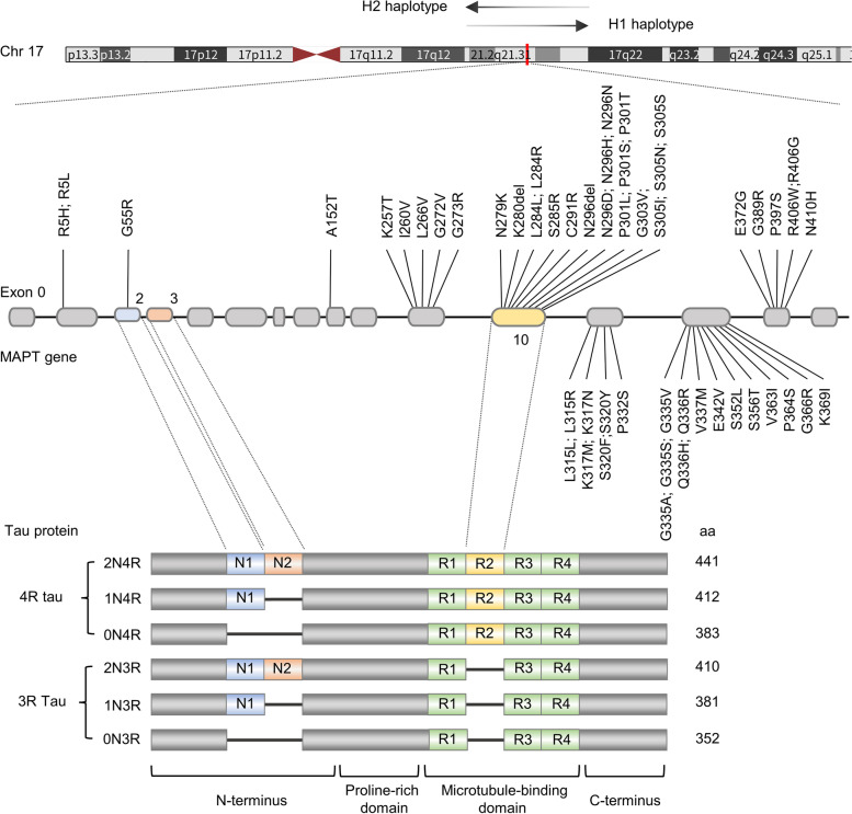
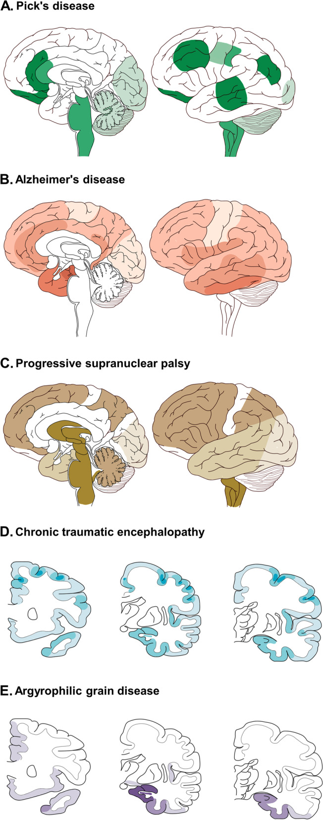
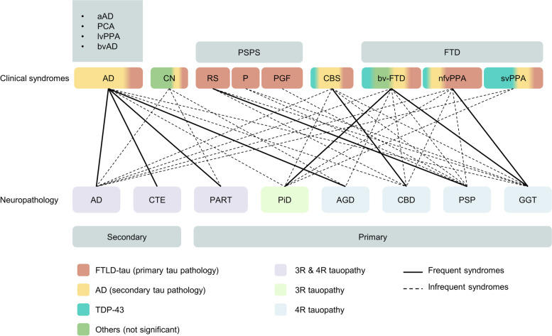
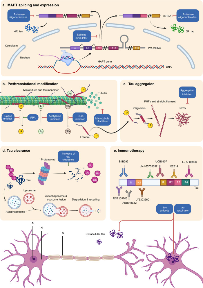
Verification
Claims the paper makes
The claims and statistics of this paper haven’t been checked yet.
Run the check — free, no account needed. It reads the open-access text and checks the sources it cites.
Claims other papers make about this one
Each is checked against this paper’s own text: does it say what the citing paper says it does?
The cited source says what the claim attributes to it.
In the citing paper
“Both 3R- and 4R-tau isoforms were detected with no noticeable difference in the 3R to 4R ratio between LOAD and HC 3D-CNs, consistent with the concept that AD is a mixed 3R/4R tauopathy (65, 66) (Fig. 3D).”
From Modeling late-onset Alzheimer’s disease neuropathology via direct neuronal reprogramming (2024) · cites this paper as [66]
Self-check
Percentages, sums and reported differences re-computed from the paper’s own numbers.
The self-check needs the paper’s full text.
Citations
Cites
1 retracted
Dementia prevention, intervention, and care: 2020 report of the Lancet Commission
Livingston, G., Huntley, J., Sommerlad, A., Ames, D., Ballard, C., Banerjee, S., Brayne, C., Burns, A., Cohen-Mansfield, J., Cooper, C., et al. (2020) · The Lancet · doi:10.1016/s0140-6736(20)30367-6 · Cited by 10,430
RetractedAlzheimer Disease Genetics Consortium (ADGC), Kunkle, B. W., The European Alzheimer’s Disease Initiative (EADI), Cohorts for Heart and Aging Research in Genomic Epidemiology Consortium (CHARGE), Genetic and Environmental Risk in AD/Defining Genetic, P. A. E. R. F. A. D. C. (., Grenier-Boley, B., Sims, R., Bis, J. C., Damotte, V., Naj, A. C., et al. (2019) · Nature Genetics · doi:10.1038/s41588-019-0358-2 · Cited by 2,946 · cited 2×
“These new risk genes were IQCK, ACE, ADAM10, ADAMTS1, and WWOX, and they also affected immunity and lipid metabolism in this study [121].”
BackgroundTau in physiology and pathology
Wang, Y., & Mandelkow, E. (2016) · Nature Reviews Neuroscience · doi:10.1038/nrn.2015.1 · Cited by 2,251
The spectrum of disease in chronic traumatic encephalopathy
McKee, A. C., Stein, T. D., Nowinski, C. J., Stern, R. A., Daneshvar, D. H., Alvarez, V. E., Lee, H.-S., Hall, G., Wojtowicz, S. M., Baugh, C. M., et al. (2013) · Brain · doi:10.1093/brain/aws307 · Cited by 2,098
Criteria for the diagnosis of corticobasal degeneration
Armstrong, M. J., Litvan, I., Lang, A. E., Bak, T. H., Bhatia, K. P., Borroni, B., Boxer, A. L., Dickson, D. W., Grossman, M., Hallett, M., et al. (2013) · Neurology · doi:10.1212/wnl.0b013e31827f0fd1 · Cited by 1,904
Jia, L., Du, Y., Chu, L., Zhang, Z., Li, F., Lyu, D., Li, Y., Li, Y., Zhu, M., Jiao, H., et al. (2020) · The Lancet Public Health · doi:10.1016/s2468-2667(20)30185-7 · Cited by 1,829
Self-citationPathology of Neurodegenerative Diseases
Dugger, B. N., & Dickson, D. W. (2017) · Cold Spring Harbor Perspectives in Biology · doi:10.1101/cshperspect.a028035 · Cited by 1,665
Palmqvist, S., Janelidze, S., Quiroz, Y. T., Zetterberg, H., Lopera, F., Stomrud, E., Su, Y., Chen, Y., Serrano, G. E., Leuzy, A., et al. (2020) · JAMA · doi:10.1001/jama.2020.12134 · Cited by 1,603
NLRP3 inflammasome activation drives tau pathology
Ising, C., Venegas, C., Zhang, S., Scheiblich, H., Schmidt, S. V., Vieira-Saecker, A., Schwartz, S., Albasset, S., McManus, R. M., Tejera, D., et al. (2019) · Nature · doi:10.1038/s41586-019-1769-z · Cited by 1,487
Clinicopathological evaluation of chronic traumatic encephalopathy in players of American football
Mez, J., Daneshvar, D. H., Kiernan, P. T., Abdolmohammadi, B., Alvarez, V. E., Huber, B. R., Alosco, M. L., Solomon, T. M., Nowinski, C. J., McHale, L., et al. (2017) · JAMA · doi:10.1001/jama.2017.8334 · Cited by 1,098
Research works printed but not linked: 1
- Boxer AL, Yu JT, Golbe LI, Litvan I, Lang AE, Höglinger GU. Advances in progressive supranuclear palsy: new diagnostic criteria, biomarkers, and therapeutic approaches. Lancet Neurol. 2017. https://doi.org/10.1016/s1474-4422(17)30157-6. · DOI · Paper page
Cited by
Sorted by citations
Items 101–200 of 329
Tau Lysine Pseudomethylation Regulates Microtubule Binding and Enhances Prion-like Tau Aggregation
Xia, Y., Bell, B. M., & Giasson, B. I. (2023) · International Journal of Molecular Sciences · doi:10.3390/ijms24098286 · Cited by 11
Bhopatkar, A. A., Bhatt, N. N., Haque, M. A., Xavier, R., Fung, L., Jerez, C., & Kayed, R. (2024) · Protein Science · doi:10.1002/pro.5099 · Cited by 9
Diagnostic and therapeutic targeting of pathological tau proteins in neurodegenerative disorders
Sahara, N., & Higuchi, M. (2023) · FEBS Open Bio · doi:10.1002/2211-5463.13711 · Cited by 9
Alhadidy, M. M., & Kanaan, N. M. (2024) · Biochemical Society Transactions · doi:10.1042/bst20230596 · Cited by 10
Kumari, S., Gupta, S., Sukhija, R., Gurjar, S., Dubey, S. K., & Taliyan, R. (2024) · European Journal of Pharmacology · doi:10.1016/j.ejphar.2024.177123 · Cited by 10
Antisense oligonucleotides: A promising advancement in neurodegenerative disease treatment
Dudzisz, K., & Wandzik, I. (2025) · European Journal of Pharmacology · doi:10.1016/j.ejphar.2025.177644 · Cited by 9
Frost, B. E., Rowe, J. B., Akinyemi, R., Abisambra, J. F., Ashton, N. J., Brendel, M., Buée, L., Butler, D., Carrillo, M. C., Chung, P. S., et al. (2025) · Alzheimer s & Dementia · doi:10.1002/alz.70078 · Cited by 9
Twenty years of therapeutic development in tauopathy mouse models: a scoping review
Langness, V. F., Simmons, D. A., McHugh, T. L. M., Butler, R. R., Zhou, J., Liu, H., Yang, T., Ellerby, L., & Longo, F. M. (2025) · Alzheimer s & Dementia · doi:10.1002/alz.70578 · Cited by 9
Singhaarachchi, P. H., Antal, P., Calon, F., Culmsee, C., Delpech, J.-C., Feldotto, M., Geertsema, J., Hoeksema, E. E., Kőrösi, A., Layé, S., et al. (2025) · Progress in Neurobiology · doi:10.1016/j.pneurobio.2025.102821 · Cited by 13
Perez-Garcia, G., Bicak, M., Haure‐Mirande, J., Perez, G. M., Otero‐Pagan, A., Sosa, M. A. G., Gasperi, R. D., Sano, M. C., Barlow, C., Gage, F. H., et al. (2023) · Neuroscience Letters · doi:10.1016/j.neulet.2023.137080 · Cited by 9
Selvarasu, K., Singh, A. K., Dakshinamoorthy, A., Sreenivasmurthy, S. G., Iyaswamy, A., Radhakrishnan, M., Patnaik, S., Huang, J., Williams, L. L., Senapati, S., et al. (2023) · Biomedicines · doi:10.3390/biomedicines12010005 · Cited by 9
Atomic Force Microscopy Applied to the Study of Tauopathies
Amorim, M. D. S. D. N., França, Á. R. S., Santos‐Oliveira, R., Sanches, J. R., Melo, T. M., Pinto, B., Barbosa, L. R. S., & Alencar, L. M. R. (2024) · ACS Chemical Neuroscience · doi:10.1021/acschemneuro.3c00819 · Cited by 7
Han, M., Saxton, A. D., Currey, H. N., Waldherr, S. M., Liachko, N. F., & Kraemer, B. C. (2024) · Disease Models & Mechanisms · doi:10.1242/dmm.050473 · Cited by 9
Rinaldi, M., Pezone, A., Quadrini, G. I., Abbadessa, G., Laezza, M. P., Passaro, M. L., Porcellini, A., & Costagliola, C. (2024) · Frontiers in Aging Neuroscience · doi:10.3389/fnagi.2024.1371745 · Cited by 9
Karabova, M. K., del Ser‐Badia, A., Hedegaard, A., Washer, S. J., Baykam, Z., O'Brien, D. P., Vendrell, I., Hester, S., Fischer, R., Johnson, E., et al. (2026) · Alzheimer s & Dementia · doi:10.1002/alz.71337 · Cited by 10
Zhang, Y., Yang, Y., Hu, Z., Zhu, M., Qin, S., Yu, P., Li, B., Xu, J., Ondrejčák, T., Klyubin, I. V., et al. (2022) · Journal of Alzheimer s Disease · doi:10.3233/jad-220351 · Cited by 8
Self-citationXia, Y., Bell, B. M., Kim, J. D., & Giasson, B. I. (2023) · Frontiers in Neuroscience · doi:10.3389/fnins.2023.1181804 · Cited by 8
Neurobiological correlation between phosphorylated tau and mood symptoms in memory clinic patients
Ozaki, T. J., Hashimoto, N., Udo, N., Narita, H., Nakagawa, S., & Kusumi, I. (2023) · Psychogeriatrics · doi:10.1111/psyg.13016 · Cited by 8
McArthur, N., Squire, J. D., Onyeachonam, O. J., Bhatt, N. N., Jerez, C., Holberton, A. L., Tessier, P. M., Wood, L. B., Kayed, R., & Kane, R. S. (2024) · Biomaterials Science · doi:10.1039/d4bm00707g · Cited by 8
Kim, Y. A., Mellén, M., Kızıl, Ç., & Santa‐María, I. (2023) · British Journal of Pharmacology · doi:10.1111/bph.16280 · Cited by 9
D’Brant, L., Rugenstein, N., Na, S. K., Miller, M. J., Czajka, T. F., Trudeau, N., Fitz, E. M., Tomaszek, L., Fisher, E. S., Mash, E., et al. (2024) · bioRxiv (Cold Spring Harbor Laboratory) · doi:10.1101/2024.05.28.596248 · Cited by 7
Non-Drug and Non-Invasive Therapeutic Options in Alzheimer’s Disease
Şovrea, A. S., Boşca, A. B., Dronca, E., Constantin, A.-M., Crintea, A., Suflețel, R. T., Lupean, R.-A., Ștefan, P.-A., Onofrei, M. M., Tschall, C., et al. (2025) · Biomedicines · doi:10.3390/biomedicines13010084 · Cited by 9
Geometry based prediction of tau protein sites and motifs associated with misfolding and aggregation
Sugiyama, M., Kosik, K. S., & Panagiotou, E. (2025) · Scientific Reports · doi:10.1038/s41598-025-93304-x · Cited by 8
Kotova, M. M., Stewart, A., Grigoryan, A., Shariff, S., Mirzaei, F., Kantawala, B., Dilbaryan, K., de Abreu, M. S., Galstryan, D. S., Cui, J., et al. (2025) · Journal of Neurochemistry · doi:10.1111/jnc.70090 · Cited by 7
Self-citationProtein quality control systems in neurodegeneration – culprits, mitigators, and solutions?
Ciechanover, A. J., & Livneh, I. (2025) · Frontiers in Neurology · doi:10.3389/fneur.2025.1604076 · Cited by 8
Pathomechanisms of depression in progressive supranuclear palsy
Jellinger, K. A. (2023) · Journal of Neural Transmission · doi:10.1007/s00702-023-02621-w · Cited by 7
Barker, R. M., Chambers, A., Kehoe, P. G., Rowe, E., & Perks, C. M. (2024) · Clinical Science · doi:10.1042/cs20230317 · Cited by 9
Imaging of Tauopathies with PET Ligands: State of the Art and Future Outlook
Conte, M., Feo, M. S. D., Sidrak, M. M. A., Corica, F., Gorica, J., Granese, G. M., Filippi, L., Vincentis, G. D., & Frantellizzi, V. (2023) · Diagnostics · doi:10.3390/diagnostics13101682 · Cited by 8
The pleiotropic architecture of human impulsivity across biological scalesPreprint
Mallard, T. T., Tubbs, J. D., Jennings, M. V., Zhang, Y., Gustavson, D. E., Grotzinger, A. D., Westwater, M. L., Williams, C. M., Fortgang, R. G., Elson, S. L., et al. (2023) · medRxiv · doi:10.1101/2023.11.28.23299133 · Cited by 7
Self-citationTargeting NOX inhibitors in neurodegeneration: a therapeutic perspective
Kaur, S., Verma, R., Sharma, V., & Singh, T. G. (2025) · Metabolic Brain Disease · doi:10.1007/s11011-025-01644-4 · Cited by 6
Murakami, K., Nguyen, T. H. V., Nagao, C., Mizuguchi, K., & Bitan, G. (2025) · JACS Au · doi:10.1021/jacsau.5c00269 · Cited by 6
Silva-Llanes, I., Smith, L. A., Abdelkader-Guillén, A., Jiménez-Villegas, J., Sarrió, D., Gema, M.-B., & Lastres‐Becker, I. (2026) · Journal of Biomedical Science · doi:10.1186/s12929-025-01210-1 · Cited by 7
PLCG2 across human disease: genetic variants, signaling mechanisms, and clinical implications
Jiang, P., Wang, M., Zhang, C., Li, X., Wang, Z. Q., Guo, Y., Zhang, J., Yan, W., & Xi, X. (2026) · Journal of Translational Medicine · doi:10.1186/s12967-025-07632-7 · Cited by 7
The O-GlcNAc dichotomy: when does adaptation become pathological?
Costa, T. J. D., Wilson, E. W., Fontes, M. T., Pernomian, L., Tostes, R. C., Wenceslau, C. F., & McCarthy, C. G. (2023) · Clinical Science · doi:10.1042/cs20220309 · Cited by 6
Time-Dependent Long-Term Effect of Memantine following Repetitive Mild Traumatic Brain Injury
Boucher, M. L., Conley, G. M., Morriss, N. J., Ospina-Mora, S., Qiu, J., Mannix, R., & Meehan, W. P., III (2024) · Journal of Neurotrauma · doi:10.1089/neu.2023.0423 · Cited by 6
Yoon, S., Kim, H. Y., Park, S., Cha, M., Kim, K., Lee, S., Kim, J., Bhang, S., & Kim, Y. (2024) · Angewandte Chemie International Edition · doi:10.1002/anie.202411942 · Cited by 6
HMGA1 deficiency: a pathogenic link between tau pathology and insulin resistance
Mirabelli, M., CHIEFARI, E. D., Arcidiacono, B., Salatino, A., Pascarella, A., Morelli, M., Credendino, S. C., Brunetti, F. S., Vito, A. D., Greco, A., et al. (2025) · EBioMedicine · doi:10.1016/j.ebiom.2025.105700 · Cited by 6
Aathira, N. S., Kaur, A., Kumar, A., Dar, G. M., Nimisha, Sharma, A. K., Bera, P., Mahajan, B., Chatterjee, A., & Saluja, S. S. (2025) · Neuroscience · doi:10.1016/j.neuroscience.2025.05.021 · Cited by 7
Smith, E. D., Torrellas, J., McKenna, R., Vo, Q., Mietzsch, M., Borchelt, D., Prokop, S., & Chakrabarty, P. (2025) · Journal of Neurochemistry · doi:10.1111/jnc.70221 · Cited by 6
Matsuda, H., & Yamao, T. (2023) · FUKUSHIMA JOURNAL OF MEDICAL SCIENCE · doi:10.5387/fms.2023-08 · Cited by 6
Rottlerin Enhances the Autophagic Degradation of Phosphorylated Tau in Neuronal Cells
Kam, M. K., Park, J.-Y., Yun, G. H., Sohn, H., Park, J., Choi, J., Koh, Y. H., & Jo, C. (2024) · Molecular Neurobiology · doi:10.1007/s12035-024-04182-9 · Cited by 6
Advances in Alzheimer’s disease: mechanistic insights and therapeutic targets
Jia, Y.-J., Ge, Y., Li, B., Yang, Y., Chen, H., Liu, J., Guo, J., Yu, J., Ye, K., Wang, J., et al. (2026) · Science China Life Sciences · doi:10.1007/s11427-025-2991-7 · Cited by 6
Self-citationReversibility and β-sheet formation are decoupled in tau condensate aging
Fischer, C. M., Edu, I. A., Šneideris, T., Baronaitė, I., Toprakcioglu, Z., Deck, L.-T., Qian, D., Scrutton, R. M., Dreyer, L. S., Wen, J., et al. (2026) · Proceedings of the National Academy of Sciences · doi:10.1073/pnas.2522993123 · Cited by 5
Malik, N., Miah, M.-U., Galgani, A., McAleese, K. E., Walker, L., LeBeau, F. E. N., Attems, J., Outeiro, T. F., Thomas, A. J., & Koss, D. J. (2024) · Acta Neuropathologica · doi:10.1007/s00401-024-02691-4 · Cited by 5
Liu, T., Ren, C., Guo, W., Zhang, X., Li, Y., Wang, Y., Zhang, Q., Chen, B., Dai, J., Yan, X., et al. (2024) · European Journal of Medicinal Chemistry · doi:10.1016/j.ejmech.2024.117046 · Cited by 6
Taha, H. B., Chawla, E., & Bitan, G. (2023) · Journal of the American Society for Mass Spectrometry · doi:10.1021/jasms.3c00065 · Cited by 4
Proteomic networks of gray and white matter reveal tissue‐specific changes in human tauopathy
Johnson, A. G., Dammer, E. B., Webster, J. A., Duong, D. M., Seyfried, N. T., & Hales, C. M. (2024) · Annals of Clinical and Translational Neurology · doi:10.1002/acn3.52134 · Cited by 5
Patterns of Tau pathology in patients with anti-IgLON5 disease visualized by Florzolotau (18F) PET
Fan, S., Jia, C., Liang, M., Ren, H., Zhang, T., Li, Q., Huang, Z., Yen, T., OuYang, C., Cui, R., et al. (2025) · Journal of Neurology · doi:10.1007/s00415-024-12874-4 · Cited by 5
Hartmann, C., Anskat, M., Ehrlich, M., Sterneckert, J. L., Pal, A., & Hermann, A. (2024) · Biomedicines · doi:10.3390/biomedicines12030641 · Cited by 5
Nesfatin-1 ameliorates pathological abnormalities in Drosophila hTau model of Alzheimer's disease
Yang, J., Baek, S.-E., Yoon, J.-W., Kim, H., Kwon, Y., & Yeom, E. (2024) · Biochemical and Biophysical Research Communications · doi:10.1016/j.bbrc.2024.150311 · Cited by 5
An Extensive Atlas of Proteome and Phosphoproteome Turnover Across Mouse Tissues and Brain RegionsPreprint
Li, W., Dasgupta, A., Yang, K., Wang, S., Hemandhar-Kumar, N., Yarbro, J. M., Hu, Z., Šalovská, B., Fornasiero, E. F., Peng, J., et al. (2024) · bioRxiv (Cold Spring Harbor Laboratory) · doi:10.1101/2024.10.15.618303 · Cited by 5
Zhou, Y., Wang, Y., Yang, H., Zhang, C., Meng, J., Zhang, L., Li, K., Huang, L.-L., Zhang, X., Luo, H., et al. (2025) · Acta Pharmaceutica Sinica B · doi:10.1016/j.apsb.2025.09.024 · Cited by 5
Self-citationKinetic Ensemble of Tau Protein through the Markov State Model and Deep Learning Analysis
Yuan, Y., Mao, X., Pan, X., Zhang, R., & Su, W. (2024) · Journal of Chemical Theory and Computation · doi:10.1021/acs.jctc.3c01211 · Cited by 4
EBP1 potentiates amyloid β pathology by regulating γ-secretase
Kim, B.-S., Hwang, I., Ko, H. R., Kim, Y. K., Kim, H. J., Seo, S. W., Choi, Y., Lim, S., Kim, Y. K., Nie, S., et al. (2025) · Nature Aging · doi:10.1038/s43587-024-00790-1 · Cited by 4
O’Day, D. H. (2025) · Biomolecules · doi:10.3390/biom15081133 · Cited by 5
Design and application of a Tau seed amplification assay for screening inhibitors of Tau seeding
Gorski, D., Evans, H., Allison, T., Barria, C., Harrison, D., Banerjee, V., Méndez, N., Shahnawaz, M., Kaalund, S. S., Folke, J., et al. (2025) · Alzheimer s Research & Therapy · doi:10.1186/s13195-025-01855-y · Cited by 5
Protein fingerprints of brain-derived extracellular vesicles predict types of tau pathology
Espourteille, J., Barve, A., Zufferey, V., Leroux, É., Perbet, R., Bégard, S., Caillierez, R., Maurage, C., Toni, N., Buée, L., et al. (2025) · Alzheimer's Research & Therapy · doi:10.1186/s13195-025-01865-w · Cited by 4
Mallard, T. T., Tubbs, J. D., Jennings, M. V., Zhang, Y., Gustavson, D. E., Grotzinger, A. D., Westwater, M. L., Williams, C. M., Fortgang, R. G., Elson, S. L., et al. (2025) · American Journal of Psychiatry · doi:10.1176/appi.ajp.20240382 · Cited by 13
Self-citationExperimental modeling for tauopathies: An isogenic panel of humanized MAPT knock-in mice
Morito, T., & Watamura, N. (2025) · Neuroscience Research · doi:10.1016/j.neures.2025.104986 · Cited by 5
Targeted tau protein degradation: a promising therapeutic approach for tauopathies
Baazaoui, N., Alfaifi, M. Y., Shati, A. A., Saad, R. B., & Garzoli, S. (2025) · Neuroscience · doi:10.1016/j.neuroscience.2025.11.041 · Cited by 5
AggrescanAI: Prediction of Aggregation-Prone Regions Using Contextualized Embeddings
Navarro, A. M., Palacios, S., Galmarini, T., Bárcenas, O., Ventura, S., & Marino‐Buslje, C. (2026) · Journal of Molecular Biology · doi:10.1016/j.jmb.2026.169643 · Cited by 4
Small molecules to target tau amyloid aggregation
Manglano-Artuñedo, Z., Peña‐Díaz, S., & Ventura, S. (2023) · Neural Regeneration Research · doi:10.4103/1673-5374.380900 · Cited by 4
Hurtado-Carneiro, V., Ayuso, Y. L. B., Velázquez, E., Lamas, C. F., de la Rosa, R. F., Gómez-Oliver, F., García, L. G., Ruiz‐Albusac, J. M., Pozo, M. A., & Fernández, E. B. (2024) · Heliyon · doi:10.1016/j.heliyon.2024.e35752 · Cited by 6
Taniguchi, D., Shimonaka, S., Ahmed, I., Elahi, M., Hatano, T., Imai, Y., & Hattori, N. (2024) · Neurobiology of Disease · doi:10.1016/j.nbd.2024.106686 · Cited by 5
Silva-Llanes, I., Madruga, E. L., Martı́nez, A., & Lastres‐Becker, I. (2025) · Frontiers in Neuroscience · doi:10.3389/fnins.2024.1530809 · Cited by 5
Mechanistic role of proteins and peptides in Management of Neurodegenerative Disorders
Awasthi, S., Tiwari, P. C., Awasthi, S., Dwivedi, A., & Srivastava, S. (2025) · Neuropeptides · doi:10.1016/j.npep.2025.102505 · Cited by 4
Cawley, N. X., Zhou, R., Farhat, N. M., Iben, J., Alexander, D. M., Luke, R., Padilla, C. J., Mohamed, H. O., Albert, O. K., Robbins, K. P., et al. (2025) · Journal of Inherited Metabolic Disease · doi:10.1002/jimd.70016 · Cited by 5
Differences in the soluble and insoluble proteome between primary tauopathies
Kavanagh, T., Balcomb, K., Trgovcevic, S., Nementzik, L. R., Kanshin, E., Halliday, G. M., Ueberheide, B. M., & Drummond, E. (2025) · Alzheimer s & Dementia · doi:10.1002/alz.70401 · Cited by 5
Mapping the Structural Landscape of Amyloid Fibrils to Guide Polymorph-Specific TherapeuticsPreprint
Sadek, A., Correia, B. E., & Lashuel, H. A. (2025) · bioRxiv (Cold Spring Harbor Laboratory) · doi:10.1101/2025.05.08.652887 · Cited by 3
Zhu, R., Kim, G., Rajewski, B. H., Angera, I. J., Valle, J. R. D., & Wang, Y. (2025) · Advanced NanoBiomed Research · doi:10.1002/anbr.202500065 · Cited by 3
NVP-BEZ235 enhances autophagy and ameliorates cognitive deficits by targeting tauopathies
Alsharif, I. (2025) · Experimental and Molecular Pathology · doi:10.1016/j.yexmp.2025.104988 · Cited by 3
Bhandari, A. K., Dhami, A. S., Hemanthlumar, R. T., Mishra, N., Joslyn, B., Roy, S., Srinivasan, J., Prajapati, K. P., Kar, K., & Anand, B. G. (2025) · Ageing Research Reviews · doi:10.1016/j.arr.2025.102879 · Cited by 4
A novel lncRNA FAM151B-DT regulates degradation of aggregation prone proteins
Renganathan, A., Minaya, M. A., Broder, M., Alfradique-Dunham, I., Miller, R. L., Dhavale, D. D., Moritz, M., Bhagat, R., Marsh, J. A., Verbeck, A. C., et al. (2025) · Molecular Psychiatry · doi:10.1038/s41380-025-03277-6 · Cited by 4
Acar, B., Pepe, N. A., Živković, A., Stark, H., & Şen, A. (2025) · Current Medicinal Chemistry · doi:10.2174/0109298673435364251002112630 · Cited by 3
Ibrahim, M., Khalil, A. M., Attia, H., Alseekh, S., Mohamed, A. F., & El-Yamany, M. F. (2026) · Journal of Neuroimmune Pharmacology · doi:10.1007/s11481-025-10270-x · Cited by 3
Shoff, T. A., Derbez-Morin, M., Cai, P., & Julian, R. R. (2026) · PNAS Nexus · doi:10.1093/pnasnexus/pgag034 · Cited by 4
Mathematical topology and geometry-based classification of tauopathies
Sugiyama, M., Kosik, K. S., & Panagiotou, E. (2024) · Scientific Reports · doi:10.1038/s41598-024-58221-5 · Cited by 4
Recent Advances in Mapping Protein Self-Assembly and Aggregation for Common Proteinopathies
Bhattacharya, S., & Thompson, D. (2024) · Acta Physica Polonica A · doi:10.12693/aphyspola.145.s37 · Cited by 3
Pandey, N., Srivastava, N. K., Kumar, A., Hussain, I., & Joshi, D. (2024) · The Egyptian Journal of Neurology Psychiatry and Neurosurgery · doi:10.1186/s41983-024-00860-6 · Cited by 3
Role of non-neuronal cells in neurorepair: a focus on proteinopathy and neurodegeneration
Rodríguez-Carreiro, S., Nogales, M. D. C., Jiménez-Galán, D., Carmona-Lorenzo, S., Caro-Martín, A., Navarro, E., & Esteras, N. (2024) · Regenerative medicine reports . · doi:10.4103/regenmed.regenmed-d-24-00005 · Cited by 2
Tau positron emission tomography in tauopathies: A narrative review
Lee, H., Gu, Y., Seo, S. W., & Moon, S. H. (2023) · Precision and Future Medicine · doi:10.23838/pfm.2023.00016 · Cited by 4
Insights to develop tau‐directed therapeutics to protect the synaptic integrity for tauopathies
Cha, E., Kim, H. J., & Lim, S. M. (2023) · Bulletin of the Korean Chemical Society · doi:10.1002/bkcs.12792 · Cited by 4
Pragati, & Sarkar, S. (2024) · Brain Research · doi:10.1016/j.brainres.2024.149347 · Cited by 3
Rierola, M., Trushina, N. I., Monteiro-Abreu, N., Conze, C., Holtmannspötter, M., Kurre, R., Holzer, M., Arendt, T., Heinisch, J. J., Brandt, R., et al. (2022) · bioRxiv (Cold Spring Harbor Laboratory) · doi:10.1101/2022.06.09.495530 · Cited by 4
The Effect of the Tau Protein on D. melanogaster Lifespan Depends on GSK3 Expression and Sex
Veselkina, E. R., Trostnikov, M. V., Roshina, N. V., & PASYUKOVA, E. G. (2023) · International Journal of Molecular Sciences · doi:10.3390/ijms24032166 · Cited by 3
Integrating visual assessments and quantification methods for tau PET staging
Gu, Y., Yun, J., Moscoso, A., Schöll, M., Shin, D., Lee, E. H., Kang, H., Yim, S., Jo, H., Kim, J. P., et al. (2025) · Alzheimer s & Dementia · doi:10.1002/alz.70352 · Cited by 2
Mohammed, S. N., Direksunthorn, T., Zwamel, A. H., Uthirapathy, S., Ballal, S., Kalia, R., Arya, R., Naidu, K. S., Ahmad, I. S., & Ahmed, J. K. (2025) · Molecular Neurobiology · doi:10.1007/s12035-025-05338-x · Cited by 3
Santiago-Ruiz, A. N., Hugelier, S., Correa, G. L., Bond, C., Lee, E. B., & Lakadamyali, M. (2025) · Advanced Science · doi:10.1002/advs.202509773 · Cited by 3
Spontaneous isomerization of tau is most prevalent in Alzheimer’s disease
Hubbard, E. E., Reagle, K., Lui, M. K., Derbez-Morin, M., Merrihew, G. E., MacCoss, M. J., & Julian, R. R. (2026) · NeuroMarkers. · doi:10.1016/j.neumar.2025.100152 · Cited by 2
Spilling the T: T cells in tauopathy mechanisms, disease progression, and therapeutic horizons
Bosch, M. E., & Ulrich, J. D. (2026) · Molecular Neurodegeneration Advances · doi:10.1186/s44477-026-00020-5 · Cited by 3
The LRRK2 G2385R Exonic Variant: A Potential Genetic Disease Modifier of Multiple System Atrophy?
Kouki, N., Traon, A. P., Chelban, V., Chanquoy, F., Fabbri, M., Bendetowicz, D., Houlden, H., Schottlaender, L., Rascol, O., Foubert‐Samier, A., et al. (2026) · Movement Disorders · doi:10.1002/mds.70219 · Cited by 2
Sharma, R., Song, B. S., Rane, R., Paul, D., Singh, B., Verma, T., Mishra, A., Karan, A., & Song, J. J. (2026) · Biomedicine & Pharmacotherapy · doi:10.1016/j.biopha.2026.119297 · Cited by 2
Demuro, S., Russo, D., Penna, I., Grabska, S., Grabski, H., Vedove, A. D., Valeri, A., Sauvey, C., Ottonello, G., Summa, M., et al. (2026) · Journal of Medicinal Chemistry · doi:10.1021/acs.jmedchem.5c01810 · Cited by 3
Using iPSC models to examine neuron–glia interactions in neurodegenerative diseases
Lopez, D. M., Keavey, L., & Bowles, K. R. (2026) · Bioscience Reports · doi:10.1042/bsr20250131 · Cited by 2
Huang, R., Ye, X., Liu, J., Yang, C., Meng, X., Dou, T., Zhuang, X., Yang, J., Zheng, N., Zhao, Y., et al. (2026) · Pharmacological Research · doi:10.1016/j.phrs.2026.108265 · Cited by 2
Rahhal, S. N., Farmer, C., Thurm, A. E., Wassif, C. A., Cawley, N. X., Perreault, J., Do, A. N. D., Bianconi, S. E., Hannah‐Shmouni, F., Guthrie, W., et al. (2023) · Molecular Genetics and Metabolism Reports · doi:10.1016/j.ymgmr.2023.101001 · Cited by 2
Potent of strategic approaches for tauopathies ranging from single-cell transcriptome to microbiome
Kim, S., Yang, S., Yang, E.-J., Kang, M., & Joo, J. (2023) · Animal Cells and Systems · doi:10.1080/19768354.2023.2285826 · Cited by 2
Mishra, S., Aziz, T., Toppo, A. J., Kumar, D., Tirkey, M. P., Beck, P., Anjum, N., Sharma, D., Khan, M. M., Kumari, S., et al. (2024) · Cureus · doi:10.7759/cureus.52423 · Cited by 2
Endothelial cellular senescence and tau accumulation: An interplay full of opportunities?
Oliveri, D., Moschetti, G., Griego, A., & Scarpa, E. (2024) · Ibrain · doi:10.1002/ibra.12154 · Cited by 2
Role of 18 F‐florzolotau PET in diagnostic and therapeutic decision‐making for cognitive impairment
Wang, J., Wang, Z., Chen, S., Wang, R., Chen, K., Lu, J., Xin, J., Huang, Y., Wang, M., Xie, F., et al. (2025) · Alzheimer s & Dementia · doi:10.1002/alz.70563 · Cited by 2
Self-citation
Metadata
- Authors
- Zhang, Y., Wu, K.-M., Yang, L., Dong, Q., & Yu, J.-T.
- Affiliations
- Shanghai Medical College of Fudan University (CN)
- Corresponding author
- Yu, Jin-Tai
- Venue
- Molecular Neurodegeneration
- Published
- 2022-04-07
- Volume
- 17
- Issue
- 1
- Pages
- 28-28
- ISSN
- 1750-1326 (ISSN-L)
- PMC
- PMC8991707
- OpenAlex
- W4223629343
- Cited by
- 323
- References
- 228
- Citations by year
- 2026: 69 · 2025: 83 · 2024: 97 · 2023: 60 · 2022: 14
- FWCI
- 8.91
- Type
- journal article
- Language
- en
- Open access
- gold
- doi.org
- Locations
- Molecular Neurodegeneration (publishedVersion, cc-by)
- PubMed Central (publishedVersion, cc-by)
- PubMed (publishedVersion)
- DOAJ (DOAJ: Directory of Open Access Journals) (submittedVersion)
- Funders
- National Natural Science Foundation of China (91849126)
- Shanghai Municipal Science and Technology Major Project (No.2018SHZDZX01)
- Topics
- Life Sciences › Neuroscience › Cellular and Molecular Neuroscience › Alzheimer's disease research and treatments
- Health Sciences › Medicine › Psychiatry and Mental health › Dementia and Cognitive Impairment Research
- Health Sciences › Medicine › Clinical Neurology › Amyotrophic Lateral Sclerosis Research
- Keywords
- tauopathy, tau protein, neurodegenerative diseases, tau isoforms, tau biomarkers, cerebrospinal fluid biomarkers, blood-based biomarkers, neuroimaging, disease-modifying therapy, early diagnosis
- MeSH terms
- Inclusion Bodies, Humans, Neurons, tau Proteins, Neurodegenerative Diseases, Protein Isoforms, Tauopathies
About these checks
Registry, retraction, Cites and statistics results are computed, not judged, with one exception: a printed reference that no index lists is read once by a model before it counts as a citation error. Claims are extracted by a model and kept only when their quote appears verbatim in the text; each cited source is then checked, with a model’s help, for whether it says what the claim relies on. Those verdicts can be wrong — read the claim’s quote and the cited source before relying on one.
Statistics and arithmetic are recomputed, not judged.
Confidence bands are shown only for fields calibrated on our annotated test set. None is calibrated yet, so no band is shown.
What counts as a claim ›How we measure confidence ›How we check this ›
Are you an author, or do you see a problem with this page? Request a review
