

The DOI matches its Crossref record; 228 of the 228 references it cites were found in a registry or an index.
Tauopathies: new perspectives and challenges
Open accessAbstract
Background: Tauopathies are a class of neurodegenerative disorders characterized by neuronal and/or glial tau-positive inclusions. Main body: Clinically, tauopathies can present with a range of phenotypes that include cognitive/behavioral-disorders, movement disorders, language disorders and non-specific amnestic symptoms in advanced age. Pathologically, tauopathies can be classified based on the predominant tau isoforms that are present in the inclusion bodies (i.e., 3R, 4R or equal 3R:4R ratio). Imaging, cerebrospinal fluid (CSF) and blood-based tau biomarkers have the potential to be used as a routine diagnostic strategy and in the evaluation of patients with tauopathies. As tauopathies are strongly linked neuropathologically and genetically to tau protein abnormalities, there is a growing interest in pursuing of tau-directed therapeutics for the disorders. Here we synthesize emerging lessons on tauopathies from clinical, pathological, genetic, and experimental studies toward a unified concept of these disorders that may accelerate the therapeutics. Conclusions: Since tauopathies are still untreatable diseases, efforts have been made to depict clinical and pathological characteristics, identify biomarkers, elucidate underlying pathogenesis to achieve early diagnosis and develop disease-modifying therapies.
Overview
The claims and statistics of this paper haven’t been checked yet.
Run the check — free, no account needed. It reads the open-access text and checks the sources it cites.
Integrity signals
Registry
Cites
Statistics
Figures
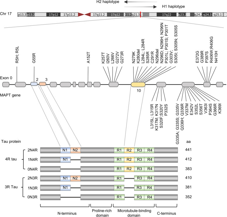
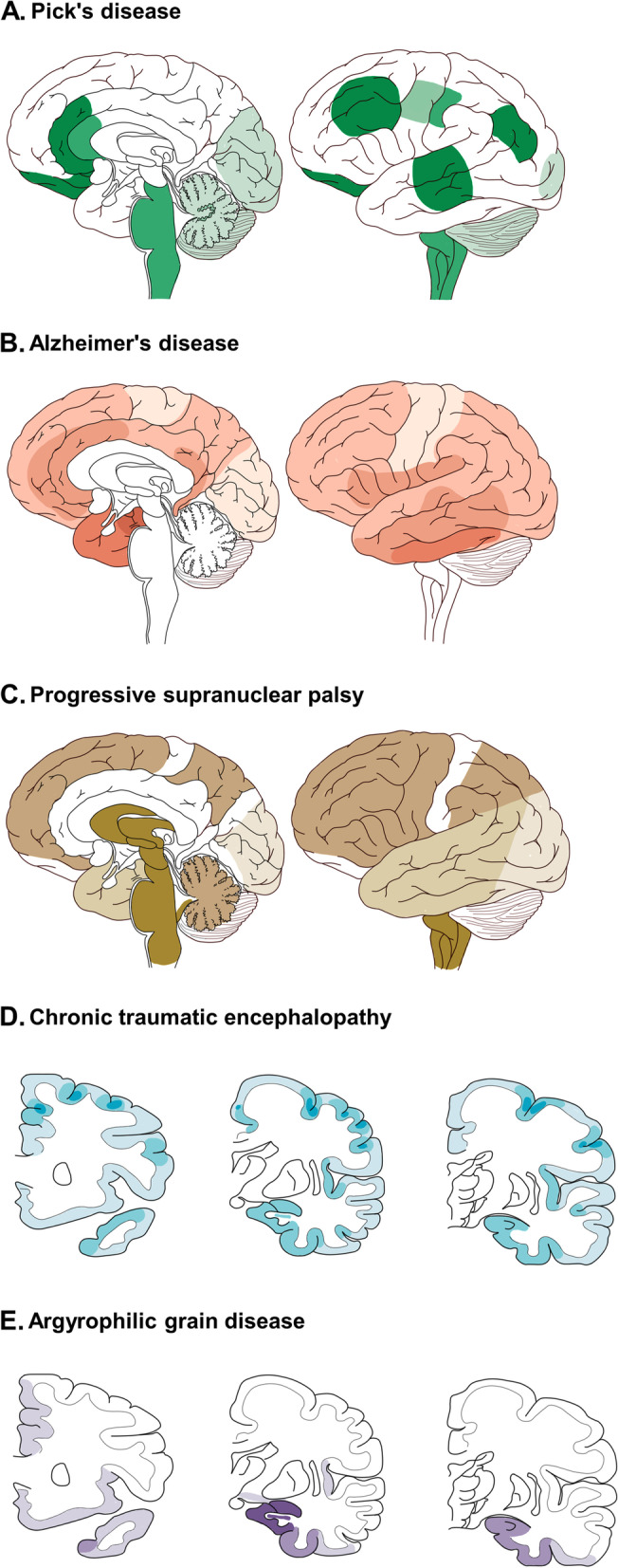
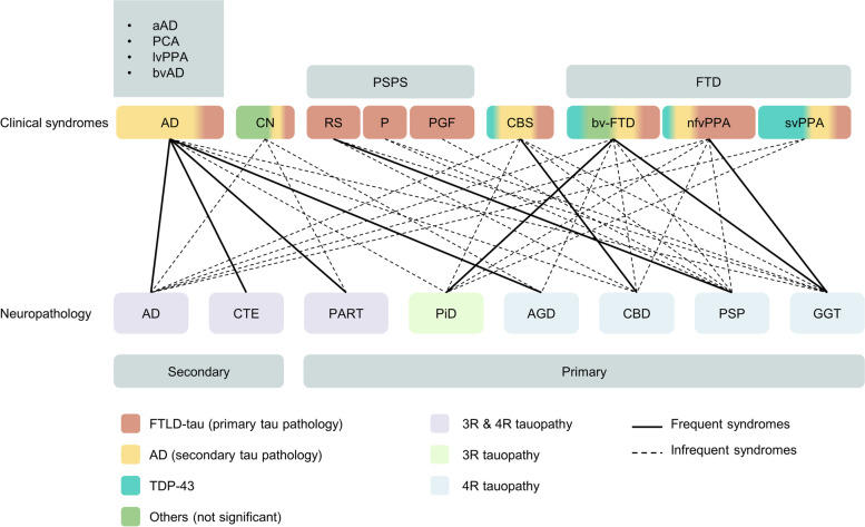
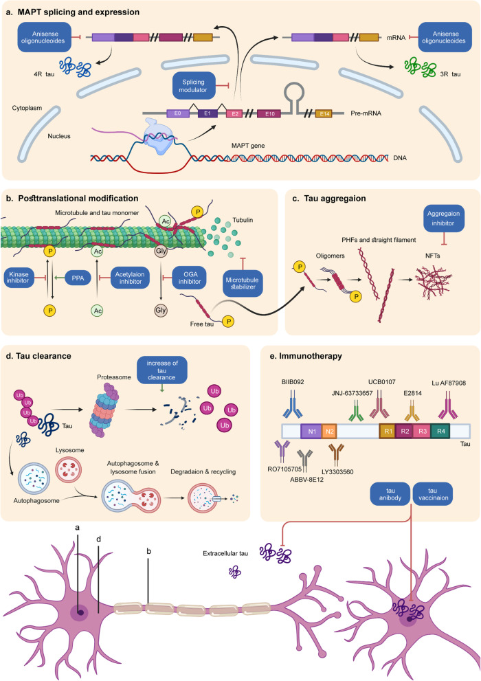
Verification
Claims the paper makes
The claims and statistics of this paper haven’t been checked yet.
Run the check — free, no account needed. It reads the open-access text and checks the sources it cites.
Claims other papers make about this one
Each is checked against this paper’s own text: does it say what the citing paper says it does?
The cited source says what the claim attributes to it.
In the citing paper
“Both 3R- and 4R-tau isoforms were detected with no noticeable difference in the 3R to 4R ratio between LOAD and HC 3D-CNs, consistent with the concept that AD is a mixed 3R/4R tauopathy (65, 66) (Fig. 3D).”
From Modeling late-onset Alzheimer’s disease neuropathology via direct neuronal reprogramming (2024) · cites this paper as [66]
Self-check
Percentages, sums and reported differences re-computed from the paper’s own numbers.
The self-check needs the paper’s full text.
Citations
Cites
1 retracted
Dementia prevention, intervention, and care: 2020 report of the Lancet Commission
Livingston, G., Huntley, J., Sommerlad, A., Ames, D., Ballard, C., Banerjee, S., Brayne, C., Burns, A., Cohen-Mansfield, J., Cooper, C., et al. (2020) · The Lancet · doi:10.1016/s0140-6736(20)30367-6 · Cited by 10,430
RetractedAlzheimer Disease Genetics Consortium (ADGC), Kunkle, B. W., The European Alzheimer’s Disease Initiative (EADI), Cohorts for Heart and Aging Research in Genomic Epidemiology Consortium (CHARGE), Genetic and Environmental Risk in AD/Defining Genetic, P. A. E. R. F. A. D. C. (., Grenier-Boley, B., Sims, R., Bis, J. C., Damotte, V., Naj, A. C., et al. (2019) · Nature Genetics · doi:10.1038/s41588-019-0358-2 · Cited by 2,946 · cited 2×
“These new risk genes were IQCK, ACE, ADAM10, ADAMTS1, and WWOX, and they also affected immunity and lipid metabolism in this study [121].”
BackgroundTau in physiology and pathology
Wang, Y., & Mandelkow, E. (2016) · Nature Reviews Neuroscience · doi:10.1038/nrn.2015.1 · Cited by 2,251
The spectrum of disease in chronic traumatic encephalopathy
McKee, A. C., Stein, T. D., Nowinski, C. J., Stern, R. A., Daneshvar, D. H., Alvarez, V. E., Lee, H.-S., Hall, G., Wojtowicz, S. M., Baugh, C. M., et al. (2013) · Brain · doi:10.1093/brain/aws307 · Cited by 2,098
Criteria for the diagnosis of corticobasal degeneration
Armstrong, M. J., Litvan, I., Lang, A. E., Bak, T. H., Bhatia, K. P., Borroni, B., Boxer, A. L., Dickson, D. W., Grossman, M., Hallett, M., et al. (2013) · Neurology · doi:10.1212/wnl.0b013e31827f0fd1 · Cited by 1,904
Jia, L., Du, Y., Chu, L., Zhang, Z., Li, F., Lyu, D., Li, Y., Li, Y., Zhu, M., Jiao, H., et al. (2020) · The Lancet Public Health · doi:10.1016/s2468-2667(20)30185-7 · Cited by 1,829
Self-citationPathology of Neurodegenerative Diseases
Dugger, B. N., & Dickson, D. W. (2017) · Cold Spring Harbor Perspectives in Biology · doi:10.1101/cshperspect.a028035 · Cited by 1,665
Palmqvist, S., Janelidze, S., Quiroz, Y. T., Zetterberg, H., Lopera, F., Stomrud, E., Su, Y., Chen, Y., Serrano, G. E., Leuzy, A., et al. (2020) · JAMA · doi:10.1001/jama.2020.12134 · Cited by 1,603
NLRP3 inflammasome activation drives tau pathology
Ising, C., Venegas, C., Zhang, S., Scheiblich, H., Schmidt, S. V., Vieira-Saecker, A., Schwartz, S., Albasset, S., McManus, R. M., Tejera, D., et al. (2019) · Nature · doi:10.1038/s41586-019-1769-z · Cited by 1,487
Clinicopathological evaluation of chronic traumatic encephalopathy in players of American football
Mez, J., Daneshvar, D. H., Kiernan, P. T., Abdolmohammadi, B., Alvarez, V. E., Huber, B. R., Alosco, M. L., Solomon, T. M., Nowinski, C. J., McHale, L., et al. (2017) · JAMA · doi:10.1001/jama.2017.8334 · Cited by 1,098
Research works printed but not linked: 1
- Boxer AL, Yu JT, Golbe LI, Litvan I, Lang AE, Höglinger GU. Advances in progressive supranuclear palsy: new diagnostic criteria, biomarkers, and therapeutic approaches. Lancet Neurol. 2017. https://doi.org/10.1016/s1474-4422(17)30157-6. · DOI · Paper page
Cited by
Sorted by citations
Items 301–329 of 329
Wiegand, T. L. T., Rubinski, A., Jung, L., Franzmeier, N., Arciniega, H., Ravanfar, P., Dewenter, A., Alosco, M. L., Tripodis, Y., Su, Y., et al. (2026) · Alzheimer s & Dementia · doi:10.1002/alz.71503
Disentangling Intrachain Folding from Interchain Assembly through Multidimensional Visualization
Sanches, M. N., Ganguly, P., Shea, J., & Leite, V. B. P. (2026) · The Journal of Physical Chemistry B · doi:10.1021/acs.jpcb.6c02030
Riboregulation: a non-canonical tau function
LeBlanc, K. R., Eck, R. J., Benbow, S. J., & Kraemer, B. C. (2026) · Molecular Neurodegeneration · doi:10.1186/s13024-026-00949-x
Omipalisib reduces hyperphosphorylated tau protein by modulating mTOR-autophagy pathway
Hwang, H., Kim, N., Jeon, S., Lee, Y., Son, J., Jeon, S. H., Lee, Y., Gee, M. S., Inn, K., & Lee, J. K. (2026) · PLoS ONE · doi:10.1371/journal.pone.0352120
Liao, K., Li, J., Jiao, F., Zhang, Z., Xu, L., Wang, Z., Li, W., & Pang, H. (2026) · EJNMMI Physics · doi:10.1186/s40658-026-00914-z
Neuroinflammation and Tauopathies
Zeenny, M.-N., Salame, I., Maatouk, L., & Akoury, E. (2026) · Molecular Biology Reports · doi:10.1007/s11033-026-12253-4
Röntgen, A., Fusco, G., Breiter, J. C., Beckwith, J. S., Lachica, J., Toomey, C. E., Singh, J., Klementieva, O. L., Gandhi, S., Lee, S. F., et al. (2026) · bioRxiv (Cold Spring Harbor Laboratory) · doi:10.64898/2026.07.05.736591
Production of Hyperphosphorylated Tau Using a Baculovirus-Insect Cell System
Min, S., Horgan, N. G., & Rochet, J.-C. (2026) · Neuromethods · doi:10.1007/978-1-0716-5234-3_1
Conformational bifurcation of tau R3–R4 oligomers underlies fibril polymorphism
Zhang, T., Dai, L., Ma, Y., Xu, S., Liu, X., Zhao, M., Li, W., Wang, J., & Liu, Y. (2026) · Biophysical Journal · doi:10.1016/j.bpj.2026.07.020
MAPT-derived circTau6 induces tau pathogenesis and neurodegeneration by encoding Tau variants
Jiang, G., Wang, D., Shao, L., Mao, X., Xiong, X., Zhang, D., Chen, W. N., Yuan, H., Wang, S., Zhang, J., et al. (2026) · Molecular Psychiatry · doi:10.1038/s41380-026-03764-4 · Cited by 1
Self-citationRNA Splicing Dysregulation in Parkinson’s Disease
Bruno, M. G., Menichetti, G., Valentino, A., Javaid, A., Belpinati, F., Ruggiero, A., Valenti, M. T., Paolone, G., Cavaliere, F., Trabetti, E., et al. (2026) · Genes · doi:10.3390/genes17080911
Aggarwal, P., & Sarkar, S. (2026) · Biochimica et Biophysica Acta (BBA) - Molecular Basis of Disease · doi:10.1016/j.bbadis.2026.168393
NanoBiT screening identifies the azaoxafluorene VT11 as potent tau interaction inhibitor
Ma, N., Stieler, J., Hilbrich, I., Vogel, T., Hilgeroth, A., Briel, D., Schaefer, M., Holzer, M., & Metelmann, M. (2026) · Scientific Reports · doi:10.1038/s41598-026-65368-w
Tau spreading coordinates intercellular lipid flux enhancing neuronal resilience to lipid toxicityPreprint
Oliveras, A., Ibatici, D., Calvo‐Noriega, M., Mastrobuoni, G., Forbes, M., Padilla, L. S., Aykul, M., Horvatović, K., Rusu, M.-C., Patsonis, M., et al. (2026) · bioRxiv (Cold Spring Harbor Laboratory) · doi:10.64898/2026.07.30.741734
Czech, J., Weyland, C. S., Neumann, M., Drzezga, A. E., & Schulz, J. B. (2026) · Springer eBooks · doi:10.1007/978-3-032-13040-2_6
Artificial Synthetic Protein Mimetics As Pathology-Targeted Therapeutics
Dohoney, R. A. (2026) · Digital Commons - DU (University of Denver) · doi:10.56902/etdcrp.2026.62
Guo, H., Savard, A., Manser, C., Dutta, K., Taylor, J. A., Rousseaux, M. W. C., Pozzi, S., Julien, J., Baird, S. D., & Gibbings, D. (2026) · Nature Biomedical Engineering · doi:10.1038/s41551-026-01774-9
Banerjee, S., Wang, K. Z. Q., DeChellis-Marks, M. R., Ross, M. R., Oury, N. H., & Chu, C. T. (2026) · Neurobiology of Disease · doi:10.1016/j.nbd.2026.107592
Arnaiz, C., Escalante, G., Donghi, L. R., Holubiec, M. I., Pedroncini, O., Paz, I. P., Gaguine, C., Bianchelli, J., Navarro, J., García, M. D., et al. (2026) · bioRxiv (Cold Spring Harbor Laboratory) · doi:10.64898/2026.09.01.745409
Lautsen, S. E., Petersen, J. K., Cornwall, C. D., Wirenfeldt, M., & Beier, C. P. (2026) · Epilepsy Research · doi:10.1016/j.eplepsyres.2026.107910
Human P301L tau expression in mice alters neuronal excitability independent of neurodegeneration
Dunton, K. L., Monchal, H., Achorn, B. E., Mandé‐Niedergang, B., Roucard, C., Roche, Y., Evrard, A., & Menniti, F. S. (2026) · Frontiers in Neuroscience · doi:10.3389/fnins.2026.1814637
When Pathologies Collide: The Intersection of Tau and Alpha-Synuclein in Neurodegenerative Diseases
Hunt, J. N., & Kortagere, S. (2026) · Molecular Neurobiology · doi:10.1007/s12035-026-06163-6
Altered Membrane Protein Abundance in the Cerebral Cortex of the JNPL3 Mouse Model of Tauopathy
Petralla, S., Hyvärinen, J., Gynther, M., Afonin, A., Lehtonen, M., Auriola, S., Panayotova, M., Kanninen, K. M., Fricker, G., & Puris, E. (2026) · Journal of Proteome Research · doi:10.1021/acs.jproteome.6c00679
Baghel, D., & Ghosh, A. (2026) · Accounts of Chemical Research · doi:10.1021/acs.accounts.6c00390
Synthetic Protein Mimetic Modulators of Tau Aggregation
Lacambra, D. (2026) · Digital Commons - DU (University of Denver) · doi:10.56902/etdcrp.2026.169
Choudhury, A., Prabha, S., Saeed, M. U., Sulaimani, M. N., & Hassan, M. I. (2026) · Expert Opinion on Therapeutic Targets · doi:10.1080/14728222.2026.2747518
Stephens, M. C., Han, R. I., Perez, A. M., Chester, H., de Haro, M., Ali, A., Hammett, A., Dere, R., Al‐Ramahi, I., & Botas, J. (2026) · Autophagy · doi:10.1080/15548627.2026.2752745
BindingDB (2024) · UC San Diego · doi:10.7270/q22r3x4x
Taouili, R., Camille, L., Oosterlynck, M., Bouillet, T., Eddarkaoui, S., Musiol, D., Formentin, C., Accart, B., Maurage, C., Colin, M., et al. (2026) · iScience · doi:10.1016/j.isci.2026.118083
Metadata
- Authors
- Zhang, Y., Wu, K.-M., Yang, L., Dong, Q., & Yu, J.-T.
- Affiliations
- Shanghai Medical College of Fudan University (CN)
- Corresponding author
- Yu, Jin-Tai
- Venue
- Molecular Neurodegeneration
- Published
- 2022-04-07
- Volume
- 17
- Issue
- 1
- Pages
- 28-28
- ISSN
- 1750-1326 (ISSN-L)
- PMC
- PMC8991707
- OpenAlex
- W4223629343
- Cited by
- 323
- References
- 228
- Citations by year
- 2026: 69 · 2025: 83 · 2024: 97 · 2023: 60 · 2022: 14
- FWCI
- 8.91
- Type
- journal article
- Language
- en
- Open access
- gold
- doi.org
- Locations
- Molecular Neurodegeneration (publishedVersion, cc-by)
- PubMed Central (publishedVersion, cc-by)
- PubMed (publishedVersion)
- DOAJ (DOAJ: Directory of Open Access Journals) (submittedVersion)
- Funders
- National Natural Science Foundation of China (91849126)
- Shanghai Municipal Science and Technology Major Project (No.2018SHZDZX01)
- Topics
- Life Sciences › Neuroscience › Cellular and Molecular Neuroscience › Alzheimer's disease research and treatments
- Health Sciences › Medicine › Psychiatry and Mental health › Dementia and Cognitive Impairment Research
- Health Sciences › Medicine › Clinical Neurology › Amyotrophic Lateral Sclerosis Research
- Keywords
- tauopathy, tau protein, neurodegenerative diseases, tau isoforms, tau biomarkers, cerebrospinal fluid biomarkers, blood-based biomarkers, neuroimaging, disease-modifying therapy, early diagnosis
- MeSH terms
- Inclusion Bodies, Humans, Neurons, tau Proteins, Neurodegenerative Diseases, Protein Isoforms, Tauopathies
About these checks
Registry, retraction, Cites and statistics results are computed, not judged, with one exception: a printed reference that no index lists is read once by a model before it counts as a citation error. Claims are extracted by a model and kept only when their quote appears verbatim in the text; each cited source is then checked, with a model’s help, for whether it says what the claim relies on. Those verdicts can be wrong — read the claim’s quote and the cited source before relying on one.
Statistics and arithmetic are recomputed, not judged.
Confidence bands are shown only for fields calibrated on our annotated test set. None is calibrated yet, so no band is shown.
What counts as a claim ›How we measure confidence ›How we check this ›
Are you an author, or do you see a problem with this page? Request a review
