

The DOI matches its Crossref record; 64 of the 64 references it cites were found in a registry or an index; 15 of the 29 sources its claims cite were checked for support.
LRP1 is a neuronal receptor for α-synuclein uptake and spread
Open accessAbstract
Background: The aggregation and spread of α-synuclein (α-Syn) protein and related neuronal toxicity are the key pathological features of Parkinson’s disease (PD) and Lewy body dementia (LBD). Studies have shown that pathological species of α-Syn and tau can spread in a prion-like manner between neurons, although these two proteins have distinct pathological roles and contribute to different neurodegenerative diseases. It is reported that the low-density lipoprotein receptor-related protein 1 (LRP1) regulates the spread of tau proteins; however, the molecular regulatory mechanisms of α-Syn uptake and spread, and whether it is also regulated by LRP1, remain poorly understood. Methods: We established LRP1 knockout (LRP1-KO) human induced pluripotent stem cells (iPSCs) isogenic lines using a CRISPR/Cas9 strategy and generated iPSC-derived neurons (iPSNs) to test the role of LRP1 in α-Syn uptake. We treated the iPSNs with fluorescently labeled α-Syn protein and measured the internalization of α-Syn using flow cytometry. Three forms of α-Syn species were tested: monomers, oligomers, and pre-formed fibrils (PFFs). To examine whether the lysine residues of α-Syn are involved in LRP1-mediated uptake, we capped the amines of lysines on α-Syn with sulfo-NHS acetate and then measured the internalization. We also tested whether the N-terminus of α-Syn is critical for LRP1-mediated internalization. Lastly, we investigated the role of Lrp1 in regulating α-Syn spread with a neuronal Lrp1 conditional knockout (Lrp1-nKO) mouse model. We generated adeno-associated viruses (AAVs) that allowed for distinguishing the α-Syn expression versus spread and injected them into the hippocampus of six-month-old Lrp1-nKO mice and the littermate wild type (WT) controls. The spread of α-Syn was evaluated three months after the injection. Results: We found that the uptake of both monomeric and oligomeric α-Syn was significantly reduced in iPSNs with LRP1-KO compared with the WT controls. The uptake of α-Syn PFFs was also inhibited in LRP1-KO iPSNs, albeit to a much lesser extent compared to α-Syn monomers and oligomers. The blocking of lysine residues on α-Syn effectively decreased the uptake of α-Syn in iPSNs and the N-terminus of α-Syn was critical for LRP1-mediated α-Syn uptake. Finally, in the Lrp1-nKO mice, the spread of α-Syn was significantly reduced compared with the WT littermates. Conclusions: We identified LRP1 as a key regulator of α-Syn neuronal uptake, as well as an important mediator of α-Syn spread in the brain. This study provides new knowledge on the physiological and pathological role of LRP1 in α-Syn trafficking and pathology, offering insight for the treatment of synucleinopathies.
Overview
AI snapshot
Written from the paper’s full text.
TL;DR
Using LRP1-knockout human iPSC-derived neurons and neuronal Lrp1 conditional knockout mice, this study identifies LRP1 as a key regulator of α-synuclein neuronal uptake and spread. LRP1 primarily mediates soluble monomeric and oligomeric α-synuclein via lysine residues in the α-synuclein N-terminus, with much weaker dependence for preformed fibrils.
Key findings
- LRP1-KO iPSNs showed significantly reduced uptake of monomeric α-synuclein (>75% reduction) and oligomeric α-synuclein (~50% reduction), while α-synuclein PFF uptake was only mildly inhibited (~5–10% reduction).
- Capping lysine residues on α-synuclein decreased its uptake by ~50%; LRP1-KO inhibited uptake of the N-terminal α-synuclein fragment similarly to full-length α-synuclein but had no effect on the ΔN fragment, and excess non-labeled N-terminal fragment competed with full-length α-synuclein uptake whereas ΔN did not.
- In Lrp1-nKO mice, h-α-synuclein spread to the cortex was significantly reduced compared with WT littermates, while hippocampal GFP and h-α-synuclein signals did not differ significantly between groups.
- As validation, tau uptake was reduced by ~90% in LRP1-KO iPSNs compared with WT.
Methods
- Human iPSCs from a healthy 83-year-old female APOE3/3 donor were edited with CRISPR/Cas9 to generate LRP1-KO isogenic lines, differentiated into iPSNs, and assayed by flow cytometry for uptake of fluorescently labeled α-synuclein monomers, oligomers, PFFs, N-terminal and ΔN fragments, lysine-capped α-synuclein, tau, and transferrin; RAP competition and electron microscopy were also used.
- Lrp1flox/flox mice crossed with CaMKII-Cre mice on an APOE3/3 background received stereotactic hippocampal injections of AAV-synapsin-GFP-synapsin-h-α-Synuclein at 6 months of age; α-synuclein spread was assessed 3 months later by immunofluorescence and Western blotting (n = 5 WT, n = 6 Lrp1-nKO for imaging analyses).
- Data are reported as mean ± s.d.; comparisons used unpaired t tests or one-way ANOVA with Tukey’s multiple comparison test, two-sided.
Limitations
- The paper states that whether other cell types, such as astrocytes and microglia, can take up and spread tau and α-synuclein via LRP1 needs further investigation, and that how LRP1, HSPGs, and APOE together affect tau and α-synuclein uptake and spread requires further study.
- Evident from the text: the iPSC experiments rely on a single parental iPSC line from one 83-year-old female APOE3/3 donor and two LRP1-KO clones, and the in vivo spread assay uses one injection site, one age, and one post-injection time point in an APOE3/3 background.
- Evident from the text: because LRP1-KO only mildly reduced PFF uptake, the strongest conclusions concern soluble monomeric and oligomeric α-synuclein rather than fibrillar α-synuclein.
Model-written summary. Check it against the paper before relying on it.
Integrity signals
Registry
Cites
Statistics
Figures
Verification
Claims the paper makes
18 Own result · 16 Own interpretation · 9 Own method · 7 Not checked yet · 3 Supported · 3 No full text of the sources · 2 Partly supported · 1 No basis given · accuracy not yet measured
Every source the paper cites for this claim was checked and supports it.
In the paper
“The aspartic acid residues in each repeat can form acidic pocket responsible for docking the lysine residues on ligands [37].”
§ LRP1 is a neuronal receptor for α-synuclein uptake and spread > Results > LRP1 regulates the uptake of α-Syn via lysine residues in the N-terminus of α-Syn
What the sources say
- Supportedcites [37] Lillis AP, Van Duyn LB, Murphy-Ullrich JE, Strickland DK. LDL receptor-related protein 1: unique tissue-specific functions revealed by selective gene knockout studies. Physiol Rev. 2008;88:887–918. source
Section IV.C states that conserved aspartic acid residues form an acidic pocket that docks lysine residues in RAP and suggests this may be a general ligand-recognition mechanism. Section IV.B describes how aspartates in complement-type repeats surround a lysine group at the docking site. Page numbers are not supplied in our copy. (LLM, conf=0.92)
plaindirect- Asserted: passes — The authors put it forward as true.
- Checkable: passes — It can be checked against evidence.
- Load-bearing: passes — The paper’s conclusions depend on it.
- Based: passes — The paper gives a reason why it holds.
- Supported
Every source the paper cites for this claim was checked and supports it.
In the paper
“Tau interacts with LRP1 through lysine residues in the microtubule-binding repeat region [29].”
§ LRP1 is a neuronal receptor for α-synuclein uptake and spread > Background
What the sources say
- Supportedcites [29] Rauch JN, Luna G, Guzman E, Audouard M, Challis C, Sibih YE, Leshuk C, Hernandez I, Wegmann S, Hyman BT, et al. LRP1 is a master regulator of tau uptake and spread. Nature. 2020;580:381–5. source
The Results report that capping lysine residues in K18, a fragment of tau’s microtubule-binding repeat region, prevented tau uptake and indicate that those residues are critical for interaction with LRP1. The Results also identify an LRP1 interaction site within that region. (LLM, conf=0.94)
plaindirect- Asserted: passes — The authors put it forward as true.
- Checkable: passes — It can be checked against evidence.
- Load-bearing: passes — The paper’s conclusions depend on it.
- Based: passes — The paper gives a reason why it holds.
- Supported
Every source the paper cites for this claim was checked and supports it.
In the paper
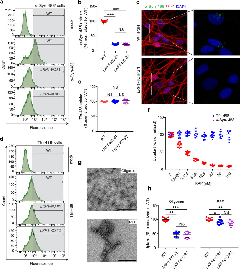
“However, unlike tau fibrils [29], LRP1-KO only had a mild inhibitory effect on the uptake of α-Syn PFFs (~ 5–10% reduction) (Fig. 2h).”
§ LRP1 is a neuronal receptor for α-synuclein uptake and spread > Results > LRP1 regulates α-Syn uptake in iPSNs
What the sources say
- Supportedcites [29] Rauch JN, Luna G, Guzman E, Audouard M, Challis C, Sibih YE, Leshuk C, Hernandez I, Wegmann S, Hyman BT, et al. LRP1 is a master regulator of tau uptake and spread. Nature. 2020;580:381–5. source
The citation is attached to the comparison with tau fibrils, not to the citing paper’s α-Syn PFF result in Fig. 2h. The source’s Results (Fig. 1c; page not available) report that LRP1 knockdown reduced uptake of sonicated tau fibrils, though it did not completely inhibit it. (LLM, conf=0.90)
plaindirect- Asserted: passes — The authors put it forward as true.
- Checkable: passes — It can be checked against evidence.
- Load-bearing: passes — The paper’s conclusions depend on it.
- Based: passes — The paper gives a reason why it holds.
- Supported
A result the paper reports from its own data — it cites no other source for it, so there is none to check. The quote shows it is in the paper.
In the paper
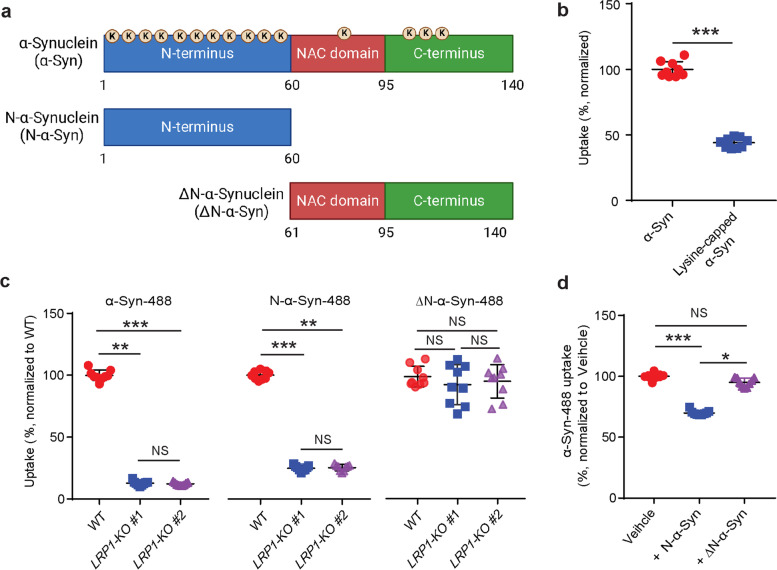
“We found that the blocking of lysine residues on α-Syn effectively decreased the uptake of α-Syn in the iPSNs (~ 50% reduction) (Fig. 3b)”
§ LRP1 is a neuronal receptor for α-synuclein uptake and spread > Results > LRP1 regulates the uptake of α-Syn via lysine residues in the N-terminus of α-Syn
plaindirect- Asserted: passes — The authors put it forward as true.
- Checkable: passes — It can be checked against evidence.
- Load-bearing: passes — The paper’s conclusions depend on it.
- Based: passes — The paper gives a reason why it holds.
- Rests on an assumption: Capping lysine residues on α-Syn with sulfo-NHS acetate specifically blocks lysine residues without otherwise altering α-Syn uptake.
A result the paper reports from its own data — it cites no other source for it, so there is none to check. The quote shows it is in the paper.
In the paper
“The blocking of lysine residues on α-Syn effectively decreased the uptake of α-Syn in iPSNs and the N-terminus of α-Syn was critical for LRP1-mediated α-Syn uptake.”
§ LRP1 is a neuronal receptor for α-synuclein uptake and spread > Abstract > Results
plaindirect- Asserted: passes — The authors put it forward as true.
- Checkable: passes — It can be checked against evidence.
- Load-bearing: passes — The paper’s conclusions depend on it.
- Based: passes — The paper gives a reason why it holds.
- Rests on an assumption: α-Syn has a similar high content (10%) of lysines as tau.
A result the paper reports from its own data — it cites no other source for it, so there is none to check. The quote shows it is in the paper.
In the paper
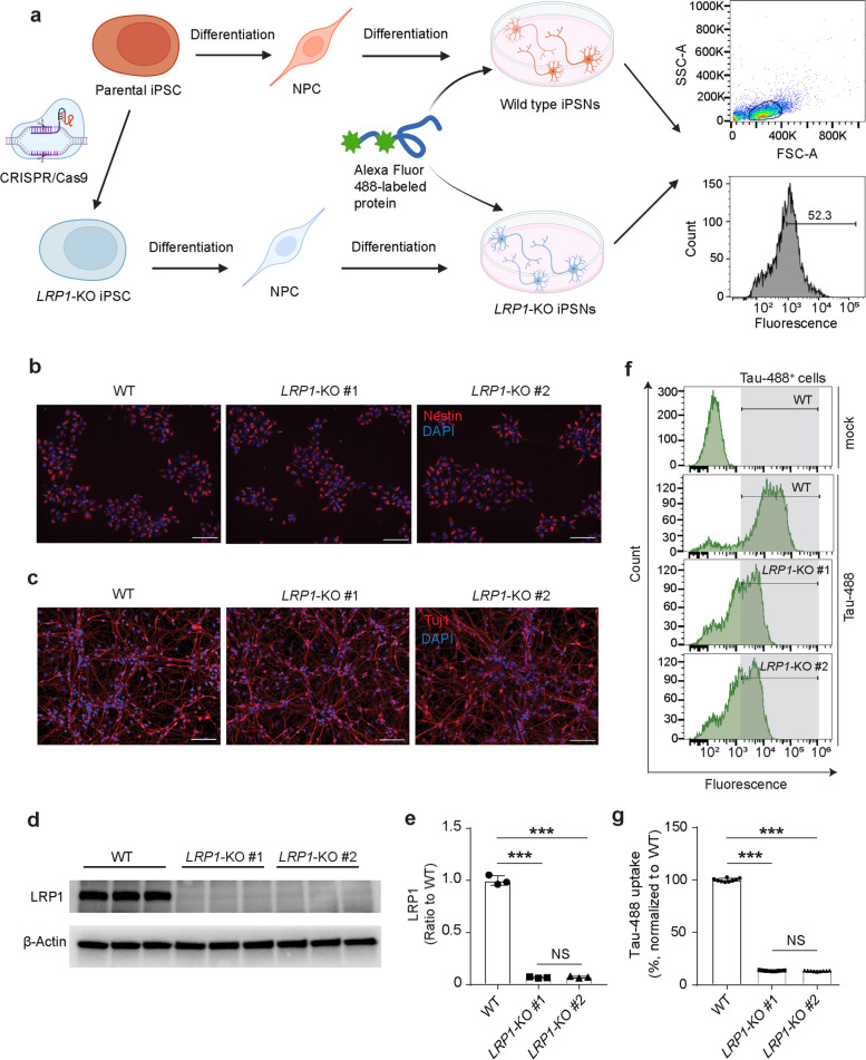
“We confirmed that the amount of internalized tau proteins was reduced by ~ 90% in LRP1-KO iPSNs compared to WT cells (Fig. 1f and g), consistent with what has been reported previously.”
§ LRP1 is a neuronal receptor for α-synuclein uptake and spread > Results > Generation and characterization of the LRP1-KO iPSNs
plaindirect- Asserted: passes — The authors put it forward as true.
- Checkable: passes — It can be checked against evidence.
- Load-bearing: passes — The paper’s conclusions depend on it.
- Based: passes — The paper gives a reason why it holds.
A result the paper reports from its own data — it cites no other source for it, so there is none to check. The quote shows it is in the paper.
In the paper
“We found that LRP1-deficiency significantly reduced neuronal α-Syn and tau uptake using LRP1-KO iPSNs generated by the CRISPR/Cas9 technique.”
§ LRP1 is a neuronal receptor for α-synuclein uptake and spread > Discussion
plaindirect- Asserted: passes — The authors put it forward as true.
- Checkable: passes — It can be checked against evidence.
- Load-bearing: passes — The paper’s conclusions depend on it.
- Based: passes — The paper gives a reason why it holds.
A result the paper reports from its own data — it cites no other source for it, so there is none to check. The quote shows it is in the paper.
In the paper
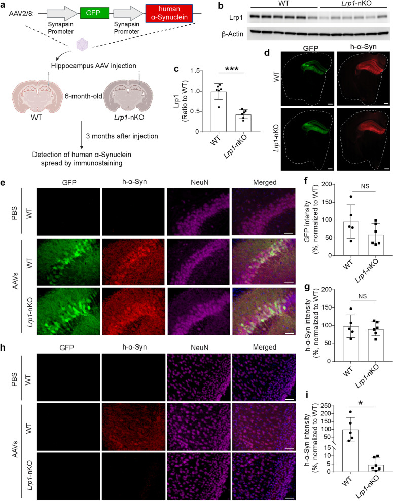
“Finally, in the Lrp1-nKO mice, the spread of α-Syn was significantly reduced compared with the WT littermates.”
- Also stated in: § Background · plain
“We found that the spreading of α-Syn was reduced in mouse brain with neuronal Lrp1 deletion.”
§ LRP1 is a neuronal receptor for α-synuclein uptake and spread > Abstract > Results
plaindirect- Asserted: passes — The authors put it forward as true.
- Checkable: passes — It can be checked against evidence.
- Load-bearing: passes — The paper’s conclusions depend on it.
- Based: passes — The paper gives a reason why it holds.
- Also stated in: § Background · plain
A result the paper reports from its own data — it cites no other source for it, so there is none to check. The quote shows it is in the paper.
In the paper
“we found neurons that had only h-α-Syn but not GFP signals”
§ LRP1 is a neuronal receptor for α-synuclein uptake and spread > Results > Neuronal Lrp1 deletion reduces α-Syn spread in mouse model
plain- Asserted: passes — The authors put it forward as true.
- Checkable: passes — It can be checked against evidence.
- Load-bearing: passes — The paper’s conclusions depend on it.
- Based: passes — The paper gives a reason why it holds.
A result the paper reports from its own data — it cites no other source for it, so there is none to check. The quote shows it is in the paper.
In the paper
“We further confirmed that the α-Syn N-terminal domain interacted with LRP1 through lysine residues.”
§ LRP1 is a neuronal receptor for α-synuclein uptake and spread > Background
boosteddirect- Asserted: passes — The authors put it forward as true.
- Checkable: passes — It can be checked against evidence.
- Load-bearing: unclear — It is unclear whether the paper’s conclusions depend on it.
- Based: passes — The paper gives a reason why it holds.
- Rests on an assumption: α-Syn has a similar high content (10%) of lysines as tau.
- Probably not a substantial claim. 9
- It may state two separate claims as one. 13
The cited sources support only part of this claim — read each source’s verdict below.
In the paper
“Particularly, the oligomeric α-Syn species appear to be prone to uptake and spread from cell-to-cell in a prion-like manner, leading to the templated misfolding of the native forms of α-Syn and formation of α-Syn aggregates throughout the brain, which are the defining pathological features of Parkinson’s disease (PD) and dementia with Lewy body (DLB) [12–19].”
§ LRP1 is a neuronal receptor for α-synuclein uptake and spread > Background
What the sources say
- Partly supportedcites [12] Brundin P, Melki R, Kopito R. Prion-like transmission of protein aggregates in neurodegenerative diseases. Nat Rev Mol Cell Biol. 2010;11:301–7. sourceExperimental — accuracy not yet measured
In “Grafted cells develop Lewy bodies” (page unavailable), the review reports uptake of α-synuclein oligomers in cultured neurons and seeding of endogenous α-synuclein aggregates. It also suggests that non-native α-synuclein can spread between cells and affect progressively greater parts of the central nervous system in Parkinson’s disease. It does not establish that oligomers specifically drive spread throughout the brain, or address dementia with Lewy bodies as the citing sentence does. (LLM, conf=0.84)
- Not checked — no full text of the sourcecites [13] Angot E, Steiner JA, Hansen C, Li JY, Brundin P. Are synucleinopathies prion-like disorders? The Lancet Neurology. 2010;9:1128–38. source
- Partly supportedcites [14] Surmeier DJ, Obeso JA, Halliday GM. Selective neuronal vulnerability in Parkinson disease. Nat Rev Neurosci. 2017;18:101–13. sourceExperimental — accuracy not yet measured
In “Does LP retrogradely spread in cPD?” (page not specified), the paper reports oligomeric α-syn spreading through the brainstem in a viral-expression model. It attributes uptake, recruitment of endogenous α-syn into aggregates, and proposed prion-like templating specifically to fibrils, however. It does not establish that oligomers drive that full sequence or produce aggregates throughout the brain. (LLM, conf=0.88)
- Partly supportedcites [15] Danzer KM, Haasen D, Karow AR, Moussaud S, Habeck M, Giese A, Kretzschmar H, Hengerer B, Kostka M. Different species of alpha-synuclein oligomers induce calcium influx and seeding. J Neurosci. 2007;27:9220–32. sourceExperimental — accuracy not yet measured
The Discussion reports that oligomer types B and C entered cells and seeded intracellular α-synuclein aggregation. It suggests that seeding could contribute to the progression of pathology in Parkinson’s brains, but the study does not demonstrate cell-to-cell spread or aggregate formation throughout the brain. (LLM, conf=0.86)
- Not checked — no full text of the sourcecites [16] Danzer KM, Krebs SK, Wolff M, Birk G, Hengerer B. Seeding induced by alpha-synuclein oligomers provides evidence for spreading of alpha-synuclein pathology. J Neurochem. 2009;111:192–203. source
- Partly supportedcites [17] Luk KC, Song C, O’Brien P, Stieber A, Branch JR, Brunden KR, Trojanowski JQ, Lee VM. Exogenous alpha-synuclein fibrils seed the formation of Lewy body-like intracellular inclusions in cultured cells. Proc Natl Acad Sci U S A. 2009;106:20051–6. sourceExperimental — accuracy not yet measured
The paper’s opening section reports that introduced α-Syn fibrils seeded conversion of endogenous α-Syn into intracellular inclusions in cultured cells. But it says oligomeric α-Syn had little effect, and the Discussion says α-Syn fibrils were not efficiently taken up without a transduction reagent. It does not show cell-to-cell spread or aggregation throughout the brain. (LLM, conf=0.88)
- Partly supportedcites [18] Hansen C, Angot E, Bergstrom AL, Steiner JA, Pieri L, Paul G, Outeiro TF, Melki R, Kallunki P, Fog K, et al. alpha-Synuclein propagates from mouse brain to grafted dopaminergic neurons and seeds aggregation in cultured human cells. J Clin Invest. 2011;121:715–25. sourceExperimental — accuracy not yet measured
The Discussion reports cell-to-cell α-syn transfer and seeding in the study’s models, and describes templated spread through progressively greater parts of the PD nervous system as a hypothesis. It does not establish that oligomers are particularly prone to uptake: the Discussion says monomeric, oligomeric, and fibrillar forms can all be internalized. Nor does it demonstrate aggregate formation throughout the brain. (LLM, conf=0.85)
- Partly supportedcites [19] Koga S, Sekiya H, Kondru N, Ross OA, Dickson DW. Neuropathology and molecular diagnosis of Synucleinopathies. Mol Neurodegener. 2021;16:83. sourceExperimental — accuracy not yet measured
Section 5.1 reports that α-synuclein can seed aggregation in cultured cells and mice, while section 4.6 describes evidence for transport of inoculated α-synuclein fibrils along the vagus nerve. The paper does not establish that oligomeric species in particular are prone to cellular uptake and cell-to-cell spread, or that this mechanism produces aggregates throughout the brain in PD and DLB. (LLM, conf=0.85)
hedgeddirect- Asserted: passes — The authors put it forward as true.
- Checkable: passes — It can be checked against evidence.
- Load-bearing: passes — The paper’s conclusions depend on it.
- Based: passes — The paper gives a reason why it holds.
Probably not a substantial claim. It may state two separate claims as one.
- Partly supported
The cited sources support only part of this claim — read each source’s verdict below.
In the paper
“LRP1 mediates and regulates the endocytosis of > 30 ligands, including apolipoprotein E (APOE) and amyloid-β (Aβ) [21–27]”
§ LRP1 is a neuronal receptor for α-synuclein uptake and spread > Background
What the sources say
- Partly supportedcites [21] Kanekiyo T, Bu G. The low-density lipoprotein receptor-related protein 1 and amyloid-β clearance in Alzheimer’s disease. Frontiers in aging neuroscience. 2014;6:93. sourceExperimental — accuracy not yet measured
The Introduction and the Structural and Functional Features section describe LRP1 as an endocytic receptor for diverse ligands. The Summary and Perspective identifies apoE as a major LRP1 ligand, and the Mechanisms of LRP1-Regulated Cellular Aβ Uptake section describes LRP1-mediated Aβ endocytosis. Our copy does not give the claimed count of more than 30 ligands. (LLM, conf=0.86)
- Partly supportedcites [22] Kanekiyo T, Cirrito JR, Liu CC, Shinohara M, Li J, Schuler DR, Shinohara M, Holtzman DM, Bu G. Neuronal clearance of amyloid-β by endocytic receptor LRP1. J Neurosci. 2013;33:19276–83. sourceExperimental — accuracy not yet measured
The Introduction (page unavailable) lists apolipoprotein E among ligands taken up through LRP1, and the Results section reports LRP1-mediated Aβ uptake in neuronal cells. Our copy does not give the claimed count of more than 30 ligands. (LLM, conf=0.87)
- Partly supportedcites [23] Yamazaki Y, Zhao N, Caulfield TR, Liu CC, Bu G. Apolipoprotein E and Alzheimer disease: pathobiology and targeting strategies. Nat Rev Neurol. 2019;15:501–18. sourceExperimental — accuracy not yet measured
The Biology of APOE section identifies LRP1 as an APOE receptor, and the Aβ clearance section describes LRP1-dependent cellular uptake of Aβ. Our copy does not support the specific claim that LRP1 mediates endocytosis of more than 30 ligands (searched: “LRP1 30 ligands” and “LRP1 more than 30 ligands endocytosis”). Page numbers were not available in the indexed passages. (LLM, conf=0.86)
- Partly supportedcites [24] Zhao N, Liu CC, Qiao W, Bu G. Apolipoprotein E, receptors, and modulation of Alzheimer’s disease. Biol Psychiatry. 2018;83:347–57. sourceExperimental — accuracy not yet measured
The ‘ApoE receptors’ section identifies LRP1 as an apoE-binding receptor, and ‘LRP1 in Aβ clearance’ reports that LRP1 affects neuronal Aβ uptake and trafficking. Our copy does not state that LRP1 mediates endocytosis of more than 30 ligands, so it does not support the claimed number. (LLM, conf=0.85)
- Supportedcites [25] Bu G. Apolipoprotein E and its receptors in Alzheimer’s disease: pathways, pathogenesis and therapy. Nat Rev Neurosci. 2009;10:333–44. source
In “ApoE receptors, members of the LDLR family” (page not provided), the paper says LRP1 binds more than 30 ligands, including apoE, and transports ligands by receptor-mediated endocytosis. In “ApoE and apoE receptors in Aβ clearance and aggregation” (page not provided), it describes LRP1-mediated Aβ clearance and says these receptors can bind Aβ directly or through chaperones. (LLM, conf=0.91)
- Partly supportedcites [26] Kanekiyo T, Bu G. The low-density lipoprotein receptor-related protein 1 and amyloid-beta clearance in Alzheimer’s disease. Front Aging Neurosci. 2014;6:93. sourceExperimental — accuracy not yet measured
The Introduction describes LRP1 as regulating endocytosis of diverse ligands, and the Summary and Perspective identifies apoE as a major LRP1 ligand. The section on mechanisms of cellular Aβ uptake says LRP1 regulates Aβ endocytosis. Our copy does not give the claimed count of more than 30 ligands. (LLM, conf=0.86)
- Not checked yetcites [27] Robert J, Button EB, Martin EM, McAlary L, Gidden Z, Gilmour M, Boyce G, Caffrey TM, Agbay A, Clark A, et al. Cerebrovascular amyloid Angiopathy in bioengineered vessels is reduced by high-density lipoprotein particles enriched in Apolipoprotein E. Mol Neurodegener. 2020;15:23. source
Checked before — its earlier verdict is held back until a new check runs.
plaindirect- Asserted: passes — The authors put it forward as true.
- Checkable: passes — It can be checked against evidence.
- Load-bearing: passes — The paper’s conclusions depend on it.
- Based: passes — The paper gives a reason why it holds.
Probably not a substantial claim.
- Partly supported
A result the paper reports from its own data — it cites no other source for it, so there is none to check. The quote shows it is in the paper.
In the paper
“We found that, similar to tau, LRP1-deficiency significantly reduced the cellular uptake of α-Syn (more than 75% reduction) in the iPSNs and consistent findings were observed from two different clones of LRP1-KO iPSNs (Fig. 2a, b, and c).”
§ LRP1 is a neuronal receptor for α-synuclein uptake and spread > Results > LRP1 regulates α-Syn uptake in iPSNs
plaindirect- Asserted: passes — The authors put it forward as true.
- Checkable: passes — It can be checked against evidence.
- Load-bearing: passes — The paper’s conclusions depend on it.
- Based: passes — The paper gives a reason why it holds.
It may state two separate claims as one.
A result the paper reports from its own data — it cites no other source for it, so there is none to check. The quote shows it is in the paper.
In the paper
“We found that RAP strongly suppressed the neuronal uptake of α-Syn in a dose-dependent manner, whereas transferrin internalization was not affected (Fig. 2f).”
§ LRP1 is a neuronal receptor for α-synuclein uptake and spread > Results > LRP1 regulates α-Syn uptake in iPSNs
boosteddirect- Asserted: passes — The authors put it forward as true.
- Checkable: passes — It can be checked against evidence.
- Load-bearing: passes — The paper’s conclusions depend on it.
- Based: passes — The paper gives a reason why it holds.
It may state two separate claims as one.
A result the paper reports from its own data — it cites no other source for it, so there is none to check. The quote shows it is in the paper.
In the paper
“we found that the addition of N-α-Syn significantly inhibited the uptake of full-length α-Syn, whereas ΔN-α-Syn had no effect (Fig. 3d).”
§ LRP1 is a neuronal receptor for α-synuclein uptake and spread > Results > LRP1 regulates the uptake of α-Syn via lysine residues in the N-terminus of α-Syn
plaindirect- Asserted: passes — The authors put it forward as true.
- Checkable: passes — It can be checked against evidence.
- Load-bearing: passes — The paper’s conclusions depend on it.
- Based: passes — The paper gives a reason why it holds.
It may state two separate claims as one.
- Rests on an assumption: The N-α-Syn and ΔN-α-Syn fragments are properly folded and their uptake reflects the role of the N-terminus.
A result the paper reports from its own data — it cites no other source for it, so there is none to check. The quote shows it is in the paper.
In the paper
“We found that LRP1-KO effectively inhibited the uptake of N-α-Syn, to a similar extent as the full-length α-Syn but had no effect on ΔN-α-Syn uptake (Fig. 3c).”
§ LRP1 is a neuronal receptor for α-synuclein uptake and spread > Results > LRP1 regulates the uptake of α-Syn via lysine residues in the N-terminus of α-Syn
plaindirect- Asserted: passes — The authors put it forward as true.
- Checkable: passes — It can be checked against evidence.
- Load-bearing: passes — The paper’s conclusions depend on it.
- Based: passes — The paper gives a reason why it holds.
It may state two separate claims as one.
- Rests on an assumption: The N-α-Syn and ΔN-α-Syn fragments are properly folded and their uptake reflects the role of the N-terminus.
What the paper did — it cites no source for this, so there is none to check. The quote shows it is in the paper.
In the paper
“After 14 days of culture, both WT and LRP1-KO cells exhibited a typical neuronal morphology and expressed the neuronal marker (Tuj1), suggesting the successful differentiation of NPCs into neurons (iPSNs) (Fig. 1c).”
§ LRP1 is a neuronal receptor for α-synuclein uptake and spread > Results > Generation and characterization of the LRP1-KO iPSNs
hedged- Asserted: passes — The authors put it forward as true.
- Checkable: passes — It can be checked against evidence.
- Load-bearing: passes — The paper’s conclusions depend on it.
- Based: passes — The paper gives a reason why it holds.
It may state two separate claims as one.
A conclusion the paper draws from its own results — it cites no other source for it, so there is none to check. The quote shows it is in the paper.
In the paper
“Together, these data indicate that our strategy of deleting LRP1 in human iPSNs was successful and tau uptake was inhibited by LRP1 deletion in these cells.”
§ LRP1 is a neuronal receptor for α-synuclein uptake and spread > Results > Generation and characterization of the LRP1-KO iPSNs
hedgeddirect- Asserted: passes — The authors put it forward as true.
- Checkable: passes — It can be checked against evidence.
- Load-bearing: passes — The paper’s conclusions depend on it.
- Based: passes — The paper gives a reason why it holds.
It may state two separate claims as one.
In the paper
“A recent study also identified LRP1 as a key regulator for the uptake and spread of microtubule-associated protein tau [29].”
§ LRP1 is a neuronal receptor for α-synuclein uptake and spread > Background
What the sources say
- Not checked yetcites [29] Rauch JN, Luna G, Guzman E, Audouard M, Challis C, Sibih YE, Leshuk C, Hernandez I, Wegmann S, Hyman BT, et al. LRP1 is a master regulator of tau uptake and spread. Nature. 2020;580:381–5. source
plaindirect- Asserted: passes — The authors put it forward as true.
- Checkable: passes — It can be checked against evidence.
- Load-bearing: passes — The paper’s conclusions depend on it.
- Based: passes — The paper gives a reason why it holds.
Probably not a substantial claim.
In the paper
“The low-density lipoprotein receptor (LDLR)-related protein 1 (LRP1) is a transmembrane receptor that belongs to the LDLR family [20].”
§ LRP1 is a neuronal receptor for α-synuclein uptake and spread > Background
What the sources say
- Not checked — no full text of the sourcecites [20] Herz J, Bock HH. Lipoprotein receptors in the nervous system. Annu Rev Biochem. 2002;71:405–34. source
plain- Asserted: passes — The authors put it forward as true.
- Checkable: passes — It can be checked against evidence.
- Load-bearing: passes — The paper’s conclusions depend on it.
- Based: passes — The paper gives a reason why it holds.
Probably not a substantial claim. It may state two separate claims as one.
A result the paper reports from its own data — it cites no other source for it, so there is none to check. The quote shows it is in the paper.
In the paper
“α-Syn has a high lysine content (15 lysines out of 140 aa, ~ 10%) with most (eleven) lysines located in the N-terminus, one lysine in NAC, and three in C-terminus (Fig. 3a).”
§ LRP1 is a neuronal receptor for α-synuclein uptake and spread > Results > LRP1 regulates the uptake of α-Syn via lysine residues in the N-terminus of α-Syn
plain- Asserted: passes — The authors put it forward as true.
- Checkable: passes — It can be checked against evidence.
- Load-bearing: passes — The paper’s conclusions depend on it.
- Based: passes — The paper gives a reason why it holds.
It may state two separate claims as one.
What the paper did — it cites no source for this, so there is none to check. The quote shows it is in the paper.
In the paper
“we confirmed that all the NPCs from the three lines were positive for neural precursor marker (Nestin) by immunostaining (Fig. 1b).”
§ LRP1 is a neuronal receptor for α-synuclein uptake and spread > Results > Generation and characterization of the LRP1-KO iPSNs
plain- Asserted: passes — The authors put it forward as true.
- Checkable: passes — It can be checked against evidence.
- Load-bearing: fails — The paper’s conclusions may not depend on it.
- Based: passes — The paper gives a reason why it holds.
Probably not a substantial claim. The paper’s conclusions may not depend on it.
None of the sources cited for this claim have been checked yet.
In the paper
“Notably, LAG3 specifically binds to α-Syn PFFs but not monomers.”
§ LRP1 is a neuronal receptor for α-synuclein uptake and spread > Discussion
What the sources say
- Not checked yetcites [41] Mao X, Ou MT, Karuppagounder SS, Kam TI, Yin X, Xiong Y, Ge P, Umanah GE, Brahmachari S, Shin JH, et al. Pathological alpha-synuclein transmission initiated by binding lymphocyte-activation gene 3. Science. 2016;353:aah3374. source
Checked before — its earlier verdict is held back until a new check runs.
plaincorrelational- Asserted: passes — The authors put it forward as true.
- Checkable: passes — It can be checked against evidence.
- Load-bearing: fails — The paper’s conclusions may not depend on it.
- Based: passes — The paper gives a reason why it holds.
Probably not a substantial claim. The paper’s conclusions may not depend on it.
- Not checked yet
What the paper did — it cites no source for this, so there is none to check. The quote shows it is in the paper.
In the paper
“Within the hippocampus, both GFP and h-α-Syn signal showed no significant differences between Lrp1-nKO and WT mice, indicating equivalent transduction of AAVs between the two groups (Fig. 4e, f and g).”
§ LRP1 is a neuronal receptor for α-synuclein uptake and spread > Results > Neuronal Lrp1 deletion reduces α-Syn spread in mouse model
hedged- Asserted: passes — The authors put it forward as true.
- Checkable: passes — It can be checked against evidence.
- Load-bearing: passes — The paper’s conclusions depend on it.
- Based: passes — The paper gives a reason why it holds.
It may state two separate claims as one.
What the paper did — it cites no source for this, so there is none to check. The quote shows it is in the paper.
In the paper
“Karyotyping in each iPSC clones revealed the preservation of normal number and appearance of chromosomes (Fig. S2a).”
§ LRP1 is a neuronal receptor for α-synuclein uptake and spread > Results > Generation and characterization of the LRP1-KO iPSNs
plain- Asserted: passes — The authors put it forward as true.
- Checkable: passes — It can be checked against evidence.
- Load-bearing: fails — The paper’s conclusions may not depend on it.
- Based: passes — The paper gives a reason why it holds.
Probably not a substantial claim. The paper’s conclusions may not depend on it.
None of the sources cited for this claim have been checked yet.
In the paper
“A recent study showed that the lymphocyte-activation gene 3 (LAG3) is a receptor for α-Syn PFFs which mediates the endocytosis and transmission of α-Syn PFFs between neurons [41].”
§ LRP1 is a neuronal receptor for α-synuclein uptake and spread > Discussion
What the sources say
- Not checked yetcites [41] Mao X, Ou MT, Karuppagounder SS, Kam TI, Yin X, Xiong Y, Ge P, Umanah GE, Brahmachari S, Shin JH, et al. Pathological alpha-synuclein transmission initiated by binding lymphocyte-activation gene 3. Science. 2016;353:aah3374. source
Checked before — its earlier verdict is held back until a new check runs.
plaindirect- Asserted: passes — The authors put it forward as true.
- Checkable: passes — It can be checked against evidence.
- Load-bearing: fails — The paper’s conclusions may not depend on it.
- Based: passes — The paper gives a reason why it holds.
Probably not a substantial claim. The paper’s conclusions may not depend on it. It may state two separate claims as one.
- Not checked yet
A conclusion the paper draws from its own results — it cites no other source for it, so there is none to check. The quote shows it is in the paper.
In the paper
“The finding may suggest that when forming the fibrils, the N-terminus of α-Syn is less exposed therefore the interaction between α-Syn and LRP1 might be limited.”
§ LRP1 is a neuronal receptor for α-synuclein uptake and spread > Discussion
hedgedconditional- Asserted: passes — The authors put it forward as true.
- Checkable: passes — It can be checked against evidence.
- Load-bearing: passes — The paper’s conclusions depend on it.
- Based: passes — The paper gives a reason why it holds.
It may state two separate claims as one.
None of the sources cited for this claim have been checked yet.
In the paper
“Monomeric α-Syn is composed of three domains: a lipid binding N-terminus domain (aa 1–60), a hydrophobic non-amyloid component (NAC) domain (aa 61–95), and a negatively charged C-terminus domain (aa 96–140) [2].”
§ LRP1 is a neuronal receptor for α-synuclein uptake and spread > Results > LRP1 regulates the uptake of α-Syn via lysine residues in the N-terminus of α-Syn
What the sources say
- Not checked yetcites [2] Meade RM, Fairlie DP, Mason JM. Alpha-synuclein structure and Parkinson’s disease - lessons and emerging principles. Mol Neurodegener. 2019;14:29. source
Checked before — its earlier verdict is held back until a new check runs.
plain- Asserted: passes — The authors put it forward as true.
- Checkable: passes — It can be checked against evidence.
- Load-bearing: fails — The paper’s conclusions may not depend on it.
- Based: passes — The paper gives a reason why it holds.
Probably not a substantial claim. The paper’s conclusions may not depend on it.
- Not checked yet
A conclusion the paper draws from its own results — it cites no other source for it, so there is none to check. The quote shows it is in the paper.
In the paper
“Our findings support the hypothesis that LRP1 is a key regulator for both α-Syn and tau uptake and spread”
§ LRP1 is a neuronal receptor for α-synuclein uptake and spread > Discussion
plaindirect- Asserted: passes — The authors put it forward as true.
- Checkable: passes — It can be checked against evidence.
- Load-bearing: passes — The paper’s conclusions depend on it.
- Based: passes — The paper gives a reason why it holds.
It may state two separate claims as one.
The paper relies on this without support.
α-Syn has a similar high content (10%) of lysines as tau.
“Notably, α-Syn has a similar high content (10%) of lysines as tau.”
The paper relies on this without support.
The underlying cellular mechanisms of α-Syn uptake and spread remain poorly understood.
“However, the underlying cellular mechanisms of α-Syn uptake and spread remains poorly understood.”
The paper relies on this without support.
Secreted soluble α-Syn can be taken up by different cells.
“Importantly, these secreted soluble α-Syn can be taken up by different cells.”
The paper relies on this without support.
Capping lysine residues on α-Syn with sulfo-NHS acetate specifically blocks lysine residues without otherwise altering α-Syn uptake.
“we capped the amines of lysines on α-Syn with sulfo-NHS acetate and then measured the internalization of α-Syn in the iPSNs.”
The paper relies on this without support.
The N-α-Syn and ΔN-α-Syn fragments are properly folded and their uptake reflects the role of the N-terminus.
“We employed the α-Syn protein fragments containing only the N-terminus (N-α-Syn) or lacking the N-terminus (ΔN-α-Syn) of full-length α-Syn (Fig. 3a).”
The paper relies on this without support.
LRP1-nKO mice have no compensatory mechanisms that restore LRP1 function or alter α-Syn spread independently of LRP1 deletion.
“we used the conditional neuronal Lrp1 knockout mice [38] by crossing Lrp1flox/flox mice with CaMKII-Cre mice to generate Lrp1-nKO (Lrp1flox/flox; Cre+/−) and WT littermate controls (Lrp1flox/flox; Cre−/−).”
Self-check
Percentages, sums and reported differences re-computed from the paper’s own numbers.
Each contradiction between two passages listed here was confirmed by an independent check.
Rated minor: it would not touch a result — a value no result rests on, a p-value that stays on the same side of the significance level, or a conflict that is clerical, resolved elsewhere in the paper, or outside its own findings.
In the paper
§ LRP1 is a neuronal receptor for α-synuclein uptake and spread > Discussion
“in the disease-associated, aggregated state of α-Syn, the N-terminal residues (37 through 97), adopts a β-sheet structure”
§ LRP1 is a neuronal receptor for α-synuclein uptake and spread > Results > LRP1 regulates the uptake of α-Syn via lysine residues in the N-terminus of α-Syn
“a lipid binding N-terminus domain (aa 1–60), a hydrophobic non-amyloid component (NAC) domain (aa 61–95), and a negatively charged C-terminus domain (aa 96–140)”
Citations
Cites
Common structure of soluble amyloid oligomers implies common mechanism of pathogenesis
Kayed, R., Head, E., Thompson, J. L., McIntire, T. M., Milton, S. C., Cotman, C. W., & Glabe, C. G. (2003) · Science · doi:10.1126/science.1079469 · Cited by 4,064
α-Synuclein Promotes SNARE-Complex Assembly in Vivo and in Vitro
Burré, J., Sharma, M., Tsetsenis, T., Buchman, V. L., Etherton, M. R., & Südhof, T. C. (2010) · Science · doi:10.1126/science.1195227 · Cited by 1,907
The many faces of α-synuclein: from structure and toxicity to therapeutic target
Lashuel, H. A., Overk, C., Oueslati, A., & Masliah, E. (2012) · Nature reviews. Neuroscience · doi:10.1038/nrn3406 · Cited by 1,684
Apolipoprotein E and Alzheimer disease: pathobiology and targeting strategies
Yamazaki, Y., Zhao, N., Caulfield, T. R., Liu, C.-C., & Bu, G. (2019) · Nature Reviews Neurology · doi:10.1038/s41582-019-0228-7 · Cited by 1,425
Self-citationApolipoprotein E and its receptors in Alzheimer's disease: pathways, pathogenesis and therapy
Bu, G. (2009) · Nature Reviews Neuroscience · doi:10.1038/nrn2620 · Cited by 1,168
Self-citationSelective neuronal vulnerability in Parkinson disease
Surmeier, D. J., Obeso, J. A., & Halliday, G. M. (2017) · Nature Reviews Neuroscience · doi:10.1038/nrn.2016.178 · Cited by 1,116
Emmanouilidou, E., Melachroinou, K., Roumeliotis, T., Garbis, S. D., Ntzouni, M., Margaritis, L. H., Stefanis, L., & Vekrellis, K. (2010) · The Journal of Neuroscience · doi:10.1523/jneurosci.5699-09.2010 · Cited by 1,115
Nemani, V. M., Lü, W., Berge‐Seidl, V., Nakamura, K., Onoa, B., Lee, M. K., Chaudhry, F. A., Nicoll, R. A., & Edwards, R. H. (2010) · Neuron · doi:10.1016/j.neuron.2009.12.023 · Cited by 1,094
Luk, K. C., Song, C., O'Brien, P., Stieber, A., Branch, J. R., Brunden, K. R., Trojanowski, J. Q., & Lee, V. M.-Y. (2009) · Proceedings of the National Academy of Sciences · doi:10.1073/pnas.0908005106 · Cited by 938
Heparan sulfate proteoglycans mediate internalization and propagation of specific proteopathic seeds
Holmes, B. B., DeVos, S. L., Kfoury, N., Li, M., Jacks, R., Yanamandra, K., Ouidja, M. O., Brodsky, F. M., Marasa, J., Bagchi, D. P., et al. (2013) · Proceedings of the National Academy of Sciences · doi:10.1073/pnas.1301440110 · Cited by 859
Research works printed but not linked: 2
- Kanekiyo T, Bu G. The low-density lipoprotein receptor-related protein 1 and amyloid-beta clearance in Alzheimer’s disease. Front Aging Neurosci. 2014;6:93. · DOI · Paper page
- Jin Y, Li F, Sonoustoun B, Kondru NC, Martens YA, Qiao W, Heckman MG, Ikezu TC, Li Z, Burgess JD, et al. APOE4 exacerbates alpha-synuclein seeding activity and contributes to neurotoxicity in Alzheimer’s disease with Lewy body pathology. Acta Neuropathol. 2022;143:641. · DOI · Paper page
Cited by
Sorted by citations
Items 101–116 of 116
miR-338-5p exerts neuroprotective effects in Parkinson’s disease via targeting LRP1
Zhang, N., Cui, R., Hua, Y., & Lu, H. (2026) · Cytotechnology · doi:10.1007/s10616-026-00978-w
Qu, M., He, Y., Yu, L., Yang, H., Lu, Y., Li, Y., Wang, R., Sun, M., Ma, M. Y., & Mi, W. (2026) · CNS Neuroscience & Therapeutics · doi:10.1002/cns.70950
Self-citationPropagation of α-synuclein pathology: models, mechanisms and future goals
Zolin, A., Weber, S. A., Xie, Y. X., & Burré, J. (2026) · Molecular Neurodegeneration Advances · doi:10.1186/s44477-026-00038-9
Han, Y., & He, Z. (2026) · Molecular Neurodegeneration Advances · doi:10.1186/s44477-026-00037-w
Luo, P., Travis, Z. D., Lenahan, C., Wu, H., Zhang, J., Yu, J., & Xu, W. (2026) · International Journal of Biological Sciences · doi:10.7150/ijbs.127431
Öz, T., Fraile-Ramos, J., Kujawski, R., Wojciechowska, O., Karaźniewicz‐Łada, M., Mikołajczak, P. Ł., Giménez‐Llort, L., & Kujawska, M. (2026) · Molecular Biology Reports · doi:10.1007/s11033-026-12195-x
Khoo, L., Ng, K. Y., Chye, C. L., Ko, Y., Lee, J. Y., Koh, Y., Tsen, M. T., & Chye, S. M. (2026) · Metabolic Brain Disease · doi:10.1007/s11011-026-01911-y
Zhou, S., Chen, X., Ni, M., Chang, W., Huang, R., Wang, Y., Zhang, X., Zhang, H., Wu, H., & Sun, Y. (2026) · Phytomedicine · doi:10.1016/j.phymed.2026.158671
脳ペリサイトによるα-Synucleinクリアランスを基盤とした新規パーキンソン病治療法構築に関する研究
横谷, み. (n.d.) · Institutional Repositories DataBase (IRDB)
Jena, S., Subham, K., Kalra, H., Naik, S., Sahu, D. D., Mishra, M., & Jha, S. (2026) · International Journal of Biological Macromolecules · doi:10.1016/j.ijbiomac.2026.154365
Karpilovsky, N. C., Goiran, T., Senkevich, K. A., Deyab, G., Twells, N. M., Cha, E. N., MacDougall, E. J., Wang, Z., Sirois, J., MacLeod, G. D. A. H., et al. (2026) · medRxiv · doi:10.64898/2026.09.09.26362627
In the wisdom of Odin: modular architecture, receptor trafficking and therapeutic implications
Pennacchietti, V., Malagrinò, F., & Gianni, S. (2026) · Expert Opinion on Therapeutic Targets · doi:10.1080/14728222.2026.2738190
High-Throughput Screening Identifies Small-Molecule Inhibitors of the Tau-LRP1 Interaction
Wang, C., Ma, C.-T., Crotty, C., Zeng, F.-Y., Bobkov, A., Covel, J. A., Rivera, E. K., Sergienko, E., Kosik, K. S., Olson, S. H., et al. (2026) · ACS Chemical Biology · doi:10.1021/acschembio.6c00599
PODXL regulates the neuronal entry of α-synuclein aggregatesPreprint
Neupane, S., Šustić, L., Maraio, L., Bouris, V., Mariutti, G., Ribeiro, R., Yin, J.-A., Melki, R., Legname, G., Cecco, E. D., et al. (2026) · bioRxiv (Cold Spring Harbor Laboratory) · doi:10.64898/2026.10.15.759470
Brain clearance of protein aggregates in degenerative diseases: the role of microglia and astrocytes
Xiong, M., Huang, Y., Jiang, H., & Zhang, Z. (2026) · Translational Neurodegeneration · doi:10.1186/s40035-026-00592-y
Novel insights into the multifaceted and tissue‐specific roles of the endocytic receptor LRP1
Yamamoto, K., Scilabra, S. D., Bonelli, S., Jensen, A., Scavenius, C., Enghild, J. J., & Strickland, D. K. (2024) · Journal of Biological Chemistry · doi:10.1016/j.jbc.2024.107521 · Cited by 47
Metadata
- Authors
- Chen, K., Martens, Y. A., Meneses, A., Ryu, D. H., Lu, W., Raulin, A. C., Li, F., Zhao, J., Chen, Y., Jin, Y., Linares, C., Goodwin, M., Li, Y., Liu, C.-C., Kanekiyo, T., Holtzman, D. M., Golde, T. E., Bu, G., & Zhao, N.
- Affiliations
- Mayo Clinic (US)
- University of Florida (US)
- Washington University in St. Louis (US)
- Corresponding author
- Bu, Guojun
- Zhao, Na
- ORCID
- Venue
- Molecular Neurodegeneration
- Published
- 2022-09-02
- Volume
- 17
- Issue
- 1
- Pages
- 57-57
- ISSN
- 1750-1326 (ISSN-L)
- PMC
- PMC9438229
- OpenAlex
- W4294168387
- Cited by
- 115
- References
- 64
- Citations by year
- 2026: 31 · 2025: 40 · 2024: 26 · 2023: 15 · 2022: 3
- FWCI
- 11.65
- Type
- journal article
- Language
- en
- Open access
- gold
- doi.org
- Locations
- Molecular Neurodegeneration (publishedVersion, cc-by)
- PubMed Central (publishedVersion, cc-by)
- PubMed (publishedVersion)
- Digital Commons@Becker (Washington University School of Medicine) (submittedVersion, cc-by)
- DOAJ (DOAJ: Directory of Open Access Journals) (submittedVersion, cc-by-sa)
- Rare & Special e-Zone (The Hong Kong University of Science and Technology) (submittedVersion)
- Funders
- NIH (P01NS074969, U19AG069701)
- Mayo Alzheimer’s Disease Research Center Developmental Grant
- Lewy Body Dementia Center Without Walls (U54NS110435)
- Topics
- Health Sciences › Medicine › Clinical Neurology › Parkinson's Disease Mechanisms and Treatments
- Life Sciences › Neuroscience › Cellular and Molecular Neuroscience › Alzheimer's disease research and treatments
- Life Sciences › Neuroscience › Neurology › Neuroinflammation and Neurodegeneration Mechanisms
- Keywords
- alpha-synuclein, Parkinson's disease, dementia with Lewy bodies, protein uptake, prion-like spread, lysine residues, N-terminus, preformed fibrils, oligomers, lysosomal degradation, iPSC-derived neurons, neurodegeneration, tau spread, isogenic iPSC lines
- MeSH terms
- Animals, Humans, Infant, Parkinson Disease, Synapsins, tau Proteins, Low Density Lipoprotein Receptor-Related Protein-1, Mice, alpha-Synuclein, Induced Pluripotent Stem Cells
About these checks
Registry, retraction, Cites and statistics results are computed, not judged, with one exception: a printed reference that no index lists is read once by a model before it counts as a citation error. Claims are extracted by a model and kept only when their quote appears verbatim in the text; each cited source is then checked, with a model’s help, for whether it says what the claim relies on. Those verdicts can be wrong — read the claim’s quote and the cited source before relying on one.
A language model drew the claims from the paper on 2026-11-10, under version 3 of the claim definition (model deepseek-v4.1-flash, prompt extract-v3).
A second model labelled each claim on 2026-11-10 (model jev-1.13-20260917, definition revision gold-guideline-v0.2).
Cited sources are checked with a language model’s help (gpt-6-sol).
Statistics and arithmetic are recomputed, not judged.
Confidence bands are shown only for fields calibrated on our annotated test set. None is calibrated yet, so no band is shown.
What counts as a claim ›How we measure confidence ›How we check this ›
Are you an author, or do you see a problem with this page? Request a review
